1216
Free Breathing R2* Mapping Using Three-Dimensional Self-Gating Motion-Compensated Stack-of-Radial MRI1MR R&D Collaborations, Siemens Healthineers, Los Angeles, CA, United States, 2Department of Radiological Sciences, University of California Los Angeles, Los Angeles, CA, United States, 3Department of Physics and Biology in Medicine, University of California Los Angeles, Los Angeles, CA, United States, 4MR Application Predevelopment, Siemens Healthcare GmbH, Erlangen, Germany, 5MR R&D Collaborations, Siemens Healthineers, Baltimore, MD, United States, 6MR R&D Collaborations, Siemens Healthineers, Austin, TX, United States
Synopsis
R2* mapping is an emerging biomarker to evaluate iron overload. Due to its relative insensitivity to motion, free-breathing 3D stack-of-radial imaging is a useful technique in patient populations with breath-hold difficulties. However, its performance for R2* quantification is still under investigation. This study proposes a new 3D self-gating motion-compensated free-breathing stack-of-radial MRI method for R2* quantification. Preliminary in vivo results demonstrated good agreement of R2* mapping in the liver with the proposed method compared to the reference results of breath-hold Cartesian MRI.
INTRODUCTION
There is an emerging need to transition from qualitative to quantitative MRI to support disease characterization and machine learning algorithm based detection. Reliability of a quantitative biomarker is essential if the method is to be used clinically. While radial approaches have demonstrated substantial motion insensitivity for qualitative imaging (1,2), the impact on quantitative imaging is not completely known.
R2* mapping is an emerging biomarker to evaluate iron overload, and has been shown to correlate with liver iron concentration (3-5). Three-dimensional (3D) breath-hold multi-echo gradient-echo (GRE) imaging has been a well validated technique for liver R2* mapping in previous studies (5,6), but conventional methods using Cartesian sampling require breath-holding to avoid artifacts. In patient populations with breath-hold difficulties, free-breathing acquisition is desired. Free-breathing multi-echo stack-of-radial imaging after gradient delay error correction has been validated for liver fat quantification (7,8), but its performance for R2* quantification is still under investigation. In this study, we propose a new self-gating motion-compensated free-breathing stack-of-radial MRI method for R2* quantification and evaluate its performance in a preliminary study.
METHODS
Sequence Design
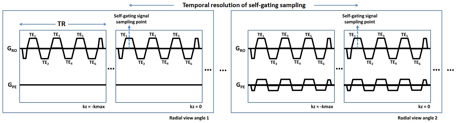
A free-breathing golden-angle-ordered multi-echo GRE stack-of-radial imaging sequence was developed to support gradient delay correction (7), self-gating and motion compensation (9,10) and fat/R2* quantification (11). The schematic diagram of the proposed sequence is illustrated in Figure 1, where the temporal footprint/resolution of the self-gating signal is determined by the number of acquired slices and the TR (Nslice x TR). The k-space origins (kx, ky and kz equivalent to zeroes) of the first echoes are used to extract the self-gating signal from different radial view angles.
In Vivo Liver MRI Experiments
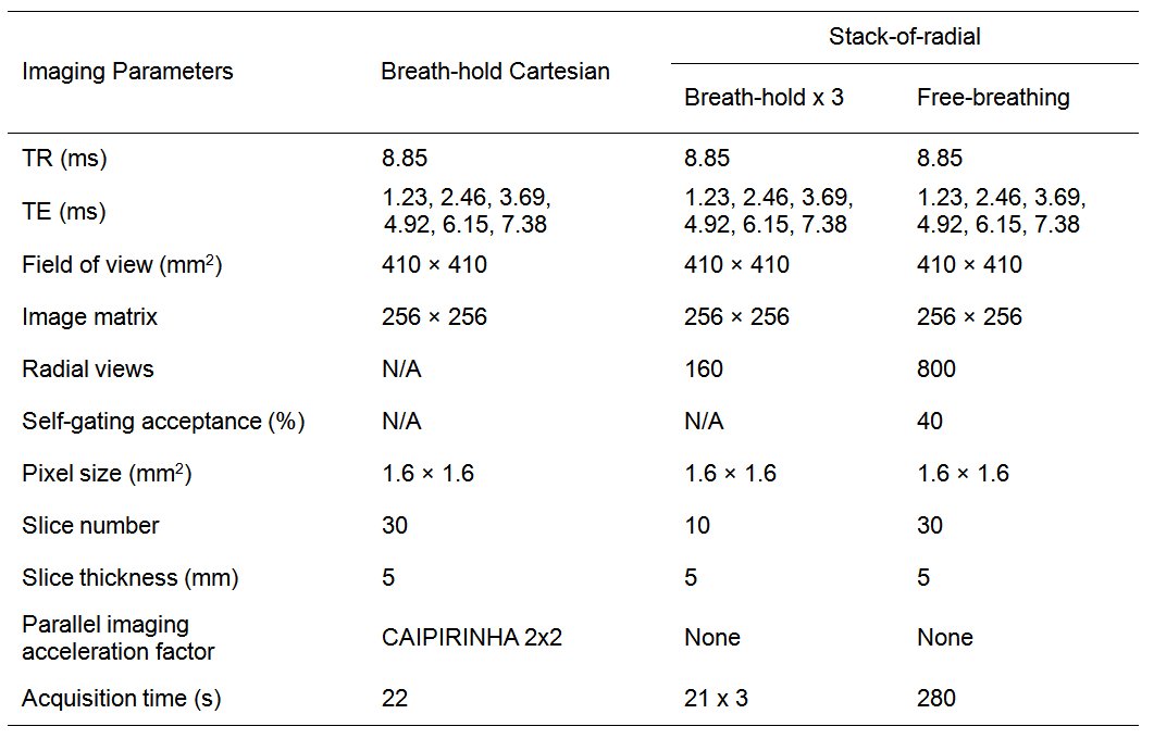
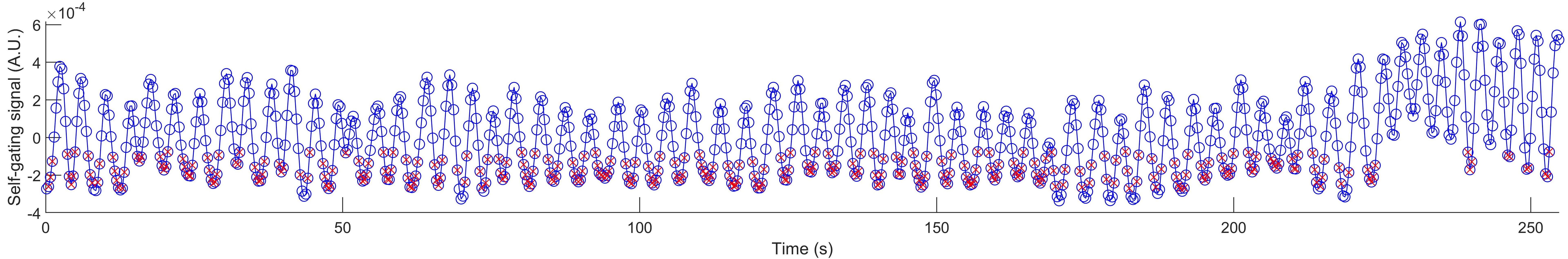
This study was HIPAA-compliant and approved by the local IRB. Written informed consent was obtained. Four subjects (all male, 36.3±3.6 yrs) were scanned at 3T (MAGNETOM Prisma, Siemens Healthcare, Erlangen, Germany). First, a single breath-hold multi-echo 3D Cartesian GRE sequence with Cartesian k-space sampling (11) was performed to cover the whole or majority of the liver volume including the liver dome. Along with the separated water and fat maps, the inline fat-corrected R2* map was generated as a reference standard (11). Second, the proposed stack-of-radial sequence was performed without self-gating correction to cover the whole liver in multiple breath-holds. Lastly, the proposed stack-of-radial sequence was performed for free-breathing acquisition to cover the whole or majority of the liver. The imaging parameters of all three sequences are listed in Table 1. Gradient delay correction was performed for all radial acquisitions (7). The self-gating signal was sampled at a temporal resolution of 318.6 ms, and was exported during scanning and plotted using Matlab (MathWorks, Natick, MA, United States).
Data Analysis
Matching slices near the liver dome and mid-liver were selected on R2* maps across four methods including breath-hold Cartesian, breath-hold stack-of-radial, free-breathing stack-of-radial without self-gating correction, and free-breathing stack-of-radial with self-gating correction. One region of interest (ROI) was placed in the liver to avoid vessels for each of these two slices in each subject, leading to 8 ROIs per method. Different stack-of-radial methods were compared to the Cartesian method using Bland-Altman analysis to determine the mean difference (MD) and limits of agreement (LoA = MD ± 1.96×standard deviation).
RESULTS AND DISCUSSION
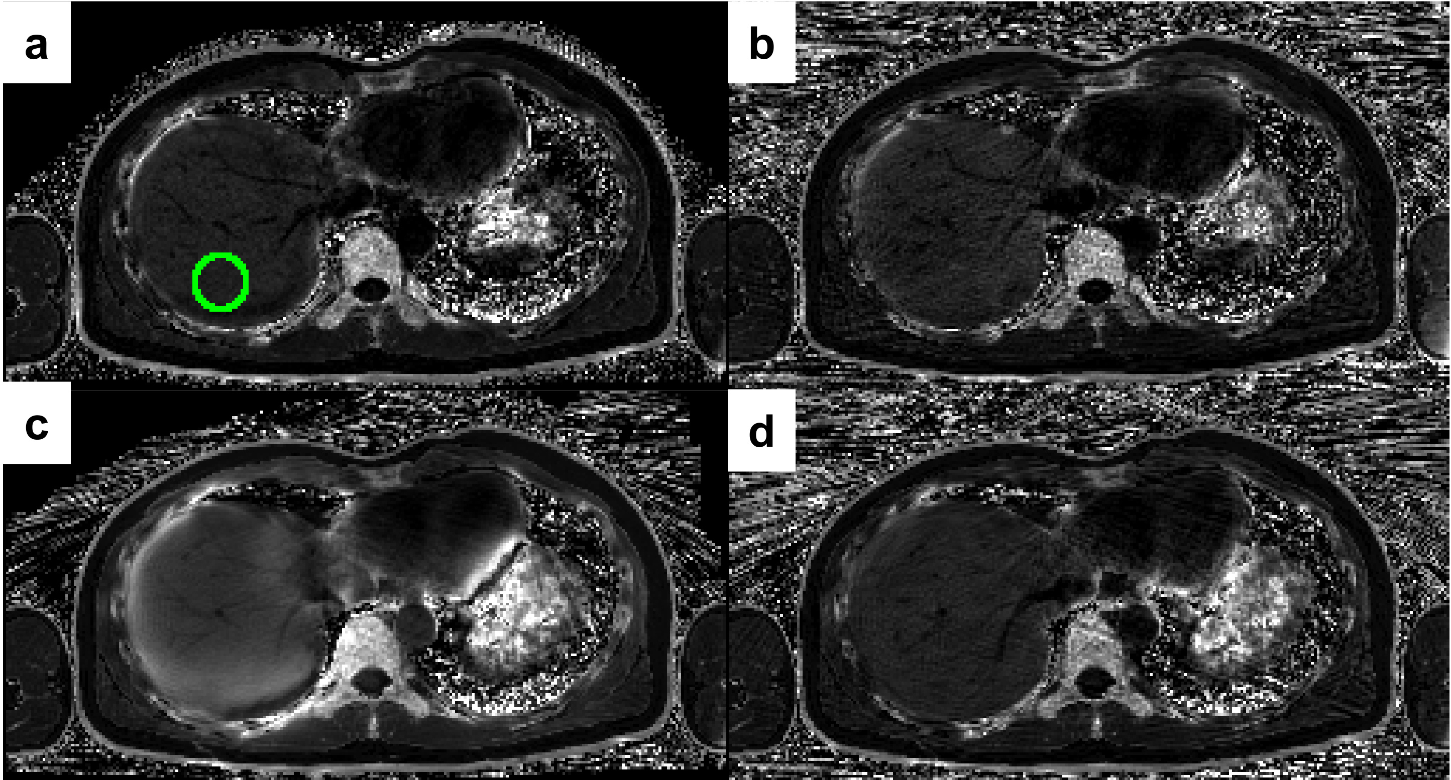
An example stack-of-radial self-gating signal curve and the selected points of data acquisition for image reconstruction are shown in Figure 2. Example R2* maps from one subject are shown in Figure 3. Free-breathing stack-of-radial without self-gating motion compensation showed elevated R2* values compared to the reference results of breath-hold Cartesian, while breath-hold radial and free-breathing radial with self-gating correction exhibit similar values, demonstrating that elevated R2* values without self-gating correction were caused by respiratory motion.
Bland-Altman analysis between the R2* maps of the breath-hold Cartesian reference and the other three stack-of-radial methods are plotted in Figure 4. One subject exhibited moderately high liver R2* values with all methods. The MD and LoA were substantially improved with free-breathing stack-of-radial with self-gating correction compared to free-breathing radial without self-gating correction. Specifically, free-breathing stack-of-radial with self-gating showed MD of 6.69 s-1, while free-breathing stack-of-radial without self-gating correction showed a high MD value of 58.48 s-1, both compared to breath-hold Cartesian.
CONCLUSION
A self-gating motion-compensated stack-of-radial imaging sequence was developed and evaluated in vivo. Preliminary results demonstrated agreement in R2* mapping in liver with the proposed method compared to the reference results of breath-hold Cartesian. Our study shows that while radial sampling may inherently provide sufficient respiratory motion insensitivity for qualitative imaging, adding motion compensation is essential for quantitative R2* mapping. This proposed method may allow free-breathing R2* mapping in patient populations with breath-hold difficulties.Acknowledgements
The authors greatly appreciate Xiaoming Bi, PhD, Jianing Pang, PhD, Robert Grimm, PhD and Berthold Kiefer, PhD for the inspiration of their related work and valuable discussion. This study was supported in part by Siemens Healthineers and the Department of Radiological Sciences at UCLA.References
1. Chandarana H, Block TK, Rosenkrantz AB, Lim RP, Kim D, Mossa DJ, Babb JS, Kiefer B, Lee VS. Free-breathing radial 3D fat-suppressed T1-weighted gradient echo sequence: a viable alternative for contrast-enhanced liver imaging in patients unable to suspend respiration. Invest Radiol 2011;46:648-653.
2. Chandarana H, Feng L, Block TK, Rosenkrantz AB, Lim RP, Babb JS, Sodickson DK, Otazo R. Free-breathing contrast-enhanced multiphase MRI of the liver using a combination of compressed sensing, parallel imaging, and golden-angle radial sampling. Invest Radiol 2013;48:10-16.
3. Wood JC, Enriquez C, Ghugre N, Tyzka JM, Carson S, Nelson MD, Coates TD. MRI R2 and R2* mapping accurately estimates hepatic iron concentration in transfusion-dependent thalassemia and sickle cell disease patients. Blood 2005;106:1460-1465.
4. Hankins JS, McCarville MB, Loeffler RB, et al. R2* magnetic resonance imaging of the liver in patients with iron overload. Blood 2009;113:4853-4855.
5. Hernando D, Levin YS, Sirlin CB, Reeder SB. Quantification of liver iron with MRI: state of the art and remaining challenges. J Magn Reson Imaging. 2014;40:1003-1021.
6. Hernando D, Kramer JH, Reeder SB. Multipeak fat-corrected complex R2* relaxometry: theory, optimization, and clinical validation. Magn Reson Med. 2013;70:1319-1331.
7. Armstrong T, Dregely I, Stemmer A, Han F, Natsuaki Y, Sung K, Wu HH. Free-breathing liver fat quantification using a multiecho 3D stack-of-radial technique. Magn Reson Med 2018;79:370-382.
8. Armstrong T, Ly KV, Murthy S, Ghahremani S, Kim GHJ, Calkins KL, Wu HH. Free-breathing quantification of hepatic fat in healthy children and children with nonalcoholic fatty liver disease using a multi-echo 3-D stack-of-radial MRI technique. Pediatr Radiol 2018;48:941-953.
9. Grimm R, Block KT, Hutter J, Forman C, Hintze C, Kiefer B, Hornegger J. Self-gating reconstructions of motion and perfusion for free-breathing T1-weighted DCE-MRI of the thorax using 3D stack-of-stars GRE imaging. Proc. Intl. Soc. Mag. Reson. Med. 20, 2012. p598.
10. Grimm R, Bauer S, Kiefer B, Hornegger J, Block T. Optimal channel selection for respiratory self-gating signals. Proc. Intl. Soc. Mag. Reson. Med. 21, 2013. p3749.
11. Zhong X, Nickel MD, Kannengiesser SAR, Dale BM, Kiefer B, Bashir MR. Liver fat quantification using a multi-step adaptive fitting approach with multi-echo GRE imaging. Magn Reson Med 2014;72:1353-1365.
Figures