1210
Repeatability Measurements of Magnetic Resonance Fingerprinting Metrics using ACR and ISMRM/NIST Phantoms: A Multi-Center Study1Medical Physics, Memorial Sloan Kettering Cancer Center, New York, NY, United States, 2Imago7 Foundation, Pisa, Italy, 3Department of Radiology, Memorial Sloan Kettering Cancer Center, New York, NY, United States, 4GE Healthcare, New York, NY, United States, 5Computer Science, Technical University of Munich, Munich, Germany, 6Global Research, GE Healthcare, Munich, Germany, 7GE Healthcare MR Workflow & Application Team, GE Healthcare, New York, NY, United States, 8Applied Physics and Applied Mathematics, Columbia University Medical Center, New York, NY, United States, 9Department of Radiology, Columbia University Medical Center, New York, NY, United States, 10Columbia Magnetic Resonance Research Program, Columbia University Medical Center, New York, NY, United States
Synopsis
Magnetic Resonance Fingerprinting (MRF) provides multiple quantitative imaging (QI) metrics within a single MR acquisition. The quantitative images must be standardized to generate MR protocols for acquisition of biomarkers. In this study both qualitative and quantitative repeatability experiments have been performed using ACR and ISMRM/NIST system phantoms at three collaborative centers (2 USA, 1 Europe) to evaluate imaging data consistency and the inherent reliability (Coefficient of Variation) of MRF. Results of both qualitative and quantitative study has shown reliability of MRF method.
Purpose
Purpose of this multi-center study was to perform repeatability measurements to compare the data consistency and inherent reliability (Coefficient of Variation) of the qualitative and quantitative (T1 and T2) imaging metrics generated using MRF technique.
Introduction
Magnetic Resonance Fingerprinting (MRF) by Dan Ma et.al [1] provides multiple Quantitative Imaging (QI) metrics (including, but not limited to, T1 and T2) within a single MR acquisition. QI metrics reflect relevant information about a biological process by measuring biophysical parameters that could be used as biomarkers, rather than solely relying on relative differences in image signal intensity. MRF method uses the highly under-sampled pseudo random acquisition method with dictionary matching. American College of Radiology (ACR) phantom an established standard phantom, consists of specific structural targets which help in the qualitative evaluation of MR images. ISMRM/NIST system phantom is required to test MRF, which consists of multiple compartment of standard T1 and T2 values for the quantitative evaluation of these metrics. This study aims to evaluate repeatability measurements of MRF method at three different centers (2 USA and 1 Europe) both qualitatively and quantitatively.Method
ISMRM/NIST phantoms were scanned with the MRF protocol at the three (3) centers on GE MRI systems (Discovery 3T MR750w at center-1 and center-2; and Signa 1.5T at center-3) using HD 8 channel high resolution brain array coil. MRF protocol parameter includes, FOV=25cm, matrix =128x128, minimum TR 14.7 ms, Flip Angle (FA) 700, number of 20 slices without gap with the total scan time of 6 minutes. The scan parameters matched TR and FA list from Jiang et.al [2]. The MRF sequence with 979-time points, 89 shot variable density spiral with 732 points was used to acquire all the data in the above mentioned MRI scanners. The dictionary resolution denoted as min:step:max was (10:2:400, 500:20:4000)ms for T1 and (20:2:400, 500:500:6000) ms for T2. The ACR phantom acquisition was performed only on the 3T scanner at the 2 sites (center-1 and center-2). ACR phantom: ACR standard acquisition protocol for Spin Echo (SE) based structural imaging consisted of TR/TE = 500/20ms; 11 slices, 5mm slice thickness, in plane resolution of 1mm x 1mm. Standard imaging was followed by the MRF prototype sequence with same slice planning and coverage as the ACR protocol. For comparison, MRF derived synthetic Spin Echo signal intensity data was utilized. The qualitative evaluation was performed based on the ACR criterion by two Board certified MR physicists. ISMRM/NIST system phantom: The manufacturer (ISMRM/NIST) provided gold standard T1 and T2 values were compared with the dictionary generated T1 and T2 maps. MRF scans were repeated five times at center-1 and center-2, and three times at center-3. The test-retest experiments required a minimum of 3-time repeatability for initial feasibility experiments and depended on available magnet time at the centers. Region of Interest (ROI) was selected on a specific sphere (14 T1 and 14 T2 Spheres) in the ISMRM/NIST system phantom to assess the metric value of the measurements. Same protocol has been used at all the three collaborative centers to maintain the data consistency. Total protocol time including the MRF scans was under fifteen minutes for the ACR phantom and six minutes for the ISMRM/NIST phantom.Result
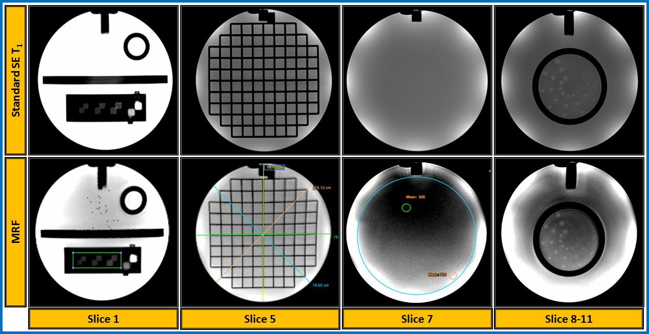
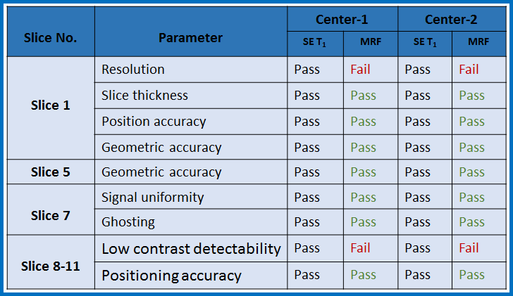
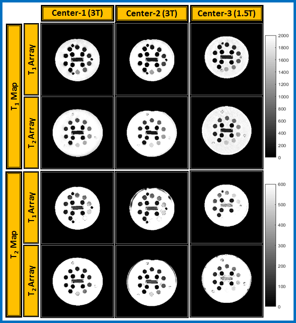
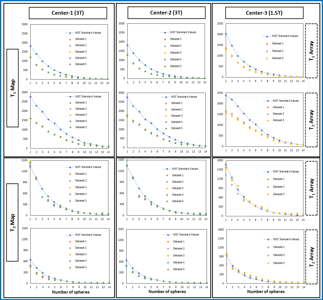
The representative image from the ACR phantom acquired at two centers (center-1 and center-2) shown in Figure 1. The qualitative ACR test of the MRF generated synthetic data passed all the criterion except the spatial resolution and low contrast detect-ability tests and results are tabulated in Table 1. Figure 2 depicts representative MRF reconstructed T1 and T2 maps from the three sites. Figure 3, shows MRF T1 and T2 values plotted for each sphere in the NIST phantom compared with the manufacturer values. Most of the MRF T1 and T2 value shows a similar trend to the ISMRM/NIST standard values along with the tendency to be consistently lower.Discussion and Conclusion
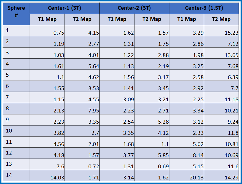
This is the first multi-center study on MRF for both qualitative and quantitative analysis using ACR and ISMRM/NIST system phantoms. The MRF T1 and T2 values show high repeatability and similar trend to manufacturer values. The coefficient of variation (in %) of less than or equal to 20% to show consistency of MRF measurements over multiple sites and repeated experiments (values are detailed in Table 2). This discrepancy may be because of inaccurate B1 estimation as reported by Buonincontri et.al [3]. In future, additional test and retest study in phantoms are needed to establish and evaluate technical performance of MRF method as a prerequisite towards utilizing MRF derived parameters as a quantitative imaging biomarker in MRI.Acknowledgements
National Institutes of Health Grant: U01 CA211205 (ASD and LHS)References
[1] Dan Ma, et. al., Nature 2013
[2] Jiang Y et. al., MRM. 2015
[3] Buonincontri et al., MRM 2016
Figures