1205
Recombinant expression and synthesis of a targeted human contrast agent for quantitative renal MRIKasey C Emoto1, Edwin Baldelomar2, Maria Veronica Clavijo-Jordan3, Jennifer R Charlton4, Courtnie Yokono5, and Kevin M Bennett6
1Biology, University of Hawaii at Manoa, Honolulu, HI, United States, 2Physics, University of Hawaii at Manoa, Honolulu, HI, United States, 3Martinos Center for Biomedical Imaging, Harvard University, Charlestown, MA, United States, 4Pediatrics, University of Virginia, Charlottesville, VA, United States, 5Bioengineering, University of Hawaii at Manoa, Honolulu, HI, United States, 6Washington University in St. Louis, Clayton, MO, United States
Synopsis
This study demonstrates the synthesis and use of a human recombinant cationic ferritin nanoparticle, synthesized in ecoli, as contrast agent for targeted renal imaging. Injected nanoparticles accumulated in the glomerular basement membrane in a mouse, allowing measurement of nephron endowment using gradient echo imaging and automated segmentation. Use of human recombinant contrast agents may allow improved biocompatibility for clinical translation.
Introduction
Chronic kidney disease (CKD) is a progressive disease that often ends in renal failure, requiring dialysis or transplant. Current clinical measures to detect renal function in CKD, such as serum or urinary markers, are indirect and insensitive to early development of the disease. Recently, cationic ferritin-enhanced MRI (CFE-MRI) has been developed to detect early microstructural changes by enabling measurements of nephron endowment in rodents and in human organs. CF binds to the glomerular basement membrane after intravenous injection, causing a detectable decrease in T2 at the site of each glomerulus (1-5). Here, we investigated the synthesis and application of a human recombinant form of cationic ferritin (HCF) as a natural iron-oxide nanoparticle contrast agent for renal imaging. We further established a general approach to forming an iron oxide core in the recombinant ferritin molecule in bacteria, allowing for rapid synthesis of a functional agent. For clinical translation, HCF may overcome limitations in agent compatibility as it is an endogenous protein regularly present in systemic circulation and in cells. To our knowledge, this is the first report of a human-based, targeted natural nanoparticle contrast agent for quantitative renal imaging. Methods
Human ferritin heavy chain (HC) and human ferritin light chain (LC) cDNA was cloned into the pVEXK-HN expression
vector with a His tag at the N-term of the heavy chain (pVEXK-HN-HC-IRES-LC,
Nature Technology Corporation). Prtotein
was expressed using the host strain BL21 (DE3) (New England Biolabs) in LB broth (Luria-Bertani broth, BD) at 37°c. Expression
was induced at OD600 ~0.5with 1mM Isopropyl-β-D-thio-galactoside (IPTG, Gold Biotechnology)after
adding ferric ammonium citrate (2mM) and cells were harvested after 24h..
Recombinat human ferritin was purified by a two step process: 1) affinity
chromatography (HisPur cobalt, ThermoFisher) and 2)size exclusion
chromatography (HiPrep 16/60 Sephacryl S-200, GE Healthcare). Ferritin
formation and iron core was confirmed using transmission electron microscopy
(TEM, Hitachi HT7700). HCF was cationized by the method of Danon (6). To verify
detection with MRI, CF was injected IV into a male B6 mouse at 5.75 mg per
100g, and the kidneys were removed and imaged in glutaraldehyde on a 7T Agilent
scanner with a 3D GRE pulse sequence (TE/TR=30/80 ms, 40x40x60 um resolution).
Glomeruli were segmented in the images and counted using in Matlab.
(Mathworks).
Results
Transformed bacteria produced human recombinant ferritin which was aqueous in solution after purification. Darkness varied by iron added to bacterial culture during synthesis (Fig 1a). The ferritin formed a ~13 nm 24mer nanoparticle with ~5 nm iron oxide core as confirmed by SDS-PAGE and TEM (Figs 1b-c). HCF was detected after intravenous injection by GRE-MRI in perfused, fixed mouse kidneys. Glomerular labeling was visible as small punctate dots in the MRI, and dots were not present in unlabeled control (Fig2). Glomeruli were identified by our custom software. Nephron number was in the mouse kidney (Nglom=18,186), consistent with previous reports.Discussion/ Conclusion
This proof of concept study demonstrates the synthesis and use of a human recombinant ferritin nanoparticle as contrast agent for targeted renal imaging. HCF was used to label the glomeruli after intravenous injection to calculate nephron endowment. Future work will focus on improving synthesis for increased iron deposition by bacteria, and in assessing toxicity in a range of animal models. HCF could be critical to translating this technology to measure nephron endowment for human allograft evaluation and potentially in vivo assessment of renal pathology.Acknowledgements
The authors gratefully acknowledge T. Carvallo and K. deRonde for technical assistance and R. Roy for help with imaging.References
Bennett et al. MRM 2008 Beeman et al. AJP Renal 2011 Beeman et al. AJP Renal 2014 Baldelomar et al. K Int. 2016 Danon et al. J. Ultrast Res 1972.Figures
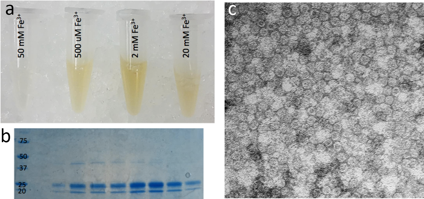
Figure
1: Human recombinant cationic ferritin (hrCF) formed a water-soluble
nanoparticle with visible iron loading that correlated with amount of iron
added to the bacteria culture. Iron content shown on tubes (a). Presence of
heavy and light chain subunits in the purified hrCF was confirmed by SDS-PAGE
(b), and formation of the 13 nm 24mer in the purified solution of hrCF, with a
~ 5 nm iron oxide core was confirmed by TEM (c).
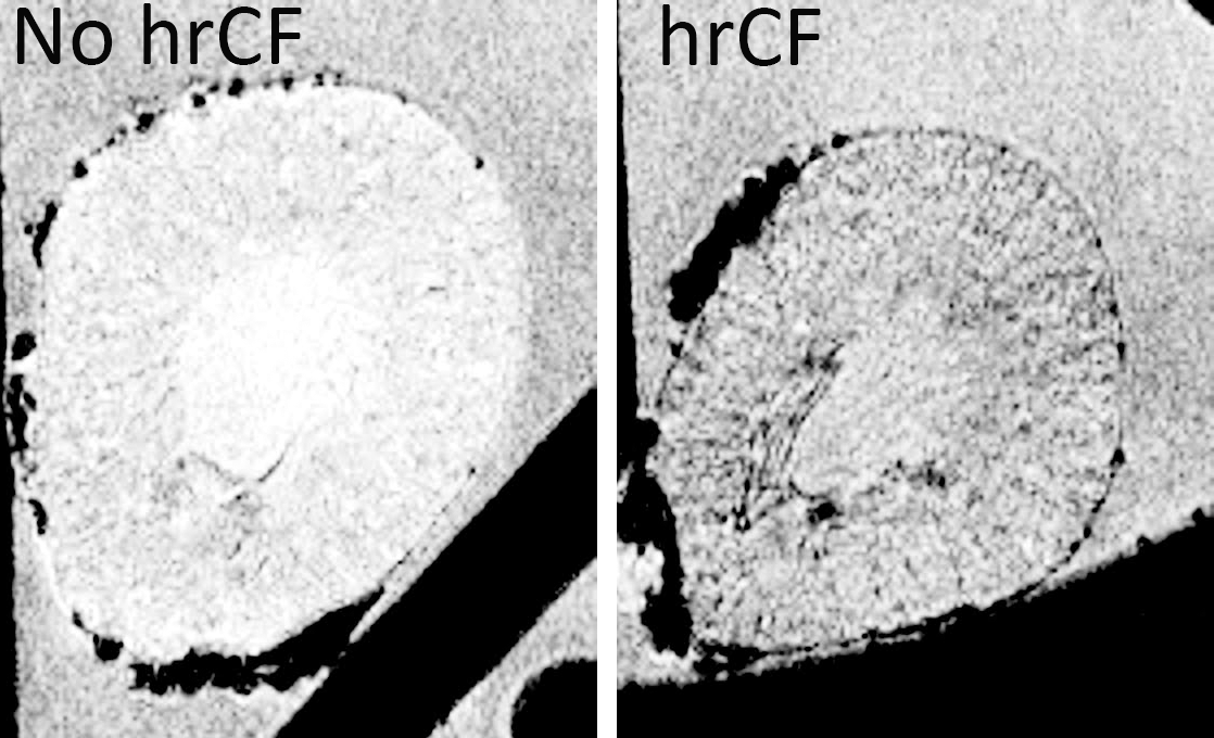
Figure 2: Single
slice from a 3D GRE-MRI image showing a perfused, fixed mouse kidney. (Left) No
addition of hrCF, (Right) with in vivo, IV hrCF administration. Dark spots
visible in the cortex were consistent with hrCF labeling of individual glomeruli,
which were counted using custom software.