1203
Fast hybrid IVIM-DK imaging in the kidney1Department of Medical Imaging, National Taiwan University Hospital, Taipei, Taiwan, 2Department of Urology, National Taiwan University Hospital, Taipei, Taiwan, 3Graduate Institute of Medical Device and Imaging, National Taiwan University, Taipei, Taiwan, 4Graduate Institute of Clinical Medicine, National Taiwan University, Taipei, Taiwan
Synopsis
The application of intravoxel incoherent motion (IVIM) and diffusion kurtosis (DK) MRI has been hindered largely by the long scan time required to sample over multiple b-values and/or diffusion-encoding directions. In this study, a novel method is described to expedite hybrid IVIM-DK imaging in the kidneys. Scan time is reduced by acquiring minimally-required non-zero b-values, while index calculation is made more time-efficient by using closed-form solution to replace nonlinear fitting. Experimental data demonstrated feasibility with b = 0/400/800/1600 s/mm2. Measurement variability was found greater among diffusion-encoding directions than between repeats, suggesting non-trivial structural anisotropy in the kidneys.
Introduction
Intravoxel incoherent motion (IVIM) imaging 1 and diffusion kurtosis (DK) imaging 2 are two MRI techniques that interrogate non-Gaussian diffusion. IVIM imaging operates in the low b-value range (<200 s/mm2) to separate the effects of microvascular blood flow and water diffusion. DK imaging operates in the high b-value range (>1000 s/mm2) to assess the degree of restricted/heterogeneous diffusion. Recently, the two methods were combined for simultaneous measurement of cerebral blood volume and diffusion heterogeneity 3. However, these methods sample over multiple b-values and require at least 15 directions of sampling to estimate the kurtosis tensor, which leads to a long scan time that is particularly problematic for application in organs subject to respiratory motion. In this study, we described a method to expedite both data acquisition and index calculation for hybrid IVIM-DK MRI in the human kidneys.Materials and Methods
Theory: According to the hybrid IVIM-DK model 3, intravascular volume fraction (f), diffusion coefficient (D), and diffusion kurtosis coefficient (K) can be simultaneously estimated by equation [1] where S(b) and S0 are the signal obtained with and without diffusion weighting, respectively. We propose to reduce the scan time by using three non-zero b values (the minimum number required to solve equation [1]) and expedite index calculation by replacing the nonlinear fitting with the following closed-form solution (equations [2] and [3]; b3 > b2 > b1 > 0).
Imaging: All MR images were obtained on a 3-Tesla clinical system, using the body coil to transmit radiofrequency pulses and the spine matrix coil and a torso matrix coil to receive signals. Respiration-gated diffusion imaging was performed with a twice-refocused spin-echo echo-planar readout (TR = 1.8 s, TE = 95 ms, field of view = 39-42 cm, voxel size = 3.0x3.0x6-3.3x3.3x6 mm3, 7 slices). b = 0, 400, 600, 800, 1200, 1600, 2000 s/mm2 (5 repetitions for b = 0; 20 non-collinear directions for non-zero b's). To test repeatability, diffusion imaging was repeated with an interval of about 10 min but without repositioning the subject. Five subjects (3 women and 2 men, age = 37-55 years) were included and each provided written informed consent before participation. The institutional review board approved this study.
Data analysis: b3 was chosen such that signal-to-noise ratio (SNR) was above two for at least 80% of the kidneys (SNR = signal minus background mean and then divided by background standard deviation). Based on equations [2] and [3], D/K/f were calculated for each diffusion-encoding direction separately and then averaged. Coefficient of variation was calculated on a voxel-wise basis to assess the variability among directions (CV_d) and between repeats (CV_r).
Results
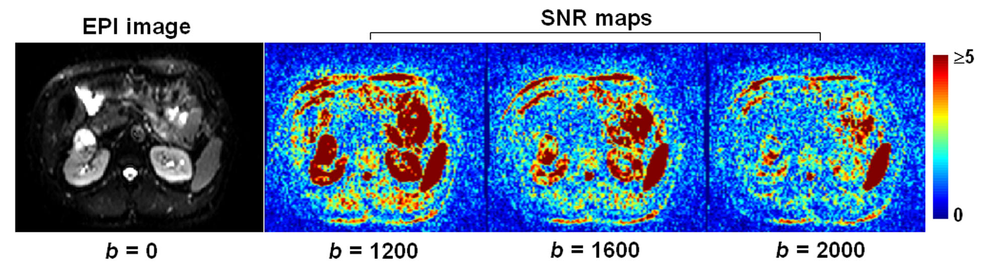
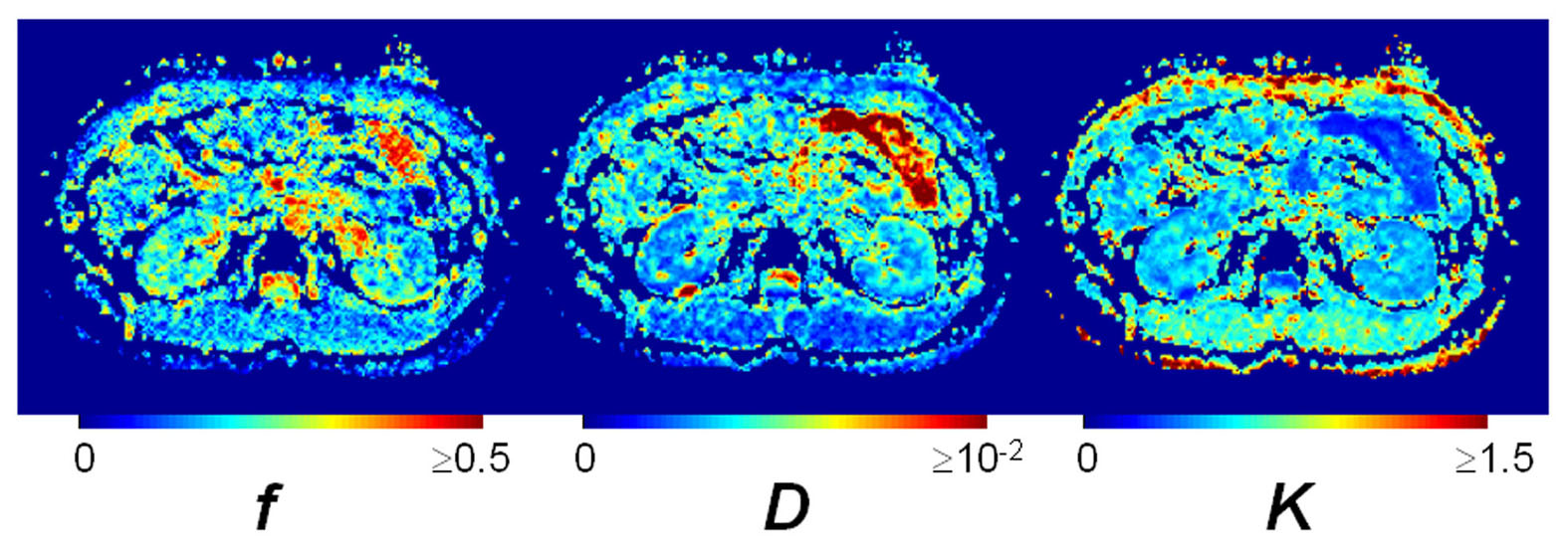
Figure 1 shows the SNR maps obtained from a representative subject. With b = 2000, the majority of the kidney voxels are indistinguishable from background noise. We thus chose 1600 to be b3 and along with b = 0/400/800 to extract D/K/f. The goodness-of-fit in terms of R2 was above 0.8 (by comparing the experimental data and the data estimated with the obtained indexes at b = 600 and 1200). Figure 2 shows the typical index maps obtained with our method. Figure 3 is the scatter plot of CV_d and CV_r (four regions of interest were drawn from each subject). CV_d is more noticeably larger than CV_r for D and K, as compared with f.Discussion
We have demonstrated the feasibility of a fast procedure for hybrid IVIM-DK imaging in the kidneys. First, scan time is reduced by using three non-zero b-values, which proved to be adequate in SNR and representing the data for b = 0-1600 s/mm2. Second, index calculation is more time-efficient by using closed-form solution. Although trace images have been used in previous body DKI studies 4,5, our data showed greater variability among diffusion-encoding directions than between repeats, which suggests non-trivial structural anisotropy in the kidneys. Diseases such as renal arterial stenosis and tumors have been known to compromise renal function during progression, which raises the concern about these patients' exposure to contrast material. Our method may serve as a useful alternative to simultaneously assess the pathological change and/or response to treatment of renal blood volume and microstructure.Acknowledgements
This work was supported by Ministry of Science and Technology, Taiwan (grant: 106-2628-E-002-003-MY3).References
1. Le Bihan D, Breton E, Lallemand D, et al. Separation of diffusion and perfusion in intravoxel incoherent motion MR imaging. Radiology 1988;168(2):497-505.2. Jensen JH, Helpern JA, Ramani A, et al. Diffusional kurtosis imaging: the quantification of non-gaussian water diffusion by means of magnetic resonance imaging. Magn Reson Med 2005;53(6):1432-1440.
3. Wu WC, Yang SC, Chen YF, et al. Simultaneous assessment of cerebral blood volume and diffusion heterogeneity using hybrid IVIM and DK MR imaging: initial experience with brain tumors. Eur Radiol 2017;27(1):306-314.
4. Mazzoni LN, Lucarini S, Chiti S, et al. Diffusion-weighted signal models in healthy and cancerous peripheral prostate tissues: comparison of outcomes obtained at different b-values. J Magn Reson Imaging 2014;39(3):512-518.
5. Nogueira L, Brandao S, Matos E, et al. Application of the diffusion kurtosis model for the study of breast lesions. Eur Radiol 2014;24(6):1197-203.
Figures