1193
High-Performance Diffusion Imaging on a 0.5T System1Research and Development, Synaptive Medical, Toronto, ON, Canada
Synopsis
Diffusion imaging is a valuable tool in the identification of neurological diseases, especially in an acute setting. Large high-field systems can be challenging to site in an acute setting. Mid-field systems have various siting advantages but historically can suffer from sub-par imaging performance. In this work we compare diffusion weighted imaging performance on a 0.5 T system with a high-performance gradient system to a typical 1.5 T clinical scanner. We demonstrate the ability to achieve comparable imaging performance both analytically as well as through imaging examples.
Introduction
Diffusion weighted MRI has been identified as a valuable tool in the diagnosis of neurological diseases, including being identified by the American Heart Association in its superiority to non-enhanced CT in the application of acute stroke1. Unfortunately in such clinical applications, access to MR imaging can be a significant barrier. The cost and installation footprint of traditional 1.5 or 3 T MR scanners are prohibitive for most institutions to install in an emergency department or location close to the acute patient population that would most benefit from access to acute diffusion imaging for neurological disorders. One possible solution to the challenges in siting MR equipment near acute patient populations would be to reduce the main field strength which enables a more compact fringe field, lower weight, and compact design systems. However, previous MR system offerings at field strengths below 1.0 T have had limited system performance and suffer from reduced SNR, resulting in poor quality diffusion imaging. Here we present an approach which enables high performance diffusion weighted imaging on a small-footprint, mid-field (0.5 T), head-only scanner.Methods
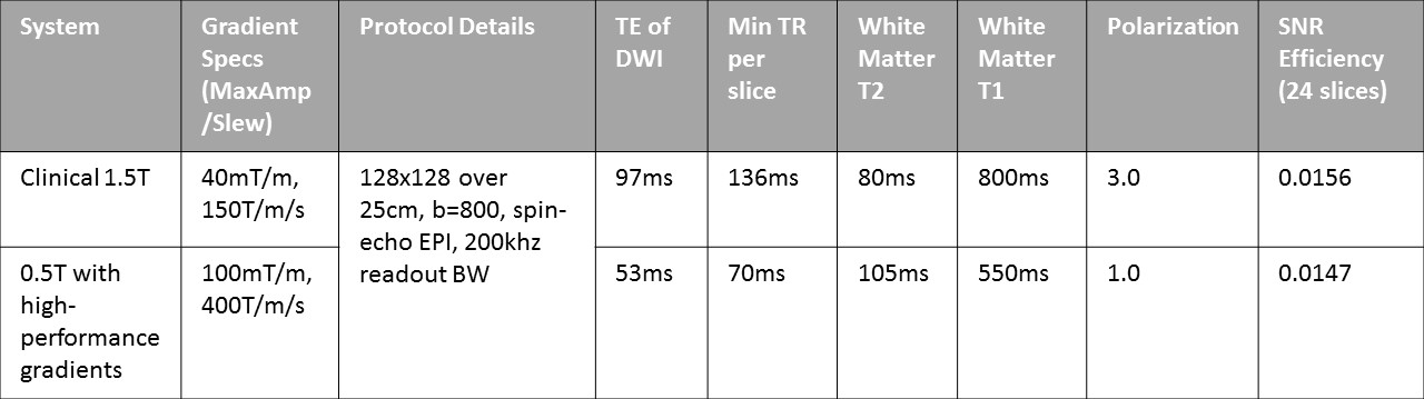
We have developed a head-only, superconducting, 0.5 T MR system which can be installed in under 300 square feet without requiring additional structural support or a quench pipe. To enable high quality routine diffusion imaging, the system is equipped with a high-performance gradient system with a maximum gradient amplitude of 100 mT/m and maximum slew rate of 400 T/m/s. We evaluated the predicted diffusion imaging performance through an SNR efficiency metric using a vendor-defined, routine protocol from a 1.5 T system. Diffusion weighted imaging on healthy volunteers was also performed to qualitatively evaluate performance at 0.5 T compared to 1.5 T with matched scan parameters (128x128 over 26 cm, no parallel imaging, 5 mm slice thickness, 200 kHz readout BW, min TE, spin echo EPI).Theory
Assuming a similar receive coil behavior for the two systems, relative SNR performance will be dictated by the relative signal levels. Using the values in Table 1 the predicted diffusion imaging performance can be evaluated using an SNR efficiency metric defined as $$ SNR_eff = {M0 * exp^{-TE \over T2} \over \sqrt{minScanTime}} $$
This expression relates the signal polarization (M0), echo time of the diffusion prescription (TE), the tissue T2 relaxation time and the minimum scan time (minScanTime) for whole brain coverage.
In addition, diffusion imaging is prone to geometric distortions in the presence of magnetic field inhomogeneity. The scale of this inhomogeneity is proportionally lower at lower field strengths, which results in significantly reduced geometric distortions. This distortion can be evaluated qualitatively on in vivo images.
Results/Discussion
The signal polarization is 3-times higher at 1.5 T. However the longer inherent T2 at 0.5 T2,3 and shorter echo time achievable with the high-performance gradient, means the relative signal at the echo time is just over 2-times higher at 0.5T. Assuming a minimum slice coverage for routine neuro protocols of 24 slices, the minimum time to acquire all 24 slices is 1.7 sec on the 0.5 T system and 3.2 sec at 1.5T. Note that these times are both greater than 3 times the T1 of white matter at the corresponding field strength, thereby allowing sufficient signal recovery between repeats and the effect of T1 recovery can be ignored. Using these values the ratio SNReff1.5T / SNReff0.5T = 1.06. Thus the SNR efficiency of a standard DWI scan on the 1.5 T system is only 6% different than what is expected on the 0.5 T system described here.
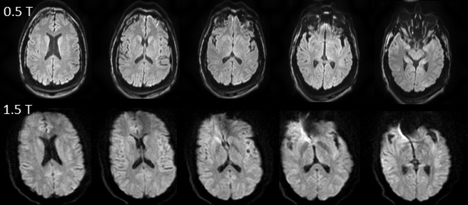
This in vivo behavior is illustrated in Figure 1 which shows mean diffusion-weighted images obtained at 1.5 T and 0.5 T on a subject with a dental implant. Significant geometric distortion in the anterior portion of the brain can be seen at 1.5 T which is not seen at lower field. The signal to noise behavior can also be seen to be qualitatively similar. Some additional image blurring can be seen in the 1.5 T images, likely a result of the effect of greater signal decay over the longer echo train duration compared to the 0.5 T system.
Conclusion
We have demonstrated that by marrying a high-performance gradient system with a mid-field, superconducting MR system designed for ease of siting; diffusion imaging performance can be achieved that is similar to a 1.5 T scanner in SNR efficiency and superior with respect to geometric distortion. Such a system may be appropriate for addressing the needs of the accessibility of MR for acute neurologic assessments.Acknowledgements
No acknowledgement found.References
- Powers W et al, Stroke (2018), 49(3).
- Bottomley P, Foster T et al (1994), Med Phys, 11:425
- Pfefferbaum A, Sullivan E el al (1999), Psychiat Res, 91:93
Figures