1190
EPIMix 3.01Neuroradiology, MR Applied Science Laboratory Europe, GE Healthcare, Stockholm, Sweden, 2Clinical Neuroscience, Karolinska Institutet, Stockholm, Sweden, 3Karolinska University Hospital, Stockholm, Sweden, 4Karolinska Institutet, Stockholm, Sweden
Synopsis
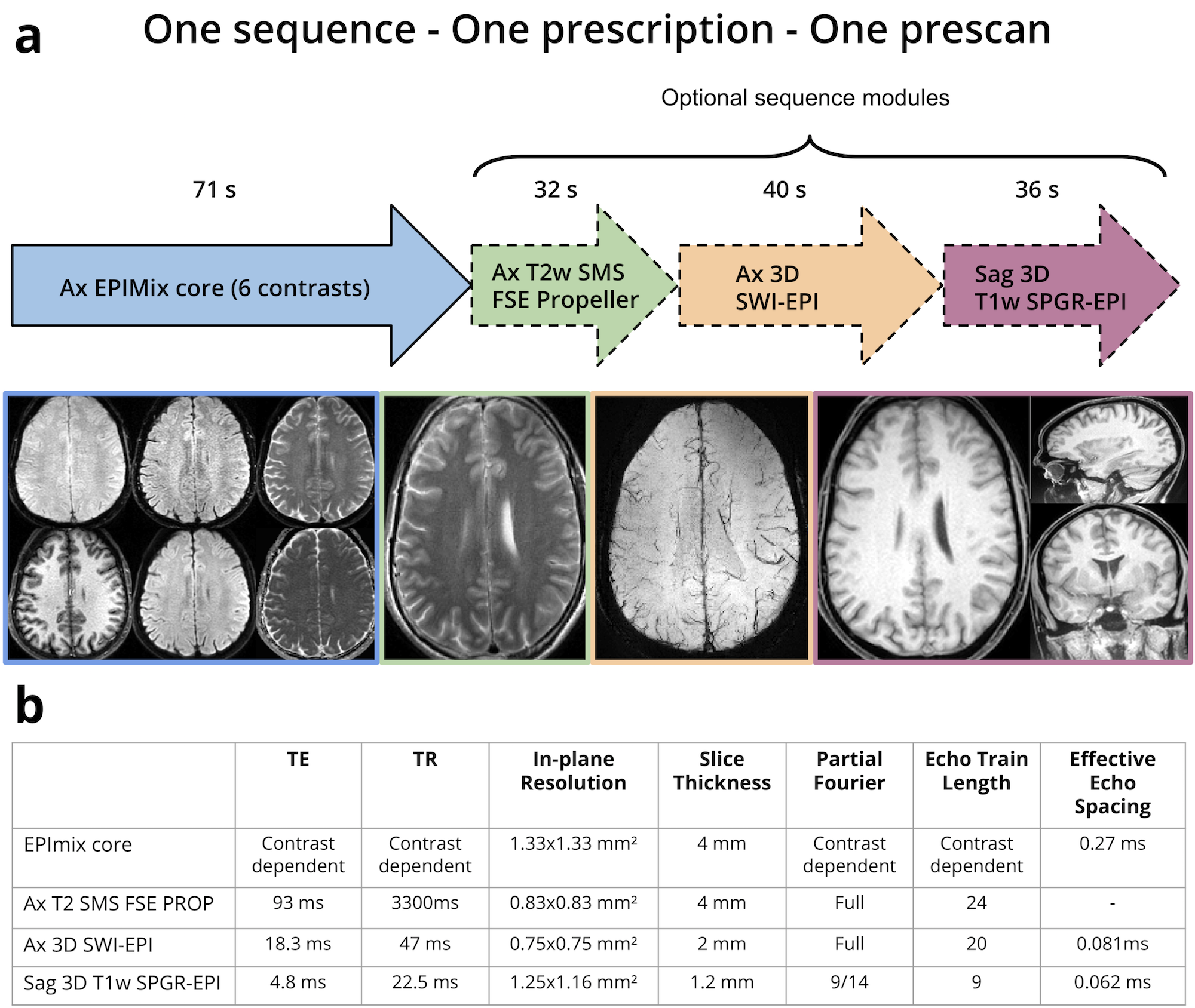
EPIMix is a multi-contrast MRI sequence that can produce T1-FLAIR, T2w, T2*w, T2-FLAIR, DWI, ADC images of the brain in about 70 s. To make EPIMix closer to a single-scan brain MRI exam, we propose optional supplemental sequence modules. The add-on sequences include T2w SMS FSE Propeller (distortion free), 3D SWI-EPI, and 3D T1w SPGR-EPI (isotropic resolution), which all have higher resolution and less distortions compared to EPIMix. With this extension to EPIMix, nine images series can be produced in 3 min total scan time.
Introduction
Various multi-contrast acquisition methods have been proposed (1–3), where the common idea is to produce multiple weighted MR contrasts (some/all of T1w/T1-FLAIR, T2w, T2*w, T2-FLAIR, DWI, ADC) in one scan. Recently, we proposed an improved version (4) of our 1-minute multi-contrast sequence ("EPIMix") (3), which can produce all of the above contrasts for 34-36 slices in about 70 seconds. With EPIMix as the only sequence in a brain MRI exam, 30 different volunteers can be scanned in one hour (5). In this work, we present our third version of EPIMix, which consists of the same 6-contrast core block, with optional high(er)-resolution 2D and 3D data. Highest on the neuroradiologists' wish list for EPIMix were distortion free FSE-based T2w images. In addition to this, we propose multi-shot 3D-EPI-driven SWI and T1w SPGR contrasts, to be executed seamlessly at the end of the core EPIMix scan, with a total maximum scan time of 3 min.Methods
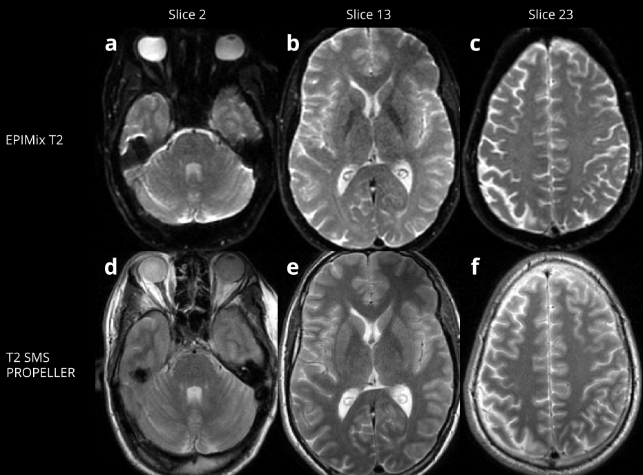
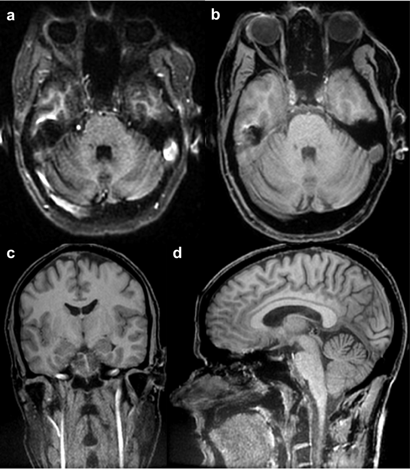
As a first step to embed high-res scans as a part of EPIMix, these sequences were for now tested in isolation. As EPIMix should work for patients not being able to remain still, we wanted at least the FSE T2w sequence block to be motion robust. Therefore, we used an in-house SMS T2w PROPELLER sequence (6), with 10 single-shot blades of size 288x48 with the same slices as for EPIMix core. The TE/TR was 93/3300 ms, and with a [MB]x[in-plane] acceleration of 2x2, the scan time became 32 s. 3D SWI-EPI used the same slice angulation and coverage as the EPIMix core, but with half the slice thickness using 68 kz encoded slices. To avoid arterial pulsation artifacts in the SWI data, an inferior sat pulse was played before each excitation. With a matrix size of 320x320, 16 shots, TE/TR = 18/39 ms, FA=18°, R=2, the scan time for this SWI block became 40 s. For the 3D T1w SPGR-EPI sequence, the logical X-Z axes were swapped to make the frequency encoding axis point in the Superior-Inferior direction. Here, a near isotropic voxel size was used (matrix 192x224, FOV=24 cm, 160 1.2 mm slices, TE/TR=4.1/22.5 ms, FA=30°, R=2 in the A/P direction), resulting in a scan time of 36 s. For 3D-EPI (SWI & SPGR), the Spectral-Spatial (SPSP) RF pulse was redesigned with better 3D volume selectivity compared to the standard SPSP RF pulse for 2D use. The SPSP pulse was designed with minimum-phase sub-pulses to improve the spatial selectivity and with minimum phase envelope to shorten TE. The EPIMix core and optional high-res scans were performed on two healthy volunteers (after informed consent) on a 3T SIGNA Premier using a 48-channel head coil. The matrix size for the EPIMix core was 180x180.Results
In Fig. 1a, the scan times of EPIMix core and optional scans are shown to scale, summing up to 2:59 min, with some relevant scan parameters summarized in Fig. 1b. The improved 3D-optimized SPSP RF pulse profile is shown in Fig. 2b and may be compared to the standard SPSP RF pulse (Fig. 2a) used for 2D EPI. The spatial selectivity is clearly improved (vertical profile Fig. 2c vs 2d), with maintained stop bands to avoid exciting the fat. The T2w SMS FSE Propeller data were acquired with 4 mm slices like the EPIMix core data, yielding distortion free motion robust images with a gridded matrix size of 288x288 (Fig. 3). In In Fig. 4, the improved resolution of T2*w 3DEPI compared to EPImix core GRE data is shown for some slice locations, the rightmost column with SWI processed minIPs. Finally, the T1w SPGR-EPI data with near-isotropic voxels are shown in sagittal and reformatted views in Fig. 5.Discussion
The core EPIMix sequence can provide T1-FLAIR, T2-w (b=0), DWI (& ADC), T2*w and T2-FLAIR MR contrasts in ~70 s with full brain coverage. We have earlier seen high correspondence of EPIMix core to conventional FSE-based images in terms of lesion detection on patients. Nevertheless, to make EPIMix more standalone for brain MRI, we proposed here to embed three optional scans with higher resolution, with no/low distortions, adding about 30-40 s each. Next, these optional modules will be integrated in EPImix core such that all contrasts can be acquired in a single scan, with only one slice prescription and prescan. This enables a complete neuro screening in less than 3 minutes drastically increasing throughput and efficiency.Acknowledgements
No acknowledgement found.References
1. Breutigam N-J, Frost R, Eickel K, Porter D. Simultaneous Multi-Contrast Imaging with Readout-Segmented EPI. In: Proceedings of the 25th Annual Meeting of the ISMRM, Honolulu, Hawaii. ; p. 520.
2. Jeong J, Nam Y, Lee J. Penta-contrast imaging: a Novel Pulse Sequence for Simultaneous Acquisition of Proton Density, T1, T2, T2* and FLAIR images. In: Proceedings of the 25th Annual Meeting of the ISMRM, Honolulu, Hawaii. ; p. 3881.
3. Skare S, Sprenger T, Norbeck O, et al. A 1-minute full brain MR exam using a multicontrast EPI sequence. Magn. Reson. Med. 2017 doi: 10.1002/mrm.26974.
4. Sprenger T, Engström M, Norbeck O, Rydén H, Avventi E, Berglund J, Skare S. Multi-contrast EPI - Towards clinical application. In: Proceedings of the Joint Annual Meeting ISMRM-ESMRMB 2018, Paris, France. ; p. 1200.
5. Skare S, Sprenger T, Norbeck O, Rydén H, Avventi E, Blomberg L, Berglund J, Skorpil M, Sandell M, Engström M. 30 brain MRI exams in 1 hour using a multi-contrast EPI sequence. In: Proceedings of the Joint Annual Meeting ISMRM-ESMRMB 2018, Paris, France. ; p. 350.
6. Norbeck O, Avventi E, Engström M, Rydén H, Skare S. Simultaneous multi-slice combined with PROPELLER. Magn. Reson. Med. 2017 doi: 10.1002/mrm.27041.
Figures