1185
5-Minute Quantitative Double-Echo in Steady-State for High-Value Diagnostic Knee MRI: Combining an Efficient Multi-Contrast Acquisition with Quantitative Imaging and Artificial Intelligence1Radiology, Stanford University, Palo Alto, CA, United States, 2Radiology, Santa Clara Valley Medical Center, San Jose, CA, United States, 3LVIS Corporation, Palo Alto, CA, United States, 4Neurology, Stanford University, Palo Alto, CA, United States
Synopsis
There exists interest in rapid diagnostic knee magnetic resonance imaging (MRI) protocols in a push towards ‘high-value radiology’. Recent efforts for expediting knee MRI involve accelerating 2D fast spin echo (FSE) sequences, which precludes multiplanar reformations, or using 3D FSE sequences, which can cause image blurring. To overcome these limitations, we show how a 5-minute quantitative double-echo steady-state (qDESS) sequence generates high-resolution and multi-contrast images using deep-learning-based super-resolution, along with automatic T2 relaxation time measurements. In a preliminary study with 25 patients, we demonstrate how qDESS can perform rapid and accurate diagnostic knee MRI using rich structural and quantitative information.
Introduction
Diagnostic magnetic resonance imaging (MRI) of the knee is typically performed using a combination of 2D fast spin-echo (FSE) sequences in different scan planes with varying contrasts, which requires approximately 20 minutes of scan time. There is great interest in expediting knee MRI protocols, especially with a push towards value-based imaging1. While many such efforts involve accelerating the 2D FSE scan or transitioning to 3D FSE acquisitions, the role of novel pulse sequences has had limited exposure. Additionally, quantitative measurements such as the T2 relaxation time that are commonplace in knee osteoarthritis imaging are relatively unexplored in diagnostic imaging2. Moreover, recent research in artificial-intelligence (AI) based deep learning methods has only seen limited clinical evaluation. To address these limitations, we seek to evaluate the diagnostic utility of the quantitative double-echo in steady-state (qDESS) sequence for diagnostic knee MRI. qDESS generates 3D high-resolution multi-contrast images and T2 relaxation time measurements in only 5-minutes. In this study, we augment DESS with deep-learning-based super-resolution (DL-SR) for enhancing slice-resolution twofold to evaluate the diagnostic utility of such a quantitative, high-resolution, multi-contrast, and AI-enhanced method in 25 patients referred for a clinical knee MRI examination.Methods
qDESS generates two echoes (S+ and S-) with T1/T2-weighted and T2-weighted contrasts respectively3–6. The two qDESS echoes are used to generate automatic T2 relaxation time by inverting the DESS signal model7. The qDESS scan parameters used in this study were: matrix=416x512, field-of-view=160mm, slice-thickness=1.6mm, TE/TR= 6/18ms, flip angle=20°, scan time=5:00 and 2x1 parallel imaging. A DL-SR algorithm described previously was used to enhance the slice-resolution of the qDESS scans to approximately 0.7mm8. Training and validation for this algorithm was performed on 34 and 10 patients referred for a clinical knee MRI respectively, who were scanned with qDESS with the scan parameters above, except with a 0.7mm slice thickness. The algorithm was also pre-trained on 124 DESS scans obtained from the Osteoarthritis Initiative9.
An additional twenty-five symptomatic patients (18 male, 7 female, age 39±19 years) referred for a routine knee MRI were scanned on one of two identical Discovery MR750 3.0T MRI scanners (GE Healthcare, Waukesha, Wisconsin) with an 8-channel transmit-receive knee coil (InVivo, Gainesville, Florida). A water-excitation sagittal 3D qDESS sequence was added to the routine knee protocol and subsequently enhanced using DL-SR.
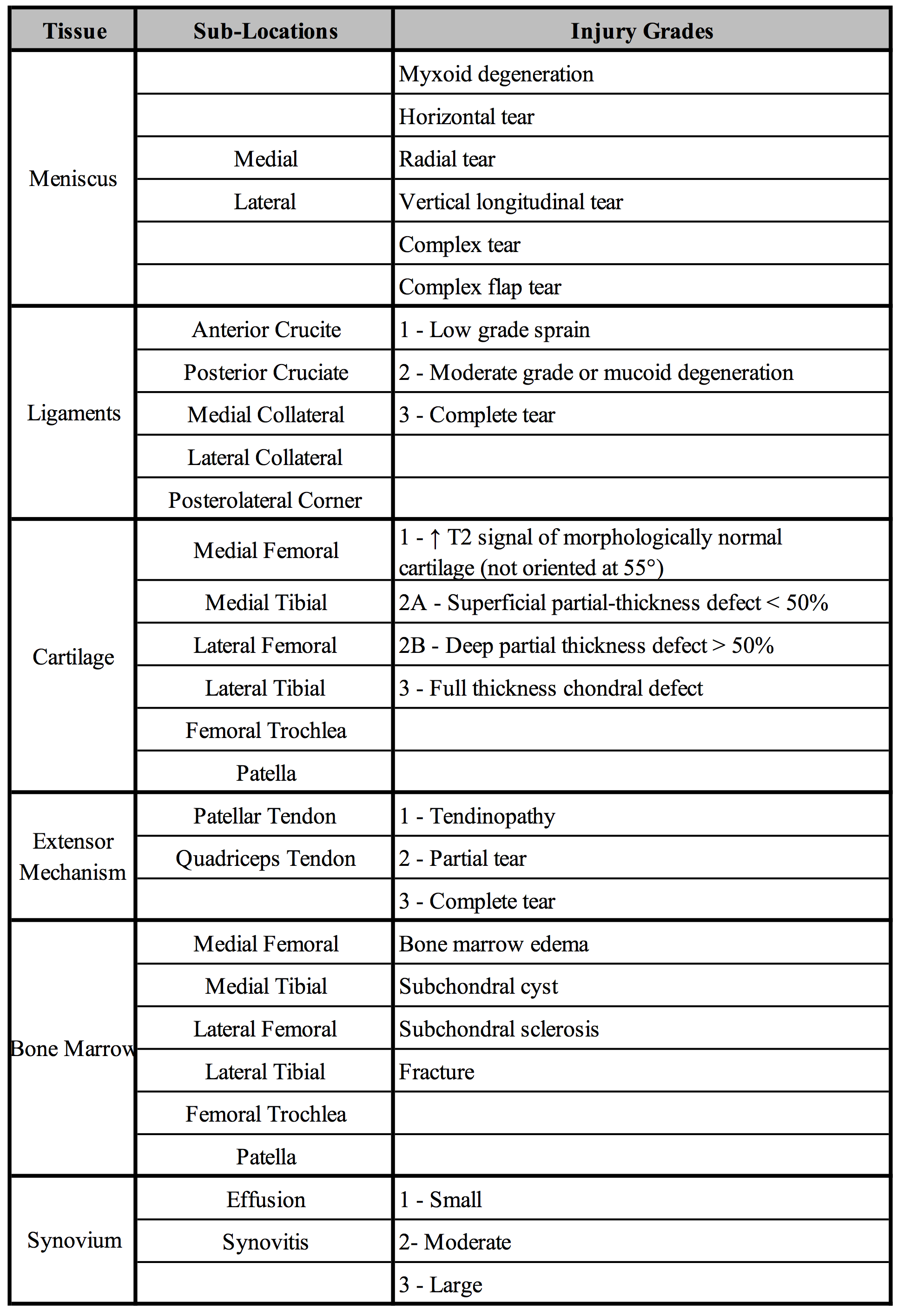
Two musculoskeletal radiologists evaluated all cases from the conventional imaging protocol and qDESS protocol (one radiologist assessed 10 qDESS scans) with a one-month washout period between the two readings. Common imaging findings in internal derangements of the knee were sub-divided into categories of ligaments (cruciate and collateral), menisci, cartilage, synovium, bone, and extensor mechanism using previously established criteria (Fig.1)10–13. The readers also scored the diagnostic quality of the protocol used (1=very poor, 2=poor, 3=acceptable, 4=good, 5=very good).
The sensitivity, specificity, and accuracy and the confidence intervals (CI) of the qDESS protocol by tissue type were calculated and compared to the conventional imaging protocol. The diagnostic quality of the qDESS and conventional protocol pooled by tissue type was also calculated. Cohen’s linearly-weighted Kappa was used to evaluate inter-observer agreement.
Results
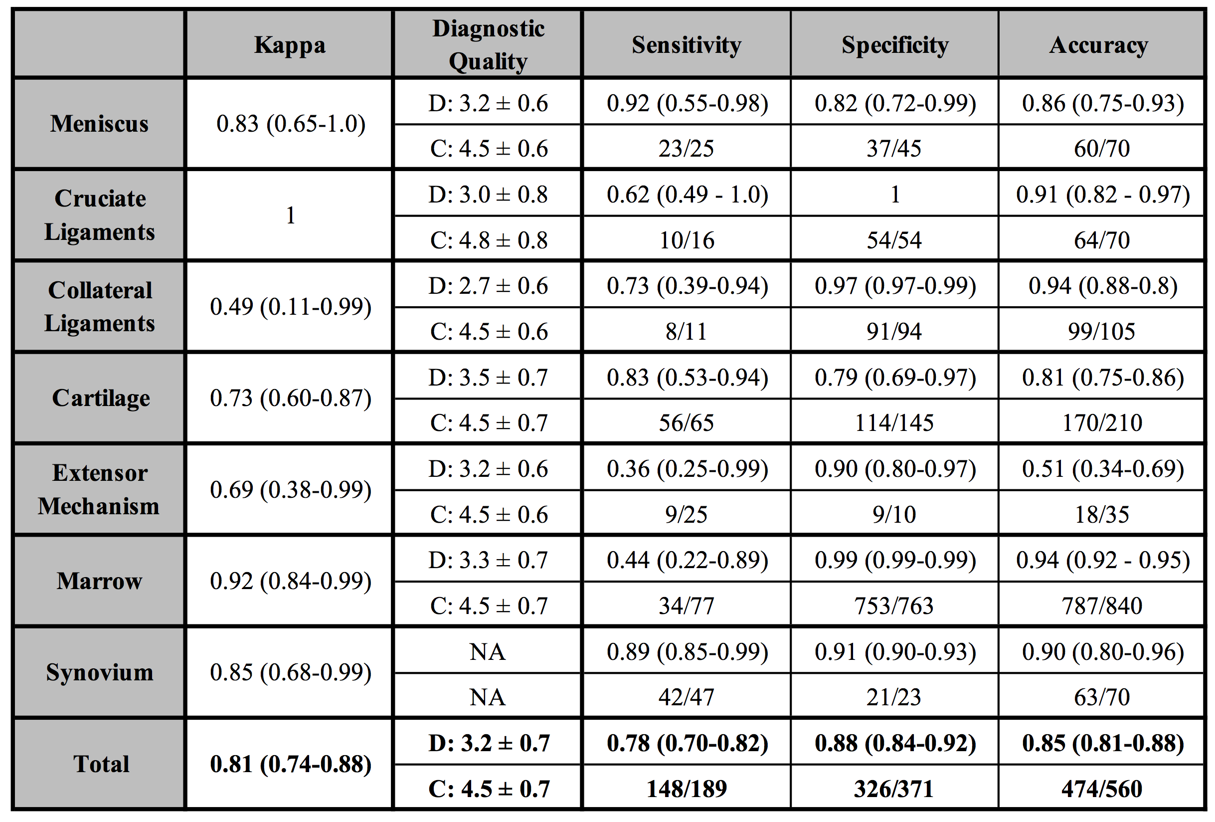
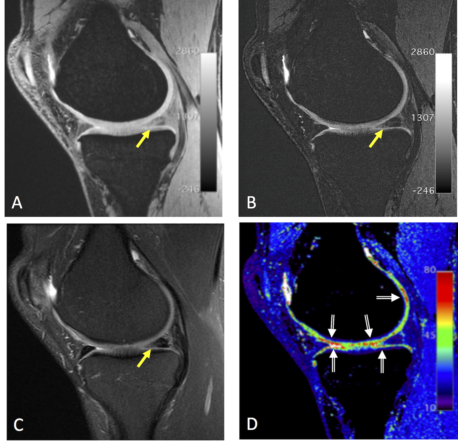
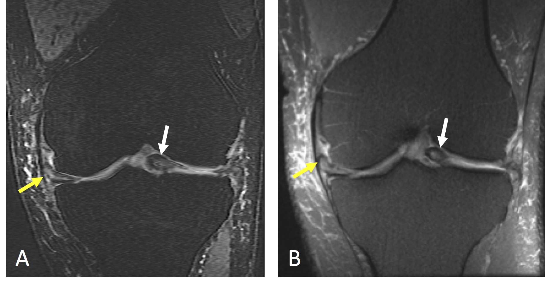
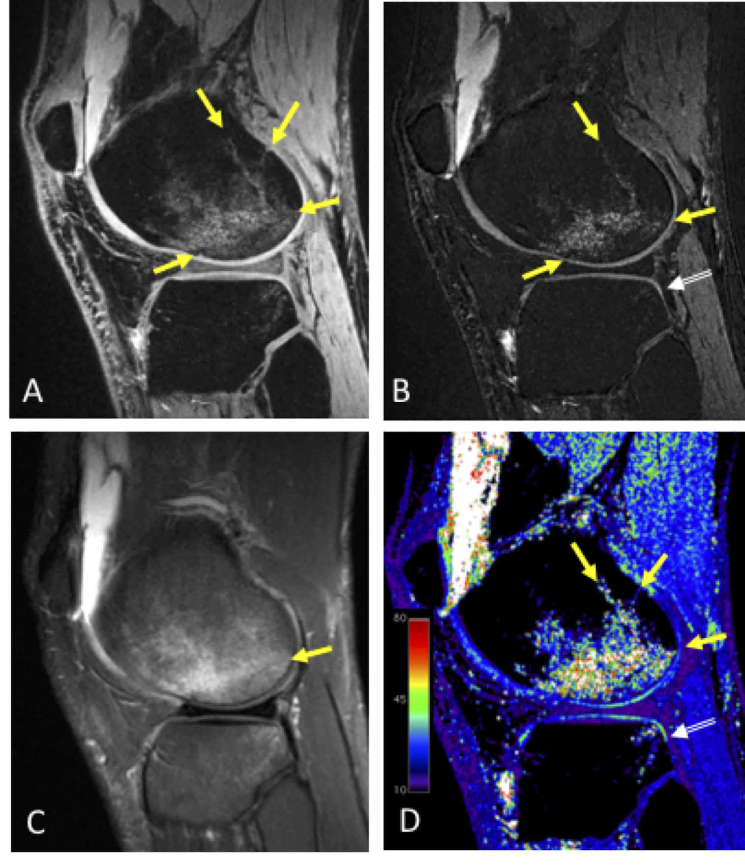
qDESS had high accuracy for evaluating injuries of the menisci, collateral ligaments, cartilage, and synovium, with lower accuracy for the extensor mechanisms and bone marrow (Fig.2). Inter-reader agreement was excellent (Kappa=0.81) and the diagnostic quality score of qDESS was consistently above acceptable. Example images of a meniscal tear and adjoining cartilage damage (Fig.3) showed high contrast for the injury and utility of the T2 map for ascertaining cartilage health. Complex flap tears of the meniscus were well depicted using DL-SR through-plane resolution enhancement in the qDESS coronal reformation (Fig.4). A tear of the anterior cruciate ligament and a corresponding fracture of the femur and tibial cartilage degeneration were well visualized with the qDESS images and the T2 map (Fig.5).Discussion
Traditional approaches to create isotropic resolution images either lead to blurring due long echo trains in 3D FSE sequences or noisy images due to parallel imaging. DL-SR overcame these limitations by acquiring images with adequate SNR and subsequently enhancing slice-resolution. The automatic T2 measurements created with qDESS demonstrated clinical utility, especially for the detection of early cartilage degenerative changes that were not depicted in the conventional or qDESS structural images. This may suggest that the routine use of T2 mapping may enhance diagnostic accuracy.Conclusion
A single 5-minute high-resolution and multi-contrast qDESS sequence with deep-learning-based super-resolution enhancement and automatic T2 mapping was able to provide high diagnostic accuracy in this pilot study, for potential use in abbreviated knee MRI protocols.Acknowledgements
We would like the acknowledge the following NIH grants for providing research support: R01 AR0063643, R01 EB002524, P41 EB015891, K24 AR062068, along with GE Healthcare.References
1. van Beek, E. J. R. et al. Value of MRI in medicine: More than just another test? J. Magn. Reson. Imaging 1–12 (2018). doi:10.1002/jmri.26211
2. Baum, T. et al. Cartilage and meniscal T2 relaxation time as non-invasive biomarker for knee osteoarthritis and cartilage repair procedures. Osteoarthritis Cartilage 21, 1474–84 (2013).
3. Staroswiecki, E., Granlund, K. L., Alley, M. T., Gold, G. E. & Hargreaves, B. A. Simultaneous estimation of T2 and apparent diffusion coefficient in human articular cartilage in vivo with a modified three-dimensional double echo steady state (DESS) sequence at 3 T. Magn. Reson. Med. 67, 1086–96 (2012).
4. Bruder, H., Fischer, H., Graumann, R. & Deimling, M. A new steady-state imaging sequence for simultaneous acquisition of two MR images with clearly different contrasts. Magn. Reson. Med. 7, 35–42 (1988).
5. Redpath, T. W. & Jones, R. A. FADE--a new fast imaging sequence. Magn Reson Med 6, 224–234 (1988).
6. Lee, S. Y. & Cho, Z. H. Fast SSFP gradient echo sequence for simultaneous acquisitions of FID and echo signals. Magn Reson Med 8, 142–150 (1988).
7. Chaudhari, A. S. et al. Five-minute knee MRI for simultaneous morphometry and T 2 relaxometry of cartilage and meniscus and for semiquantitative radiological assessment using double-echo in steady-state at 3T. J. Magn. Reson. Imaging 47, 1328–1341 (2018).
8. Chaudhari, A. S. et al. Super-resolution musculoskeletal MRI using deep learning. Magn. Reson. Med. 80, 2139–2154 (2018).
9. Peterfy, C. G., Schneider, E. & Nevitt, M. The osteoarthritis initiative: report on the design rationale for the magnetic resonance imaging protocol for the knee. Osteoarthr. Cartil. 16, 1433–1441 (2008).
10. Noyes, F. R. & Stabler, C. L. A system for grading articular cartilage lesions at arthroscopy. Am. J. Sports Med. 17, 505–513 (1989).
11. Nguyen, J. C., De Smet, A. A., Graf, B. K. & Rosas, H. G. MR Imaging–based Diagnosis and Classification of Meniscal Tears. RadioGraphics 34, 981–999 (2014).
12. Schweitzer, M. E., Tran, D., Deely, D. M. & Hume, E. L. Medial collateral ligament injuries: evaluation of multiple signs, prevalence and location of associated bone bruises, and assessment with MR imaging. Radiology 194, 825–829 (1995).
13. Hong, S. H. et al. Grading of anterior cruciate ligament injury. Diagnostic efficacy of oblique coronal magnetic resonance imaging of the knee. J. Comput. Assist. Tomogr. 27, 814–9
Figures