1174
DANTE Prepared EPI for Fast Whole Brain VASO Detection at 3T1National Institutes of Mental Health, National Institutes of Health, Bethesda, MD, United States
Synopsis
Vascular space occupancy (VASO) fMRI may provide more quantifiable and localized hemodynamic measures of brain activitythan conventional BOLD measurements. Due to application of inversion recovery pulse, conventional approach for VASO measurements suffers from low imaging efficiency, high SAR and interference of inflow and CBF effect. We demonstrated that DANTE prepared single echo or multi-echo EPI can be used to acquire robust full brain multi-slice VASO activation with benefit of high image efficiency, low SAR without complication of inflow and CBF effect.
Introduction:
Vascular space occupancy (VASO) fMRI is a microvascular-weighted method that may provide more quantifiable and localized hemodynamic measures of brain activitythan conventional BOLD measurements [1]. The need for using inversion recovery in VASO (IR-VASO) detection, however, reduces temporal resolution due to the inversion time delay required for blood signal null. In addition, IR-VASO only allows limited imaging coverage due to its short time period of blood null. When more imaging slices are acquired, image signal may potentially be confounded by in-flow effects and cerebral blood flow (CBF). Situation can be partially improved based on application of multiple inversion recovery pulses at the cost of compromised imaging SNR and dramatically increased SAR [2]. DANTE (a rapid series of low flip angle RF pulses interspersed with gradients) pulse trains has been demonstrated as a black blood (BB) MR preparation, naturally adapted to readout sequences for fast high-resolution BB imaging acquisition. Its efficiency for generating BB images can be several times greater than conventional blood suppression modules, such as inversion recovery [3]. Here we demonstrate that a DANTE prepared multi-echo EPI (DANTE-mEPI) can be used to provide whole brain vascular space occupancy (VASO) image measurement.Theory and Methods:
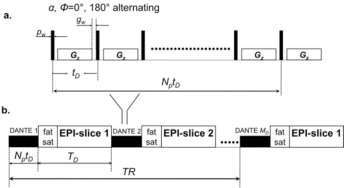
The proposed DANTE-prepared imaging sequence is shown in Fig. 1, indicating both the DANTE preparation module itself (Fig. 1a ), as well as the proposed method for embedding it within a multi-slice single echo or multi-echo EPI sequence (Fig. 1b). Three healthy volunteers (male, age 35-45 years) underwent fMRI scans. Written informed consent was obtained from all subjects. Tasks of fMRI,flashing checkerboard 8Hz accompanied with right-hand finger-tapping (1 Hz), 0.5 minutes ON-OFF blocks were performed. Siemens Prisma scanner with a high-performance gradient was employed to study subjects. A Siemens 32-channel head coil was employed. Common parameters used for single echo and multi echo EPI were as follows: FAEPI=70°, GRAPPA (R=3), resolution 2.1x2.1 mm in-plane (92x92 pixels) with field of view 192 mm, 2 mm slice thickness with 50% gap, bandwidth 2074 Hz/pixel. For imaging of DANTE single echo EPI with echo time TE=9 ms, 38 slices were acquired within 3s TR. For imaging of DANTE multi echo EPI with echo time TE=11 and 29 ms, respectively, 32 slices were acquired. When running conventional GE-EPI for comparison, the DANTE flip angle was set to zero .DANTE parameters: Number of pulses = 44; Interpulse delay = 700 us, Flip angle = 7.Results and Discussion:
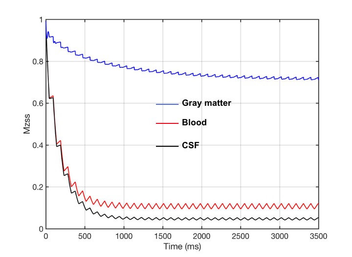
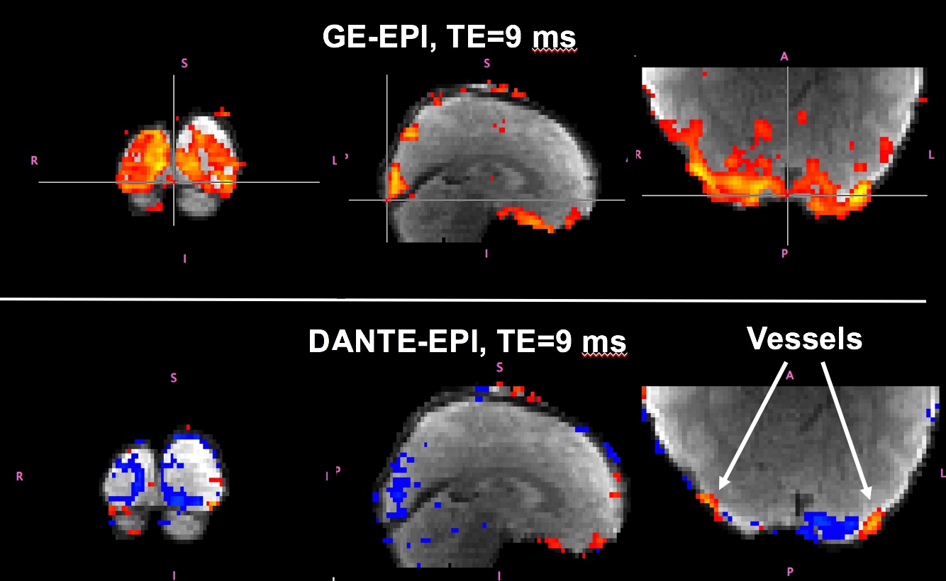
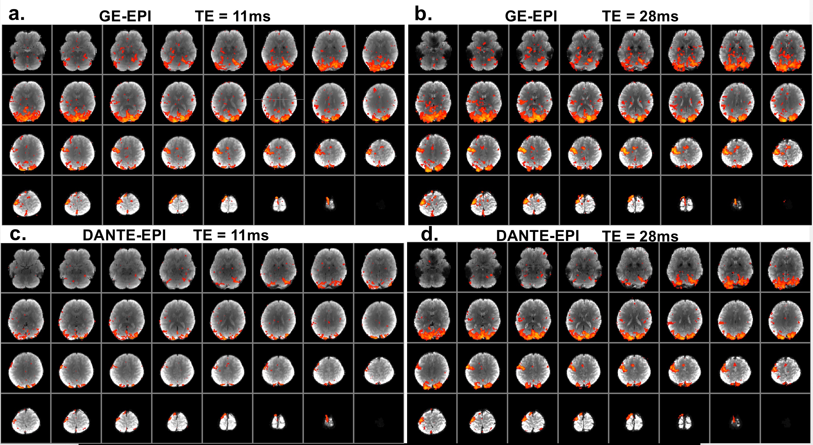
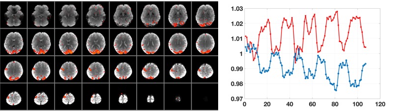
It was shown in Bloch simulation of Fig. 2 that moving blood can be suppressed down to less than 10-15% residual signal level. Major blood travelling faster than 2-3 mm/s in the intravascular including blood in artery, arteriole, venule, vein remains suppressed all the time after short transition time. With short echo time, 9 ms, the VASO activation can be directly observed upon signal decreasing in the fMRI images from DANTE single echo EPI, lower panel of Fig. 3. However, at some of location, where the blood is in the larger veins, the BOLD effect is still stronger than VASO effect. The positive signal from BOLD upon activation can still be observed in DANTE-EPI images.Due to the restriction of the minimal available echo time in EPI sequence, the VASO effect from the DANTE single echo EPI may be compromised as shown in Fig.3. To solve the problem, DANTE prepared multi-echo EPI can be employed for extrapolating the images with echo time to zero. Multi-echo DANTE-EPI with echo time 11 ms and echo 28 ms were shown in Fig. 4c and Fig. 4d, respectively. By comparison of BOLD effect in images from GE-EPI, Fig. 4a and DANTE-EPI, Fig. 4b, it is very clear that when echo time is short BOLD activation of DANTE-EPI is largely decreased, which was caused by the cancellation between BOLD and VASO effects. Ideally, the strongest VASO effect can be observed when echo time of read-out sequence is equal to zero, which minimizes the T2*weighted BOLD effect. Multi-echo images of DANTE-EPI at each individual TR time were extrapolated to achieve spin density images with zero echo time. The activation map, calculated from these spin density images, was shown in Fig. 5, (left panel) as pure VASO fMRI. The normalized average activation of BOLD (blue line, calculated from GE-EPI, Fig. 5b) and VASO (red line, calculated from Fig.5 left panel) are shown in Fig. 5 (right panel). Signal differences of BOLD and VASO are approximately to be 2% and 1%, respectively. Note, the signal decrease corresponds to an increase in activity in VASO).Conclusion:
We demonstrated that DANTE prepared multi-echo EPI can be used to acquire robust full brain VASO activation with high image SNR, low SAR without complication of inflow and CBF effect.Acknowledgements
This work was supported by the Intramural Research Program of the National Institute of Mental Health, USA.References
1. H Lu, et al., Magnetic Resonance in Medicine 50 (2), 263-274, 2003.
2. Donahue MJ, Magnetic Resonance in Medicine, 2009 Feb;61(2):473-80.
3. L Li, et al., Magnetic Resonance in Medicine 68 (5), 2012.
Figures