1169
High-resolution segmented-accelerated EPI using Variable Flip Angle FLEET with tailored slice profiles1Athinoula A. Martinos Center for Biomedical Imaging, Massachusetts General Hospital, Boston, MA, United States, 2Department of Radiology, Harvard Medical School, Boston, MA, United States, 3Vanderbilt University Institute of Imaging Science, Nashville, TN, United States, 4Department of Biomedical Engineering, Vanderbilt University, Nashville, TN, United States, 5Harvard-MIT Division of Health Sciences and Technology, Massachusetts Institute of Technology, Cambridge, MA, United States
Synopsis
New evidence suggests that fMRI has spatial specificity at scales far below current voxel sizes, but encoding limits preclude single-shot EPI at sufficient spatial resolution. Segmented EPI can help overcome these limits, but is well-known to be temporally unstable. Here we propose a reordering of the EPI segments, known as FLEET, combined with
Introduction
There is growing evidence that hemodynamic responses to neuronal activation are far more spatially specific than previously believed,1 motivating$$$\;$$$fMRI$$$\;$$$acquisitions$$$\;$$$with$$$\;$$$higher spatial resolutions.$$$\;$$$However, we are limited by the spatial encoding capabilities$$$\;$$$of$$$\;$$$MRI to reach these higher resolutions$$$\;$$$while$$$\;$$$maintaining whole-brain coverage.2 Increasing the acceleration factor results in $$$\sqrt(R)$$$ and $$$g$$$-factor signal-to-noise ratio (SNR) penalties, and increasing the readout duration to sample further in k-space results$$$\;$$$in increased $$$T_2^*$$$-induced spatial blurring and B0 distortions. Segmented EPI is a possible solution to this problem, but it suffers from spurious ghosting resulting from inter-segment phase differences arising from motion- and respiration-induced B0 changes. It was recently shown that the temporal SNR (tSNR) of BOLD time-series based on accelerated single-shot EPI can be dramatically increased by swapping the slice-segment ordering of the multi-shot-EPI-based autocalibration scan using the fast low-angle excitation echo-planar technique (FLEET) to minimize the inter-segment delay.3,4 This method used constant, low flip angles with added dummy scans to achieve equal magnetization across segments, which led to an overall loss in tSNR—which is tolerable for ACS data acquisition. Here, we extend this approach to acquire reordered segmented, multi-shot EPI for the fMRI acquisition itself, using a variable-flip-angle (VFA) scheme (VFA-FLEET) to maximize the SNR of the fMRI data and Shinnar-Le Roux (SLR) pulses to produce consistent magnetization between segments.Theory
By accounting for the pseudo-steady-state of
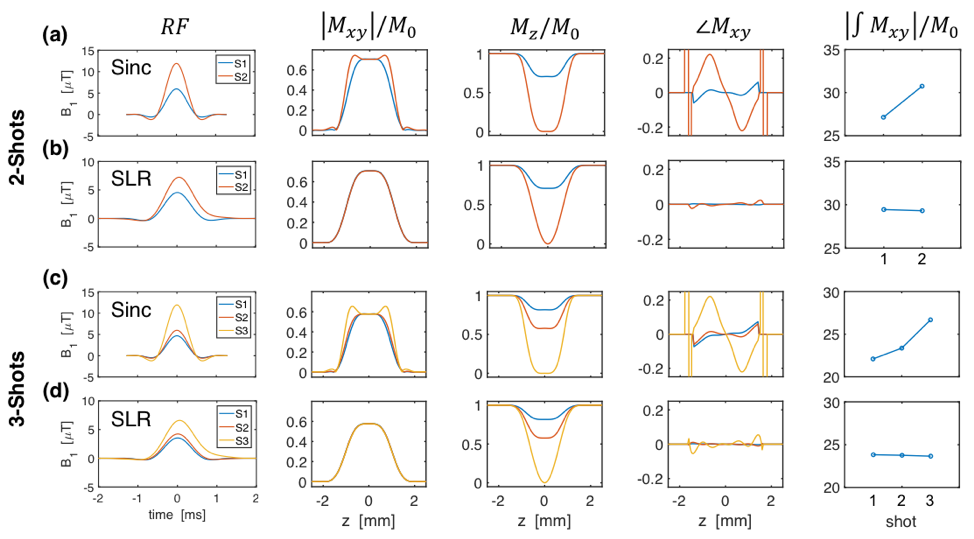
VFA-FLEET, target flip angles are determined recursively:3$$ \alpha_{i-1}=\tan^{-1}(\sin(\alpha_i)).$$ To
maximize magnetization, the final excitation is set to 90°, giving $$$\alpha_i$$$={45°,90°}
or {35°,45°,90°} for 2- or 3-shots, respectively (with no dummies). Owing to
the non-square slice profiles, using Hann-windowed sinc RF pulses results in
non-uniform slice profiles from shot-to-shot;5-7 figure
1 shows how tailored SLR pulses overcome this.Methods
Acquisition: All experiments were conducted at 3 T using a 32-channel receive coil. Four subjects (3F, 29±4-years) were scanned with conventional-segmented EPI, and VFA-FLEET EPI with sinc (VFA-FLEET-sinc) and SLR (VFA-FLEET-SLR) pulses, using 2 or 3 segments (Nseg), unaccelerated, matrix=96x96, 2.1-mm isotropic resolution, 30/33 slices (Nseg=2/3), 20% slice gap, TE=30ms, TR=Nseg×2.4s, 62 repetitions. The flip angle of VFA-FLEET that determines the image signal level is the first flip angle; to dissociate the role of segment reordering from flip angle, the conventional-segmented was repeated for each Nseg with α=90° and 45°(Nseg=2) or 35°(Nseg=3). Combined segmented-accelerated was tested on two subjects (1F, 32±5-years) using matrix=128x128, 1.5-mm isotropic resolution, 33/31 slices (Nseg=2/3), no slice gap, TE=30ms, TR=Nseg×2.2s, and all combinations of Nseg=2/3 and R=3/4 resulting in an effective acceleration of Nseg×R for each segment. Images were reconstructed offline using navigator-based ghost-correction within each segment then navigator-based ghost-correction between segments. Optionally, to account for differences in shot-to-shot signal, a scaling factor that minimized the mean-square error between navigators was applied to the segments. GRAPPA reconstruction with ACS-FLEET4 was applied to the combined accelerated segments. Analysis: For each time-series, the first two volumes were discarded, and the remaining volumes were motion-corrected and linear-drift-corrected. tSNR$$$\;$$$and$$$\;$$$skew—the$$$\;$$$deviation$$$\;$$$of$$$\;$$$a$$$\;$$$voxel’s temporal intensity distribution from normality—were used to characterize the various acquisitions.Results
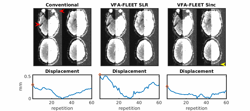
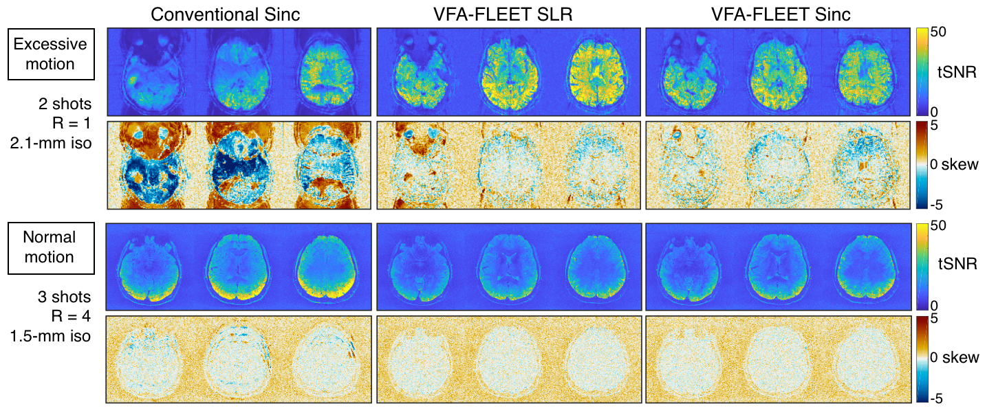
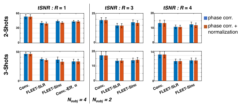
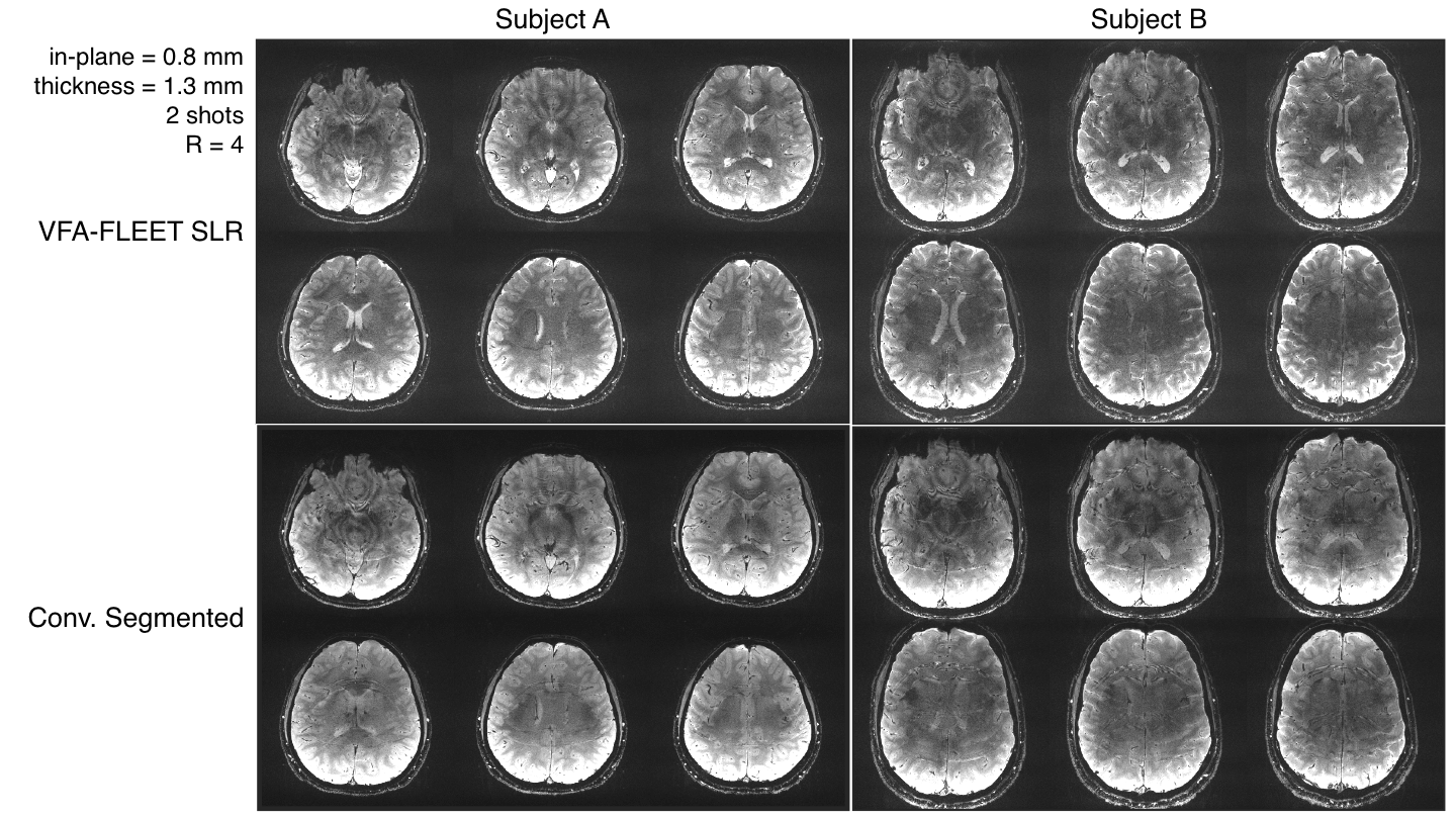
Figure 2 illustrates how spurious ghosts in the conventional-segmented images are effectively eliminated in both VFA-FLEET sequences and stable ghosts in the VFA-FLEET-sinc images are reduced in the VFA-FLEET-SLR images. In Figure 3, the impact of spurious ghosting is reflected in the tSNR and temporal skew maps. In Figure 4, the group-averaged whole-brain tSNR was highest for the conventional-segmented acquisitions and lowest for VFA-FLEET-SLR although differences in flip angle and slice profile explain some of these differences. The intersegment-normalization did remove stable ghosts in both VFA-FLEET acquisitions, but at the cost of decreased tSNR. Figure 5 shows the feasibility of acquiring sub-mm3 resolutions with just R=4 VFA-FLEET-SLR.Discussion
While conventional-segmented had the highest tSNR, it was marred by nonuniformity that would make its sensitivity to activation spatially heterogeneous. Although VFA-FLEET-SLR had reduced group tSNR, its homogeneity and its ability to faithfully excite the same slice profile make it the more attractive option since it inherently reduces ghosting. As we translate this to 7T, where B0-fluctuations are longer-ranging, spurious ghosting will be exacerbated, further motivating this technique. Care will need to be taken to account for the increased B1-inhomogeneity at ultra-high fields. Finally, segmentation decreases the temporal resolution by Nseg; however, VFA-FLEET is compatible with simultaneous multi-slice imaging, and the RF phase can be designed to eliminate the CAIPI blips.8Conclusions
VFA-FLEET offers a solution to the encoding limits on single-shot EPI and to spurious ghosting of conventional-segmented EPI. While stable ghosts remained in the VFA-FLEET-sinc acquisition, using tailored-SLR RF pulses to improve the signal uniformity between segments nearly completely eliminated them. With improved spatial homogeneity of tSNR relative to conventional-segmented, VFA-FLEET, combined with acceleration, may provide more reliable detection of brain activity at ultra-high-resolution while maintaining whole-brain coverage.Acknowledgements
References
1 Uludag, K. & Blinder, P. Linking brain vascular physiology to hemodynamic response in ultra-high field MRI. Neuroimage 168, 279-295, doi:10.1016/j.neuroimage.2017.02.063 (2018).
2 Polimeni, J. R. & Wald, L. L. Magnetic Resonance Imaging technology-bridging the gap between noninvasive human imaging and optical microscopy. Curr Opin Neurobiol 50, 250-260, doi:10.1016/j.conb.2018.04.026 (2018).
3 Mansfield, P. Spatial mapping of the chemical shift in NMR. Magn Reson Med 1, 370-386 (1984). 4 Polimeni, J. R. et al. Reducing sensitivity losses due to respiration and motion in accelerated echo planar imaging by reordering the autocalibration data acquisition. Magn Reson Med 75, 665-679, doi:10.1002/mrm.25628 (2016).
5 Pauly, J., Leroux, P., Nishimura, D. & Macovski, A. Parameter Relations for the Shinnar-Leroux Selective Excitation Pulse Design Algorithm. IEEE transactions on medical imaging 10, 53-65, doi:Doi 10.1109/42.75611 (1991).
6 Kim, S. G., Hu, X., Adriany, G. & Ugurbil, K. Fast interleaved echo-planar imaging with navigator: high resolution anatomic and functional images at 4 Tesla. Magn Reson Med 35, 895-902 (1996).
7 Kang, D. H., Chung, J. Y., Kim, D. E., Kim, Y. B. & Cho, Z. H. in 20th International Society of Magnetic Resonance in Medicine Annual Meeting. 4175.
8 Kang, D. H. et al. in 19th International Society of Magnetic Resonance in Medicine Annual Meeting. 4575.
9 Polimeni, J. R. et al. in 20th International Society of Magnetic Resonance in Medicine Annual Meeting. 2222.
Figures