1168
Ultra-high spatial resolution TURBINE fMRI at 7T1Wellcome Centre for Human Neuroimaging, UCL Institute of Neurology, London, United Kingdom, 2Wellcome Centre for Integrative Neuroimaging, University of Oxford, Oxford, United Kingdom
Synopsis
Ultra-high spatial resolution fMRI often uses 3D-EPI for data acquisition. However, 3D-EPI can be susceptible to artefacts from inter-shot variability, such as motion and physiological noise. Here, we present an improved hybrid radial-Cartesian 3D EPI sampling strategy, TURBINE, to generate ultra-high spatial resolution fMRI data at conventional scan times at 7T. TURBINE enables intrinsic correction of global and z-dependent shot-to-shot phase variations, and self-navigated motion correction. Using these features, along with a temporally regularized reconstruction, we demonstrate robust BOLD activation in 0.67mm isotropic resolution (16mm slab, TRvol = 2.32s) and 0.8x0.8x2.0mm (whole-brain, TRvol = 2.4s) acquisitions.
Introduction
3D acquisition schemes in fMRI have been shown to be more beneficial than conventional 2D multi-slice EPI in a number of different contexts, including tSNR efficiency1 and for high-resolution applications2. Layer-specific fMRI, for example, often employs multi-shot 3D Cartesian EPI (3D-EPI) readouts to enable millimeter or sub-millimeter resolution. Several drawbacks are present with 3D-EPI approaches, however, including:
- susceptibility to motion and physiological noise across shots
- separate reference scans for parallel imaging calibration
- limited acceleration due to the spatio-temporal coherence of the trajectory
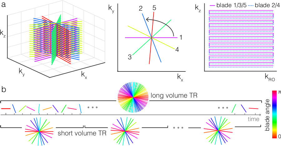
To address some of the weaknesses of 3D-EPI, we recently introduced the TURBINE3 acquisition scheme for fMRI, which uses a multi-shot 3D hybrid radial-Cartesian readout4-6. In TURBINE, kx,y-kz EPI “blades” are rotated about the kz axis in golden angle increments7 resulting in a Cartesian sampling of kz, and radial sampling in the kx-ky plane (Figure 1). The golden-angle sampling enables post-hoc selection of spatial and temporal resolution, allowing for intrinsic physiological noise and motion correction4 via self-navigation, and self-calibration of coil sensitivity information. Furthermore, its distributed spatio-temporal point-spread-function facilitates k-t accelerated reconstructions8-9. Here we demonstrate ultra-high resolution TURBINE fMRI at 7T, enabling 0.67mm isotropic resolution over 16mm slab acquisitions at TRvol=2.32s , and a 0.8x0.8x2.0mm3 whole-brain acquisition at TRvol=2.4s, both using sensitivity-encoded, temporally regularized k-t reconstructions.
Methods
Data Acquisition: Data was acquired at 7T on a healthy volunteer using two different TURBINE protocols:
- 16mm slab: 0.67mm isotropic, TR/TE=58/23ms, blade EPI matrix 288x24, TRvol = 2.32s (40 blades/volume), Reff= 7.2x$$$\frac{\pi}{2}$$$
- whole-brain: 0.8x0.8x2.0mm3, TR/TE=50/22ms, blade EPI matrix 240x66, R=2 within-blade, TRvol = 2.4s (48 blades/volume), Reff= 10x$$$\frac{\pi}{2}$$$
Protocol-1 was acquired twice, on slabs centered over the visual cortex and motor cortex respectively. All fMRI acquisitions were performed with a 30s/30s on/off flashing checkerboard and finger-tapping task. A 0.7mm isotropic T1-MPRAGE was acquired for structural comparison.
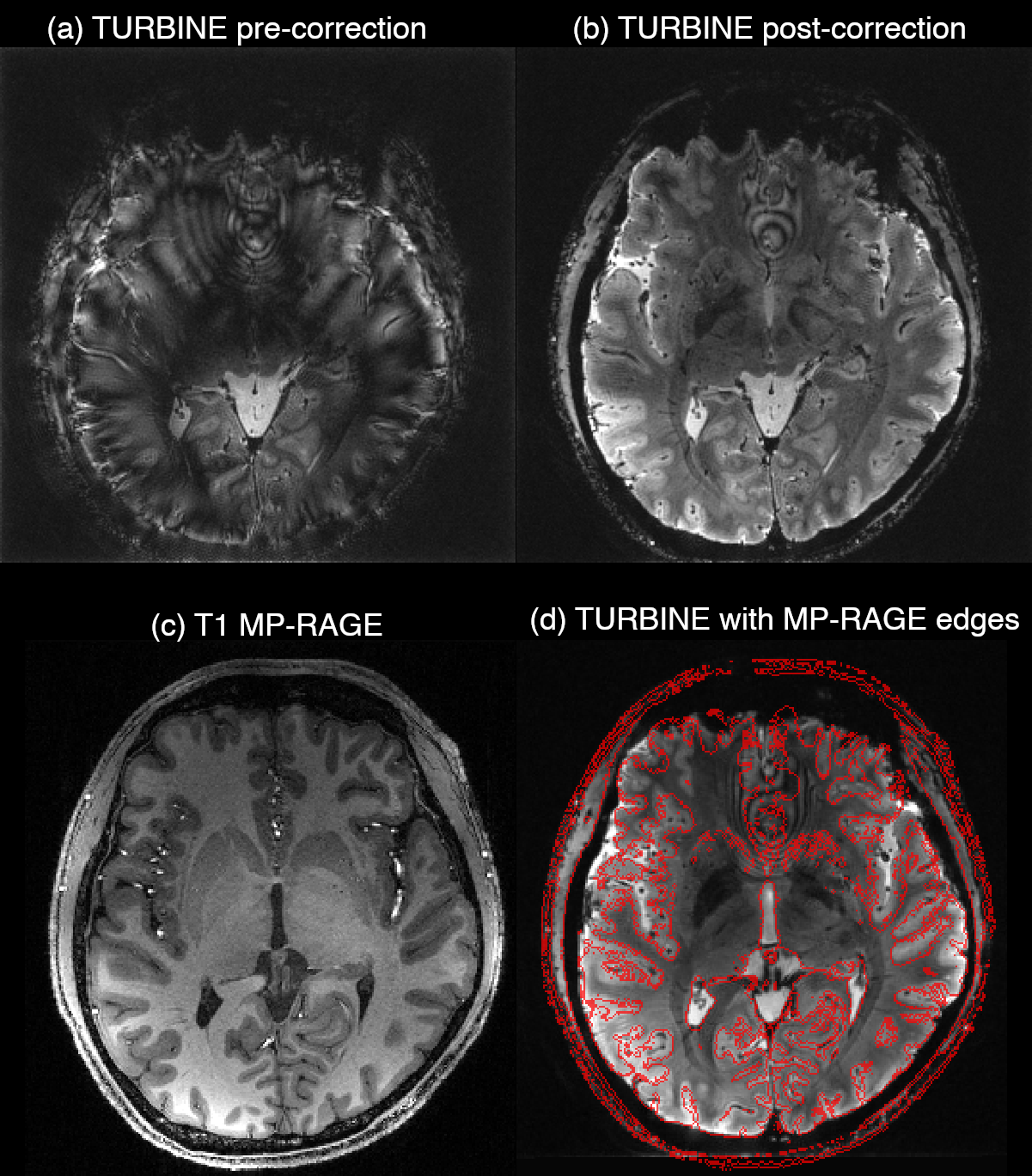
Corrections: Global and z-dependent phase corrections, EPI Nyquist ghosting and radial trajectory corrections were applied blade-by-blade prior to image reconstruction. For the whole-brain data, individual blades were also corrected for rotation about the z-axis using an initial high temporal resolution, low-spatial resolution navigator (TRvol = 0.8s) reconstruction (see ref 4 for details).
Image Reconstruction: Coil sensitivities (S) were estimated from the temporal mean of the data using the adaptive combine method10, and the sampling operator (F) was modelled with the NUFFT11. Images were linearly reconstructed using solving the regularized least squares problem:
$$\min_{x}\|FSx-k\|^2_2+\lambda\|\nabla_{t}x\|^2_2$$
with $$$x$$$ as the image time-series, $$$\nabla_{t}$$$ as the temporal finite difference operator, and the regularization parameter $$$\lambda=10^{5}$$$. Data were analyzed using a GLM and the output z-stat images were threshold-free cluster enhanced for visualization12.
Results
Figs. 2a,b show the impact of phase and trajectory corrections on reconstructed TURBINE images, although some residual artefacts can be seen in the anterior regions due to uncorrected B0 inhomogeneity. Figs. 2c,d highlight the distortion-free x-y plane provided by TURBINE, with excellent correspondence to the boundaries defined by the T1-MPRAGE structural image.
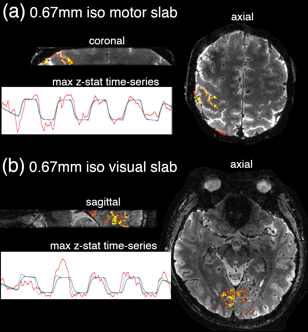
Figure 3 shows the results from the 0.67mm isotropic slab acquisitions, over the motor cortex (3a) and visual cortex (3b) respectively. The activation can be seen to be highly localised to cortical gray matter regions, showcasing the excellent specificity provided by the 0.67mm isotropic resolution. The underlays are T2*-weighted TURBINE images. Time-courses from the max z-stat voxels show excellent BOLD contrast.
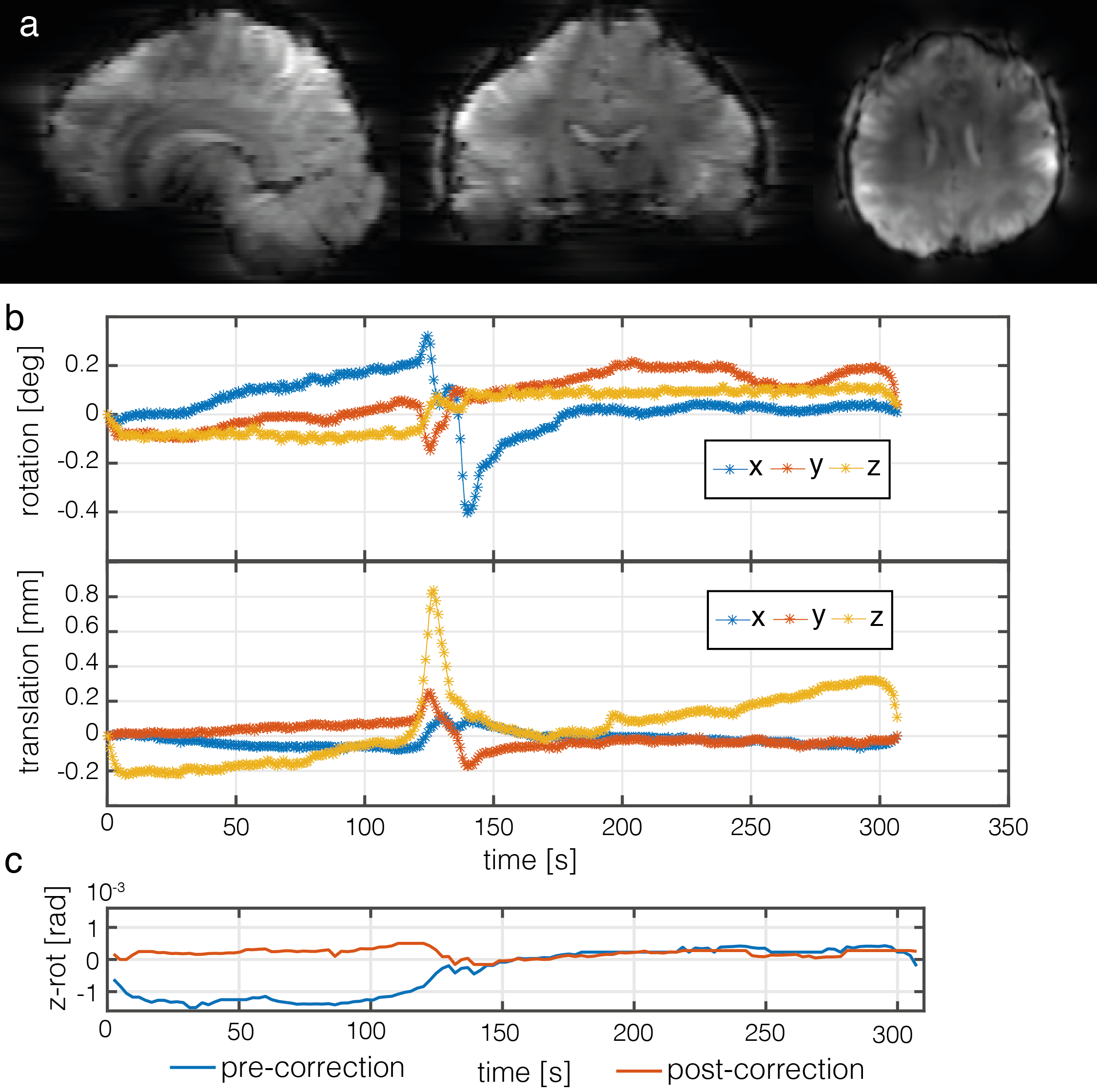
Figure 4a shows an example frame from the high temporal resolution
in the whole-brain acquisition, along with
estimated rotation and translations (Fig. 4b). In Fig. 4c, we see the result of
correcting for z-rotation, with the post-correction approximately 3.4x less RMS
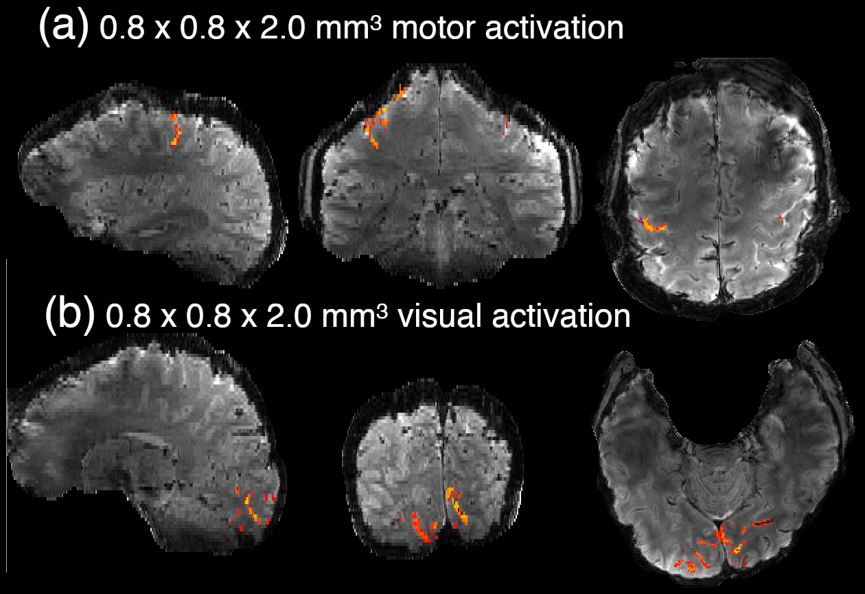
deviation from baseline. Fig. 5 shows the high resolution motor (5a) and visual
(5b) activation overlaid on the 0.8x0.8x2.0mm3 TURBINE images, demonstrating
again excellent spatial specificity to cortex. Some z-direction distortion is visible in the coronal image, due to poor shimming near the top of the head.
Discussion
We have demonstrated ultra-high resolution TURBINE fMRI at 7T using 0.67mm isotropic and 0.8x0.8x2.0mm3 whole-brain acquisitions. Unlike previous work6, which relied on extremely long volume TRs and spatial smoothing, these datasets were reconstructed at nominal TRvol of 2.32s and 2.4s respectively, and did not require any spatial smoothing. Instead, the highly accelerated datasets relied on temporal regularization to facilitate robust image reconstruction.
The TURBINE sequence provides minimal in-plane distortion, and facilitates intrinsic correction of shot-to-shot phase variation, motion, and trajectory errors for robust high-resolution BOLD imaging. Future work will include B0 correction to mitigate residual artefacts and remaining z-direction distortion.
Conclusion
We have shown highly spatially specific BOLD activation can be characterized by ultra-high resolution TURBINE imaging, which can enable more robust imaging in applications such as layer-specific fMRI.Acknowledgements
The Wellcome Centre for Human Neuroimaging is supported by core funding from the Wellcome [203147/Z/16/Z]. The Wellcome Centre for Integrative Neuroimaging is supported by core funding from the Wellcome Trust (203139/Z/16/Z). KM is supported by the Wellcome Trust (202788/Z/16/Z), and MC is supported by the Royal Academy of Engineering (RF201617\16\23).References
1. B.A. Poser, P.J. Koopmans, T. Witzel, L.L. Wald, M. Barth. “Three dimensional echo-planar imaging at 7 Tesla.” Neuroimage. 2010;51(1):261–266
2. L. Huber, D. H. Y. Tse, C. J. Wiggins, K. Uludağ, S. Kashyap, D. C. Jangraw, P. A. Bandettini, B. A. Poser, and D. Ivanov. “Ultra-high resolution blood volume fMRI and BOLD fMRI in humans at 9.4 T: Capabilities and challenges.” NeuroImage 178 (2018), pp. 769-779.
3. J. A. McNab, D. Gallichan, and K. L. Miller. “3D steady-state diffusion-weighted imaging with trajectory using radially batched internal navigator echoes (TURBINE).” Magnetic Resonance in Medicine 63.1 (2010), pp. 235–242.
4. N. N. Graedel , J. A. McNab, M. Chiew, and K. L. Miller. “Motion correction for functional MRI with three-dimensional hybrid radial-Cartesian EPI” Magnetic Resonance in Medicine 78.2 (2017), pp. 527-540
5. Ehses et al., Proceedings of the 22nd Annual Meeting of ISMRM (2014)
6. Bollman et al. Proceedings of the 26th Annual Meeting of ISMRM (2018)
7. S. Winkelmann, T. Schaeffter, T. Koehler, H. Eggers, and O. Dössel. “An Optimal Radial Profile Order Based on the Golden Ratio for Time-Resolved MRI.” IEEE Transactions on Medical Imaging 26.1 (2007), pp. 68–76.
8. M. Chiew, S. M. Smith, P. J. Koopmans, N. N. Graedel, T. Blumensath, and K. L. Miller. “k-t FASTER: Acceleration of functional MRI data acquisition using low rank constraints.” Magnetic Resonance in Medicine 74.2 (2015), pp. 353-364
9. M. Chiew, N. N. Graedel, J. A. McNab, S. M. Smith, and K. L. Miller. “Accelerating functional MRI using fixed-rank approximations and radial-cartesian sampling.” Magnetic Resonance in Medicine, 76.6 (2016), pp. 1825-1836
10. D. O. Walsh, A. F. Gmitro, and M. W. Marcellin. “Adaptive reconstruction of phased array MR imagery.” Magnetic Resonance in Medicine 43.5 (2000), pp. 682-690
11. J. A. Fessler, and B. P. Sutton. “Nonuniform fast Fourier transforms using min-max interpolation.” IEEE Transactions on Signal Processing 51.2 (2003), pp. 560-574
12. S. M. Smith, and T. E. Nichols. "Threshold-free cluster enhancement: addressing problems of smoothing, threshold dependence and localisation in cluster inference." NeuroImage 44 (2009), pp. 83–98
Figures