1167
Full-FOV, Whole-brain, Half-millimetre Resolution fMRI at 7T using Accelerated multi-band EPIK with TR-external Phase CorrectionSeong Dae Yun1 and N. Jon Shah1,2,3,4
1Institute of Neuroscience and Medicine 4, INM-4, Forschungszentrum Juelich, Juelich, Germany, 2Institute of Neuroscience and Medicine 11, INM-11, JARA, Forschungszentrum Juelich, Juelich, Germany, 3JARA - BRAIN - Translational Medicine, Aachen, Germany, 4Department of Neurology, RWTH Aachen University, Aachen, Germany
Synopsis
Ultra-high spatial resolution fMRI can identify brain activations with precise spatial localisation. There have been numerous attempts to achieve a sub-millimetre resolution in fMRI by using reduced- or full-FOV imaging. Although the reduced-FOV scheme can achieve more enhanced resolution than the full-FOV scheme, the restricted FOV often limits its use for more general functional studies. This work aims to present a novel half-millimetre resolution fMRI method which can also provide full-FOV and whole-brain coverage. The method was developed based on EPIK and a TR-external EPI phase correction scheme. Here, the above configuration was employed for exemplary finger-tapping fMRI at 7T.
Introduction
Ultra-high spatial resolution fMRI is of great interest in the fMRI community as it can reveal activated brain regions with precise spatial localisation. There have been numerous attempts to achieve sub-millimetre resolution in fMRI using reduced-FOV (rFOV) imaging methods.1-3 The rFOV approach can be realised using inner-volume selection, outer-volume suppression or 2D selective excitation pulses4 and can achieve a substantially higher spatial resolution (e.g. 0.65 mm × 0.65 mm)1 than the full-FOV scheme (e.g. 0.8 mm × 0.8 mm)5. However, the restricted FOV often limits its use for more general functional studies. This work aims to present a novel, half-millimetre resolution fMRI method which can also provide full-FOV and whole-brain coverage. The method was developed based on EPI with keyhole (EPIK) and a TR-external EPI phase correction scheme.6-10 Here, the performance of the proposed method was verified with finger-tapping fMRI at 7T.Methods
To achieve a high-resolution in fMRI, many research works employ acceleration techniques such as parallel imaging, partial Fourier or multi-band techniques. In this work, in order to achieve an even higher resolution, EPIK was additionally employed. When compared to EPI, EPIK has been proven to provide a higher temporal resolution, a sharper PSF and enhanced robustness against susceptibility distortions, while maintaining the capability of tracking dynamic signal changes for fMRI or perfusion MRI.6-9 In particular, a sharper PSF in EPIK leads to less image blurring artefacts, which is an essential feature for more spatially localised mapping. Figure 1 depicts a schematic representation of the k-space trajectory of EPIK. Its acquisition strategy resembles three-shot EPI but differs from it in that the central k-space region (keyhole) is fully sampled with the Nyquist criterion (Δky = 1/FOV). The peripheral k-space is sparsely sampled with Δky = 3/FOV, where the missing lines are reconstructed using the sliding window technique. In this way, the total number of phase encoding lines to be sampled can be largely reduced. This work employed 48 lines for the keyhole region, which was determined to be an optimal value by the MRI simulator, JEMRIS.11 In addition to EPIK, this work also combines a TR-external EPI phase correction scheme to further reduce the 'minimum TE required'.10 As depicted in Fig. 2, this scheme allocates the navigator echoes of EPI to a separated TR loop, which precedes the main EPI readout loop. The scheme has been proven to be effective in reducing the minimum TE required while maintaining the comparable performance of eliminating EPI ghost artefacts. The proposed method was verified with block-based finger-tapping fMRI. Data from a healthy male-volunteer were acquired on a Magnetom Terra 7T scanner (Siemens, Germany) under the following settings: TR/TE = 3000/25 ms, FOV = 210 × 210 mm2, matrix = 408 × 408 (0.51 × 0.51 mm2), partial Fourier = 5/8, 3-fold in-plane/2-fold inter-plane (multi-band) acceleration, αPC/αMain = 10°/90° and 60 slices with 2.0 mm thickness.Results
Figure 3a shows representative reconstructed slices from the fMRI scan. It is observed that the images are well reconstructed without any severe artefacts and clearly reveal a detailed spatial representation of the anatomical structures. Specific brain regions were chosen (marked by coloured rectangles) and displayed with an enlarged scale in Figs. 3b and c. The figure delineates microstructures such as the stria of Gennari in the visual cortex and the cortical layers well. Activated voxels were identified with an uncorrected p-value < 0.001, which are overlaid on the MP2RAGE12 scan (see Fig. 4). The whole slices are displayed with three different views (axial, sagittal and coronal), each of which shows its representative results. For the axial view, four representative slices are presented, showing that the identified voxels are distributed precisely along the cortical ribbon. Figure 4d shows the time-course data examined at the voxel showing the maximum t-score. Its signal behaviour shows excellent agreement with the given paradigm input.Discussion and conclusions
This work demonstrated half-millimetre resolution mapping of functional quantities with full-FOV and whole-brain coverage at 7T. The achieved resolution (0.51 mm) is substantially better than levels achieved in recent high-resolution fMRI studies, which suggests its potential use for columnar or layer-specific fMRI. The method used relatively small acceleration factors (i.e. 3-fold in-plane/2-fold inter-plane), which do not induce a significant SNR degradation and thus ensures reliable image reconstruction. For the same acceleration factors, matrix size and TR as above, the maximally possible performance of the method is listed in Fig. 5. Investigation on a more optimal setting remains as a further work.Acknowledgements
We gratefully acknowledge assistance from Elene Iordanishvili with inspection of reconstructed images and delineation of microstructures (e.g. stria of Gennari and cortical layers).References
- Heidemann RM, Ivanov D, Trampel R, Fasano F, Pfeuffer J, Turner R. Zoomed GRAPPA (ZOOPPA) for Functional MRI. In Proceedings of the 18th Annual Meeting of ISMRM, Stockholm, Sweden, 2010. Abstract 2889.
- Zimmermann J, Goebel R, De Martino F, van de Moortele PF, Feinberg D, Adriany G, Chaimow D, Shmuel A, Uğurbil K, Yacoub E. Mapping the organization of axis of motion selective features in human area MT using high-field fMRI. PLoS One. 2011;6(12):e28716.
- Kemper VG, De Martino F, Vu AT, Poser BA, Feinberg DA, Goebel R, Yacoub E. Sub-millimeter T2 weighted fMRI at 7 T: comparison of 3D-GRASE and 2D SE-EPI.Front Neurosci. 2015 May 5;9:163.
- Feinberg DA, Yacoub E. The rapid development of high speed, resolution and precision in fMRI. Neuroimage. 2012 Aug 15;62(2):720-5.
- Kendrick Kay, Keith Jamison, Luca Vizioli, Ruyuan Zhang, Eshed Margalit, Kamil Ugurbil. A critical assessment of data quality and venous effects in ultra-high-resolution fMRI. bioRxiv. 2018. doi: 10.1101/337667.
- Zaitsev M, Zilles K, Shah NJ. Shared k-space echo planar imaging with keyhole. Magn Reson Med. 2001;45(1):109-117.
- Zaitsev M, D'Arcy J, Collins DJ, Leach MO, Zilles K, Shah NJ. Dual-contrast echo planar imaging with keyhole: application to dynamic contrast-enhanced perfusion studies. Phys Med Biol. 2005 Oct 7;50(19):4491-505.
- Yun S, Reske M, Vahedipour K, et al. Parallel imaging acceleration of EPIK for reduced image distortions in fMRI. NeuroImage. 2013;73:135-143.
- Yun S, Shah NJ. Whole-brain high in-plane resolution fMRI using accelerated EPIK for enhanced characterisation of functional areas at 3T. PLoS One. 2017;12(9):e0184759.
- Yun S, Shah NJ. On the analysis of EPI phase correction with small tip angle excitation to reduce minimum required TE: application to whole-brain submillimetre resolution fMRI at 3T. In Proceedings of the 26th Annual Meeting of ISMRM, Paris, France, 2018. Abstract 4245.
- Stöcker T, Vahedipour K, Pflugfelder D, Shah NJ. High-performance computing MRI simulations. Magn Reson Med. 2010 Jul;64(1):186-93.
- Marques JP, Gruetter R. New developments and applications of the MP2RAGE sequence - focusing the contrast and high spatial resolution R1 mapping. PLoS One. 2013 Jul 16;8(7):e69294.
Figures
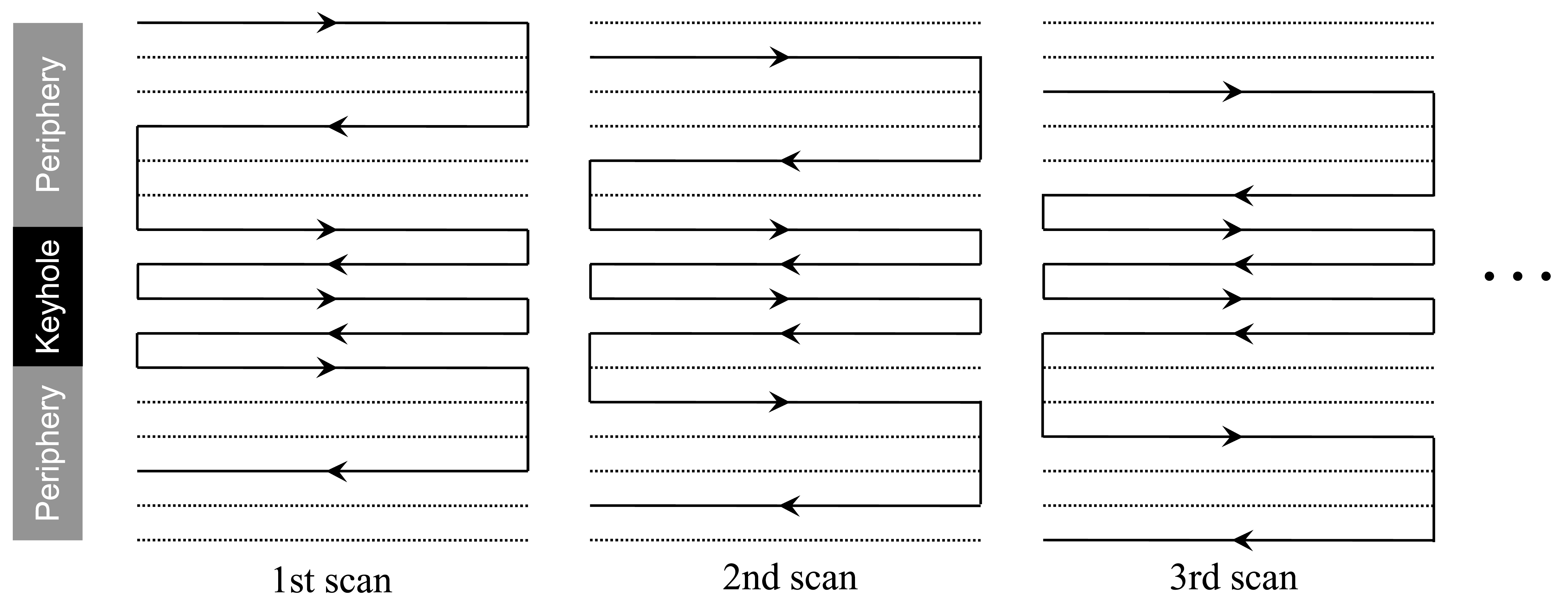
Figure 1. Schematic
representation of the k-space trajectory of EPIK. In every temporal scan, the
central k-space (keyhole) region is fully sampled whilst the peripheral k-space
is sparsely sampled resembling three-shot EPI. The solid and dotted lines
indicate the k-space lines to be sampled and skipped, respectively. The sampling
lines of the subsequent scans (4th, 5th, …) follow the above sampling
configuration starting from the 1st scan case.
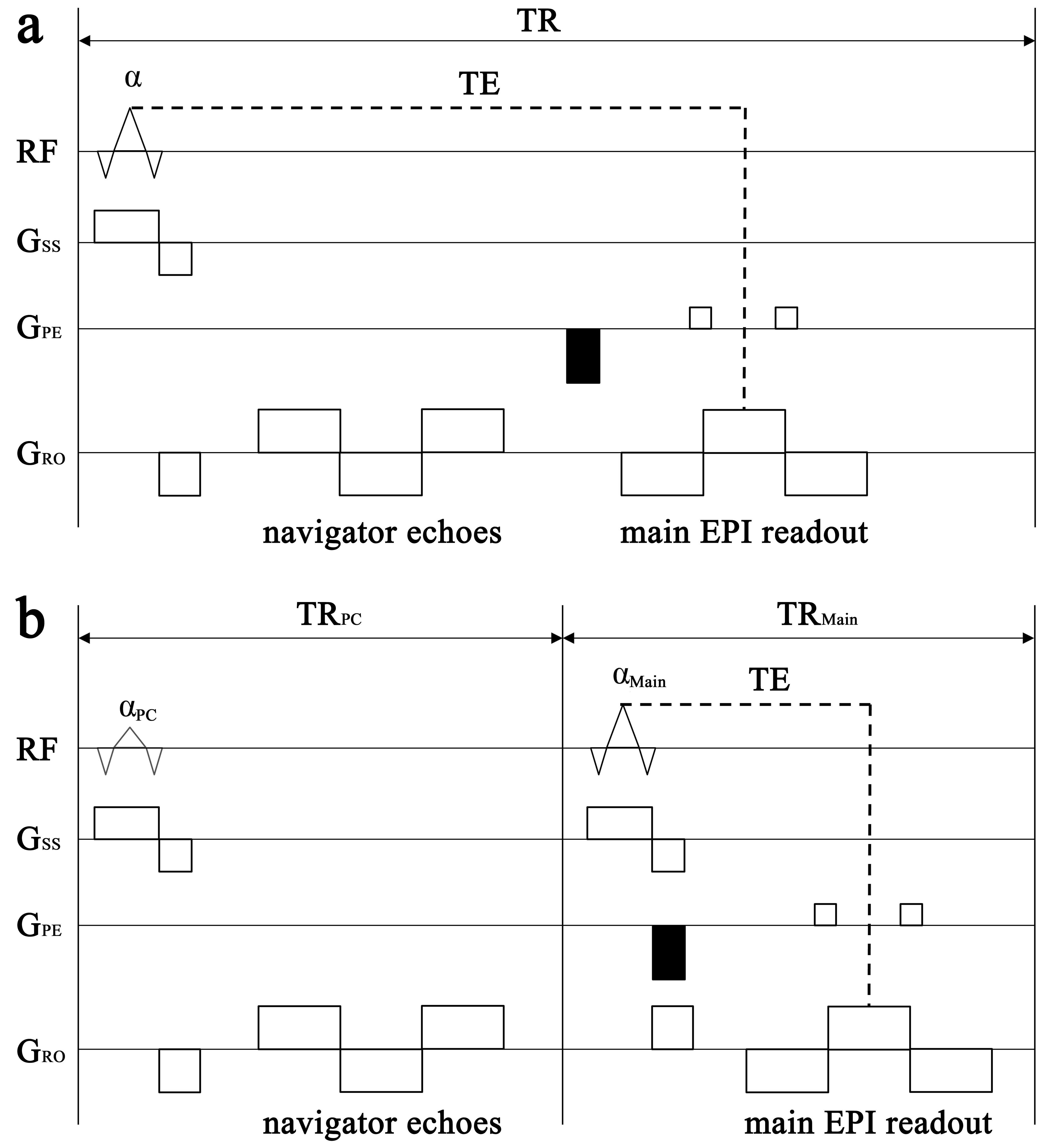
Figure 2. Schematic representation of an EPI
sequence with (a) a TR-internal and
(b) a TR-external navigator echo schemes
for EPI phase correction. In the TR-external scheme, the navigator echoes are
located in a separated TR loop (TRPC), which precedes the main EPI
TR loop (TRMain). As illustrated in the figure, the TR-external
scheme is expected to have a reduced 'minimum TE required'. Furthermore, this
scheme can locate the phase encoding gradient (marked with black shading) together
with other encoding gradients (see Fig. b), which leads to an additional reduction
of TE.
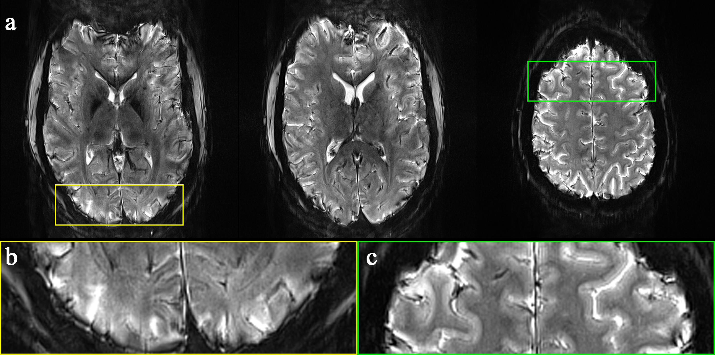
Figure 3. (a) Three representative reconstructed slices (0.51 × 0.51 mm2)
from the proposed method, (b) enlarged
plot of the region marked by the yellow rectangle, where the stria of Gennari
(myelinated band) in the visual cortex can be observed and (c) enlarged plot of the region marked
by the green rectangle, where cortical layers can be observed.
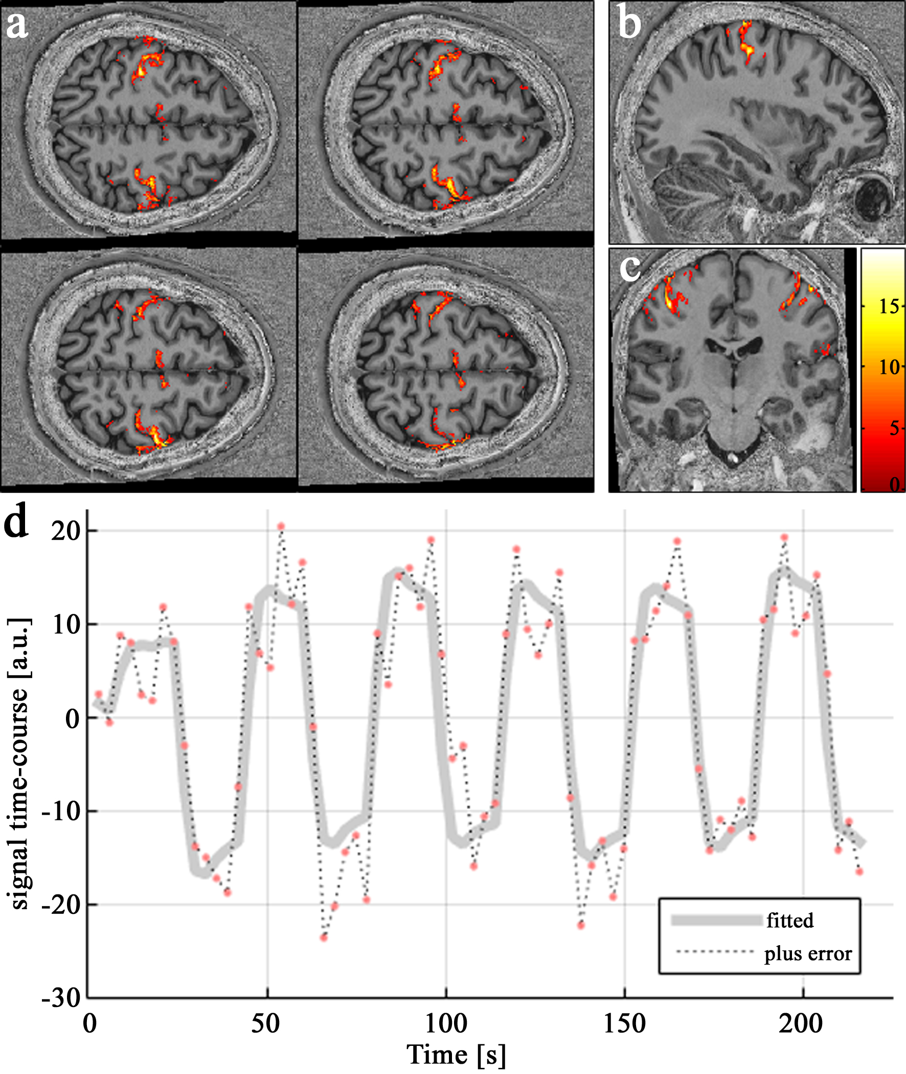
Figure 4. Results of finger-tapping fMRI. Activated
voxels overlaid on the MP2RAGE scan presented with the (a) axial, (b) sagittal
and (c) coronal views. For the axial
view, four representative slices are presented, showing that the identified
voxels are distributed precisely along the cortical ribbon. (d) Time-course data examined at the
voxel showing the maximum t-score. Its signal behaviour shows excellent agreement
with the given paradigm input.
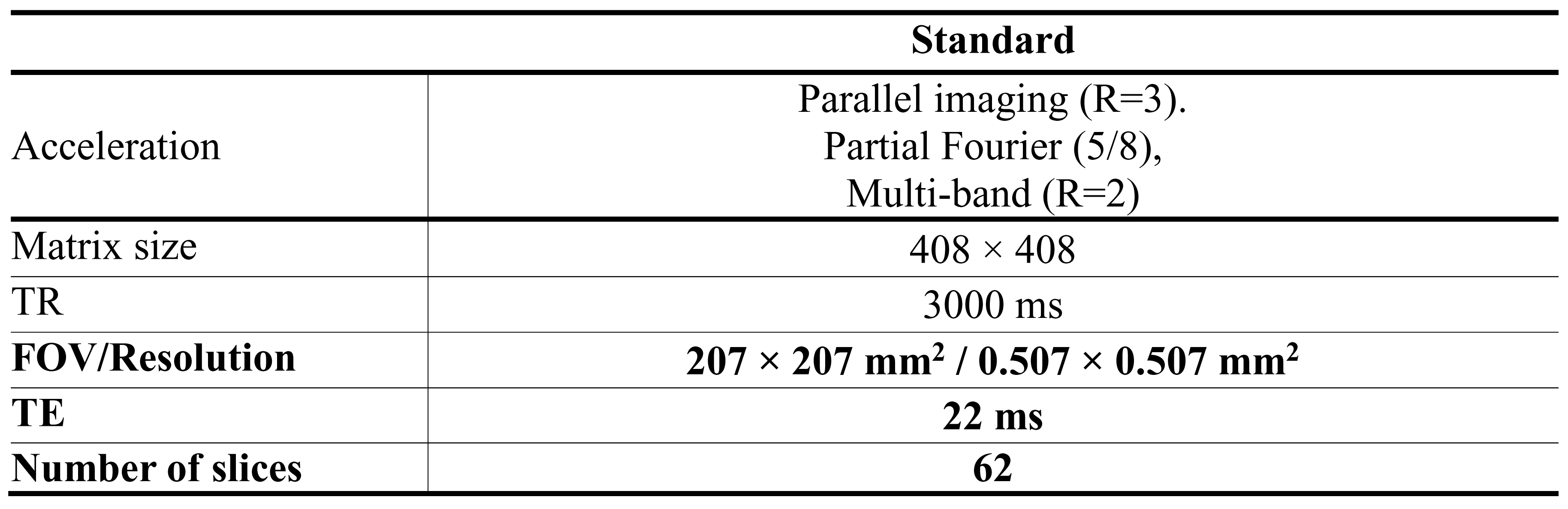
Figure 5. A possible imaging configuration
of the proposed method when pushing its limit toward highest possible
resolution and slice coverage whilst other parameters (acceleration, matrix
size and TR) were kept identical to the present study. A smaller voxel size, a shorter
TE and a slightly larger number of slices can be achieved with the above
setting. Investigation on a more optimal setting for a particular fMRI
study remains as a further work.