1165
Accelerated spin-echo fMRI using Multisection Excitation by Simultaneous Spin-echo Interleaving (MESSI) with ‘complex-basis’ RF-encoded generalized SLIce Dithered Enhanced Resolution Simultaneous Multi-Slice (MESSI-gSlider-SMS)1Athinoula A. Martinos Center for Biomedical Imaging, Massachusetts General Hospital, Charlestown, MA, United States, 2Department of Radiology, Harvard Medical School, Boston, MA, United States, 3Harvard-MIT Division of Health Science and Technology, Cambridge, MA, United States
Synopsis
High spatiotemporal resolution spin-echo fMRI is challenging as it requires a long echo time to generate BOLD-contrast, resulting in longer repetition times. We propose a new technique, Multisection Excitation by Simultaneous Spin-echo Interleaving (MESSI), which utilizes the dead time in long TE acquisitions to improve the slice-coverage of SE-fMRI. For further accelerations, we combine the MESSI with both ‘complex-basis’ RF-encoded gSlider and conventional-SMS. Compared with standard SE-EPI acquisition with the same TR, the proposed MESSI-gSlider acquisition shows comparable tSNR but with eight-fold increase in slice-coverage. This method should be beneficial for applications requiring high spatiotemporal resolution SE-fMRI with whole-brain coverage.
Introduction
Spin-echo(SE)-fMRI has shown to provide improved BOLD spatial specificity when compare to gradient-echo(GE)-fMRI1-4. However, high spatiotemporal SE-fMRI is difficult to achieve due to the long echo times(TE) to generate BOLD-contrast, resulting in long repetition times(TR). Simultaneous Multi-Slice(SMS) provides increased temporal efficiency of fMRI5-9, and faster temporal sampling has been beneficial in many domains8,10-11; however, MB acceleration is limited by the total acceleration-factor (Rtotal=Rinplane×MB), which is typically set to 6–8 to avoid large g-factor noise. To achieve SE-fMRI with high-spatial specificity, high Rinplane-acceleration is used to limit distortion and image blurring, thereby limiting MB-acceleration capability. To overcome this limitation, we previously developed ‘complex-basis’ RF-encoded gSlider12 that achieves a 2× gain in slice-coverage without temporal smoothing. Here, we propose a new technique, Multisection Excitation by Simultaneous Spin-echo Interleaving (MESSI), which utilizes the dead time in long-TE acquisitions13-14 to improve the slice-coverage of SE-fMRI. MESSI is then combined with both ‘complex-basis’ gSlider12 and conventional-SMS to provide 8× slice-acceleration for SE-fMRI at Rinplane=4. Using this technique, SE-fMRI experiments with sensory stimulation and with a breath-hold task were performed and compared with conventional-SMS at MB-2 to demonstrate that a 4× increase in slice-coverage can be achieved with minimal penalty.Methods
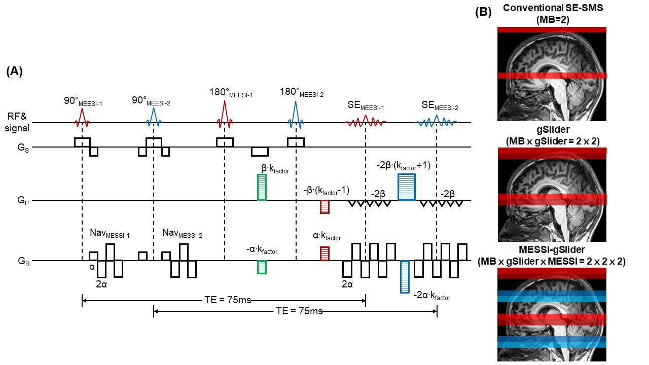
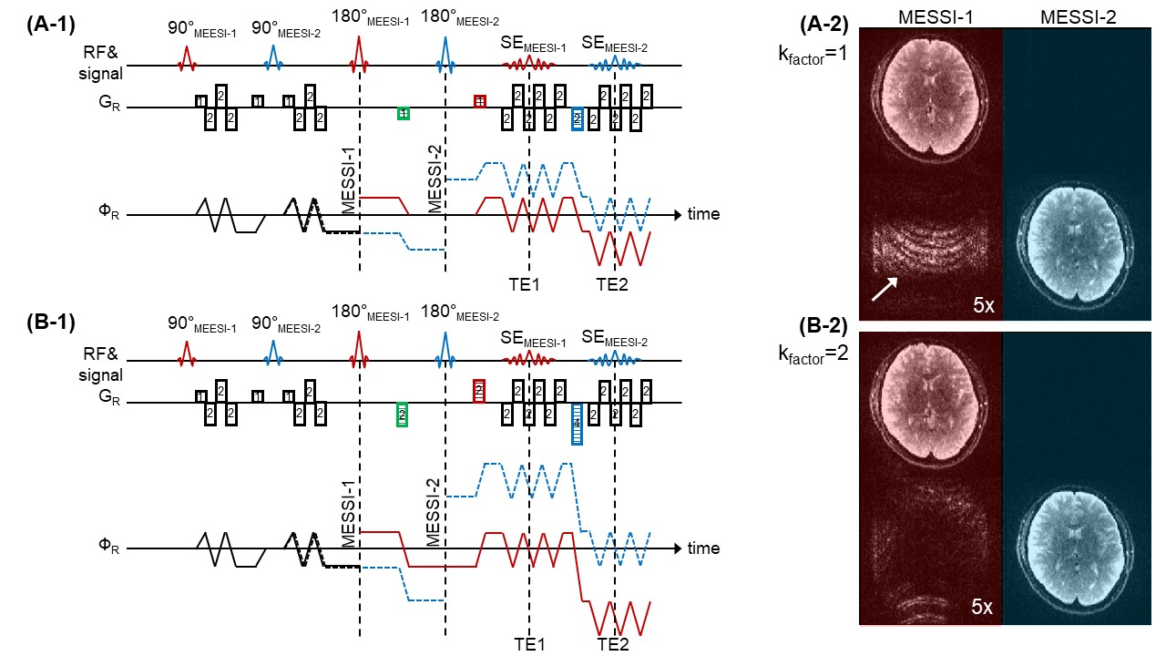
The MESSI-gSlider sequence diagram and excitation scheme are shown in Fig.1. To excite two different slice-groups simultaneously in the MESSI sequence (denoted as MESSI-1 and MESSI-2), the following components were added to the conventional-SE-EPI sequence as depicted in Fig.1A: i) an additional readout and 90° and 180° pulses for MESSI-2-group (blue-colored pulses and SE) with a TE matched to the MESSI-1-group, ii) to separate the k-spaces of the two MESSI-groups, two dephasing-gradients (green-colored gradients) that dephase signal pathways for both MESSI-groups, iii) two rephasing-gradients for MESSI-1-group (red-colored gradients), and iv) two rephasing-gradients for MESSI-2-group (blue-colored gradients). The effect of the spins of the MESSI sequence in the cases where kfactor is set to 1 or 2 is illustrated in Figs.2A and 2B, respectively. Rephasing-gradients for MESSI-1-group rephase spins in the readout for MESSI-1-group while dephasing magnetization from MESSI-2-group. The same is true for the rephasing gradients for the MESSI-2-group. Increasing the value of kfactor increases the signal dephasing between the MESSI-slice-groups and reduces potential high k-space signal-leakage. Data were collected from three healthy subjects using a 3T Siemens Prisma scanner with the vendor-supplied 32-channel coil. To directly investigate the level of signal-leakage between MESSI slice-groups, either MESSI-1 or MESSI-2 pulses were set to 0° with kfactor set to 1 or 2. To compare MESSI-gSlider against conventional-SMS and gSlider, two fMRI experiments were conducted, visual stimulation and a breath-hold task. Scans for all methods were acquired at the same TR. For visual stimulation, the subject viewed four 36s (12-s on, 20-s off) runs of flashing “checkboard” stimulus. For the timed breath-hold task, the subject was cued to hold their breath for 12-s followed by 30-s of free breathing with four-trials/run. The scan protocols for the fMRI experiments were as follows: TR/TE=2000/75ms, FOVxy=210×210mm2, p.f.=6/8, 1.5mm isotropic resolution, including (i) conventional-SMS with Rinplane×MB=4×2, 22-slices, FOVz=33mm, (ii) gSlider with Rinplane×MB×gSlider=4×2×2, 44-slices, FOVz=66mm, and (iii) MESSI-gSlider with Rinplane×MB×gSlider×MESSI=4×2×2×2, 84-slices, FOVz=126mm, kfactor=2.Results
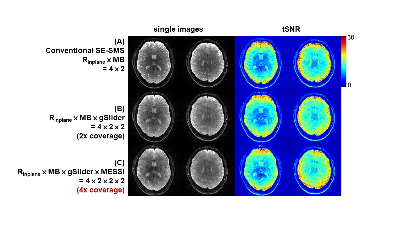
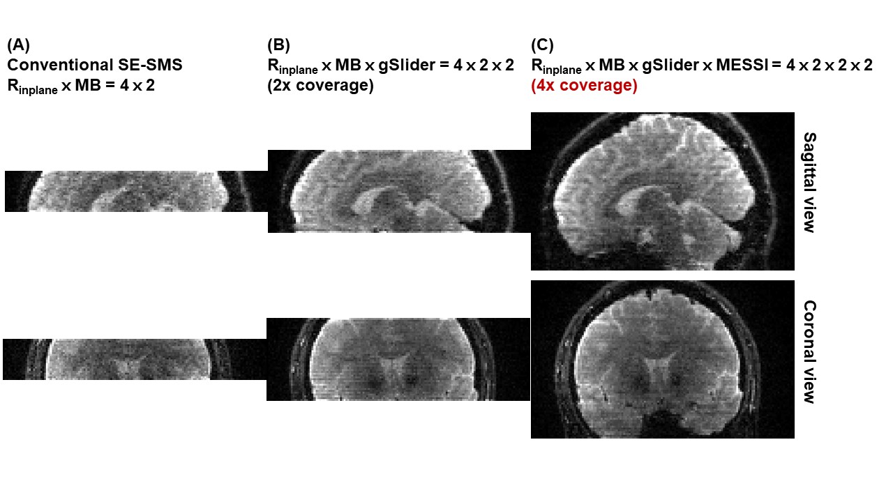
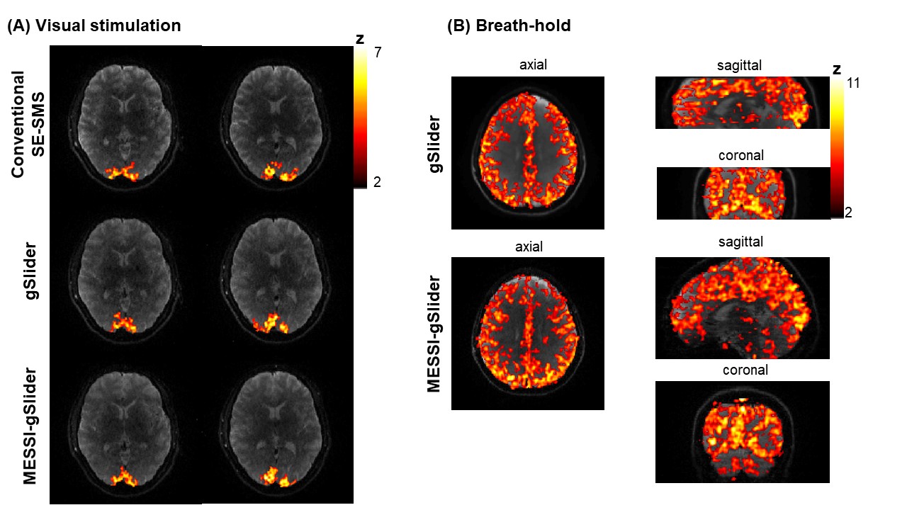
Figs.2A-2 and 2B-2 show the signal-leakage between the MESSI-groups for acquisitions with kfactor=1 and 2. Compared to the kfactor=1 case, the kfactor=2 case shows substantially less signal-leakage, as expected. Moreover, the leakage from MESSI-1 into MESSI-2 slice-group is small in both kfactor=1 and 2 cases as the leakage has undergone large T2 and T2* decays. Fig.3 shows that comparable image-quality and tSNR can be obtained from conventional-SMS, gSlider, and MESSI-gSlider acquisitions. Fig.4 demonstrates the extended slice-coverage in sagittal and coronal views, where gSlider and MESSI-gSlider exhibit similar image-quality to conventional-SMS, while achieving 2× and 4× slice-acceleration, respectively. In particular, the MESSI-gSlider acquisition enables whole-brain coverage at 1.5mm isotropic resolution. Finally, Fig.5 shows the feasibility of fMRI using MESSI-gSlider, with z-statistic maps (FSL-Feat) of conventional-SMS, gSlider and MESSI-gSlider. For both fMRI experiments, the MESSI-gSlider method maintains high-temporal resolution at 2× brain coverage when compared to gSlider alone, while exhibiting comparable activation patterns. The similarity of activation maps between the acquisitions indicates that fMRI sensitivity is not compromised from the addition of MESSI.Discussion and Conclusion
We proposed a MESSI-gSlider-SMS sequence to accelerate SE-fMRI, which enables 4× increased slice-coverage with minimal penalty compared with the conventional-SMS acquisition. Although SE-BOLD reduces sensitivity compared to GE-BOLD15, future work will focus on applying this method to ultra-high field SE-fMRI, where the T2-weighting should improve microvascular specificity. Additionally, the higher-resolution imaging at ultra-high field may greatly benefit from the 4-fold increase in slice-coverage.Acknowledgements
This work was supported in part by the NIH NIBIB (grants R01-MH116173, R01-EB020613, P41-EB015896, U01-EB025162 and R01-EB019437), by the BRAIN Initiative (NIH NIMH grant R01-MH111419), by the shared instrumentation (grants S10RR023401, S10RR019307, S10RR019254, S10RR023043) and by the MGH/HST Athinoula A. Martinos Center for Biomedical Imaging. We thank Anna I. Blazejewska for helping with fMRI experiments and Daniel Park for helping with implementing MESSI-gSlider sequence.References
[1] E. Yacoub, T. Q. Duong, P.F. Van De Moortele et al. Spin-echo fMRI in humans using high spatial resolutions and high magnetic fields. Magn. Reson. Med. 2003; 49(4):655–664.
[2] N. Harel, J. Lin, S. Moeller et al. Combined imaging-histological study of cortical laminar specificity of fMRI signals. Neuroimage. 2006; 29(3):879–887.
[3] S. P. Lee, A. C. Silva, K. Ugurbil et al. Diffusion-Weighted Spin-Echo fMRI at 9.4 T : Microvascular / Tissue Contribution to BOLD Signal Changes. Magn. Reson. Med. 1999; 928(5):919–928.
[4] S. Han, J. P. Son, H. Cho et al. Gradient-echo and spin-echo blood oxygenation level-dependent functional MRI at ultrahigh fields of 9.4 and 15.2 Tesla. Magn. Reson. Med. 2018; DOI: 10.1002/mrm.27457.
[5] D. J. Larkman, J. V. Hajnal, A. H. Herlihy et al. Use of multicoil arrays for separation of signal from multiple slices simultaneously excited. J. Magn. Reson. Imaging 2001;13(2):313–317.
[6] F. A. Breuer, M. Blaimer, R. M. Heidemann et al. Controlled aliasing in parallel imaging results in higher acceleration (CAIPIRINHA) for multi-slice imaging. Magn. Reson. Med. 2005;53(3):684–691.
[7] S. Moeller, E. Yacoub, O. A. Cheryl et al. Multiband multislice GE-EPI at 7 tesla, with 16-fold acceleration using partial parallel imaging with application to high spatial and temporal whole-brain FMRI. Magn. Reson. Med. 2010; 63(5):1144–1153.
[8] D. A. Feinberg, S. Moeller, S. M. Stephen et al. Multiplexed echo planar imaging for sub-second whole brain fmri and fast diffusion imaging. PLoS One. 2010; 5(12).
[9] K. Setsompop, B. A. Gagoski, J. R. Polimeni et al. Blipped-controlled aliasing in parallel imaging for simultaneous multislice echo planar imaging with reduced g-factor penalty. Magn. Reson. Med. 2012; 67(5): 1210–1224.
[10] L. D. Lewis, K. Setsompop, B. R. Rosen et al. Fast fMRI can detect oscillatory neural activity in humans. Proc Natl Acad Sci. 2016;113(43):6679-6685.
[11] A. K. Sahib, K. Mathiak, M. Erb et al. Effect of temporal resolution and serial autocorrelations in event-related functional MRI. Magn Reson Med. 2016;76(6):1805-1813.
[12] S. Han, C. Liao, M.K. Manhard et al. Accelerated spin-echo fMRI using generalized SLIce Dithered Enhanced Resolution Simultaneous MultiSlice (gSlider-SMS) with 'complex-basis' RF-encoding. ISMRM 2018, #3406
[13] P. van Gelderen, J.H. Duyn, N.F. Ramsey et al. The PRESTO technique for fMRI. Neuroimage. 2012;62:676-681.
[14] B. Bilgic, H. Ye, L. L. Wald et al. Simultaneous Time Interleaved MultiSlice (STIMS) for Rapid Susceptibility Weighted acquisition. Neuroimage. 2017;155:577-586.
[15] K. Boyacioǧlu, J. Schulz, N. C. J. Müller et al. Whole brain, high resolution multiband spin-echo EPI fMRI at 7T: A comparison with gradient-echo EPI using a color-word Stroop task. Neuroimage. 2014;97:142-150.
Figures