1163
Radiomics analysis of peritumoral tissue on gadoxetic acid-enhanced MR imaging for prediction of early recurrence in hepatocellular carcinoma1Department of Radiology, West China Hospital,Sichuan University, Chengdu, China
Synopsis
Postoperative recurrence has become the main obstacle in prognosis of HCC patients, even after successful curative therapy. Our study analyzed the radiomics features derived from peritumoral tissue on gadoxetic acid-enhanced MR images, combined with clinical characteristics and subjective imaging findings, to evaluate their ability to preoperatively predict early recurrence of HCC after surgical resection. An AUC of 0.882 for the radiomics signature and an improved AUC of 0.926 for integrated radiomics nomogram were obtained. These results suggest that radiomics features can accurately and objectively predict early recurrence of HCCs after curative resection from preoperative MR images.
Introduction
Hepatocellular carcinoma (HCC) is the sixth leading cause of cancer-related death worldwide.1 Hepatic resection is recommended as the first treatment for patients with well-preserved liver function, while long-term prognosis still remains poor due to a high recurrence rate (68%-96%) within 5 years.2, 3
Qualitative radiological features reported to be predictive of early recurrence were related to peritumoral tissue, such as arterial enhancement and hypointensity on hepatobiliary phase, which suggests that imaging features involving peritumoral tissue may reveal a direct association with tumor prognosis and recurrence.4, 5 Moreover, Radiomics analysis has been proposed as a robust and effective imaging assessment method to quantify tumor phenotypic characteristics by extracting multiple quantitative features from traditional medical images.6
Texture features of peritumoral tissue based on CT images have been reported as predictors for MVI of HCC in recent studies.7, 8 However, few studies have focused on the potential ability of peritumoral radiomics features extracted from MR images in predicting early recurrence. Therefore, the aim of this study was to determine whether the peritumoral radiomics features from gadoxetic acid-enhanced MR images can predict early recurrence (<1year) in patients with hepatocellular carcinoma (HCC) after surgical resection.
Methods
39 patients with surgically confirmed HCC were enrolled in this IRB-approved prospective study. A total of 1925 texture features within peritumoral region (a 5-pixel-wide radius surrounding the tumor boundary) were obtained from multiple phases (T2-weighted, non-enhanced T1-weighted, arterial phase, portal venous phase and hepatobiliary phase). The most predictive radiomics features were selected to build a radiomics signature using the least absolute shrinkage and selection operator (LASSO) algorithm with a 10-fold cross validation applied. Then, the radiomics signature, clinical variables and subjective imaging findings were assessed using multivariable logistic regression analysis to construct a radiomics nomogram. Receiver operation characteristic (ROC) analysis and decision curve analysis were performed to evaluate the accuracies of the predictive models.Results
Nineteen patients (48.7%) experienced early recurrence by imaging follow-up. Four radiomics features were identified as independent predictors of early recurrence, including Correlation, InverseDifferenceMoment (IDM) from T2-weighted images and IDM, Histogram MinIntensity from arterial phase images, showed a favorable discriminatory ability with an area under the ROC curve (AUC) of 0.882 (95%CI:0.753-0.921), sensitivity of 84.6% and specificity of 88.0%.The radiomics signature (odds ratio [OR]=7.501,95%CI[1.406,40.014];p=0.018), AFP level>400ng/ml(OR=0.002,95%CI[0.000,0.696];p=0.037) and non-smooth tumor margin (OR=177.425,95%CI[1.355,39.14];p=0.070) were identified as independent predictive factors for early recurrence. The integrated radiomics nomogram comprising all predictive factors showed an improved predictive performance than the radiomics signature (AUC, 0.926 vs.0.882, p<0.05), with incremental clinical usefulness.Discussion
In our study, we found that quantitative radiomics features from peritumoral region are promising predictors of early recurrence in HCCs.Previous studies revealed that peritumoral tissue is the first to be affected by microvascular invasion (MVI), a known risk factor of early recurrence in HCC, vessels of which serves as the main blood dissemination path from which intrahepatic and extrahepatic metastasis originate.9 In addition, radiographic imaging findings from the literature also showed that the peritumoral region was the most informative region, including tumor margin, peritumoral enhancement and hypointensity on hepatobiliary phase.4, 5, 10 In the present study, the integrated radiomics nomogram comprising all predictive factors showed an improved predictive performance, which suggest that radiomics features may have incremental value for conventional approaches.Hui et al11 demonstrated the prognostic capability of texture features derived from MR images for predicting early recurrence of HCC patients, with accuracy values range from 0.78 to 0.84. In this study, the radiomics signature based on routine MR images demonstrated satisfactory discriminating power with an AUC of 0.882 and proved to be an independent predictor for early recurrence in the radiomics nomogram (p=0.018). Therefore, imaging features involving peritumoral tissue may reveal a direct association with early recurrence and reflect more prognostic information compared to those of tumor itself. Further studies are warranted to reproduce and assess our reported findings.Conclusion
Peritumoral radiomics features from preoperative MR images had the potential of predicting early recurrence in patients with HCC after resection.Acknowledgements
No acknowledgement found.References
1. Torre LA, Bray F, Siegel RL, et al., Global cancer statistics, 2012. CA Cancer J Clin, 2015. 65(2): p. 87-108.
2. Poon RT, Fan ST, Lo CM, et al., Long-term survival and pattern of recurrence after resection of small hepatocellular carcinoma in patients with preserved liver function: implications for a strategy of salvage transplantation. Ann Surg, 2002. 235(3): p. 373-382.
3. Lau WY and Lai EC, Hepatocellular carcinoma: current management and recent advances. Hepatobiliary Pancreat Dis Int, 2008. 7(3): p. 237-257.
4. An C, Kim DW, Park YN, et al., Single Hepatocellular Carcinoma: Preoperative MR Imaging to Predict Early Recurrence after Curative Resection. Radiology, 2015. 276(2): p. 433-443.
5. Lee S, Kim SH, Lee JE, et al., Preoperative gadoxetic acid-enhanced MRI for predicting microvascular invasion in patients with single hepatocellular carcinoma. J Hepatol, 2017. 67(3): p. 526-534.
6. Vial A, Stirling D, Field M, et al., The role of deep learning and radiomic feature extraction in cancer-specific predictive modelling: a review. Translational Cancer Research, 2018.
7(3): p. 803-816.7. Zheng J, Chakraborty J, Chapman WC, et al.,Preoperative Prediction of Microvascular Invasion in Hepatocellular Carcinoma Using Quantitative Image Analysis. J Am Coll Surg, 2017. 225(6): p. 778-788 e771.
8. Armato SG, Petrick NA, Chakraborty J, et al., Preoperative assessment of microvascular invasion in hepatocellular carcinoma. 2017. 10134: p. 1013410.
9. Roayaie S, Blume IN, Thung SN, et al., A system of classifying microvascular invasion to predict outcome after resection in patients with hepatocellular carcinoma. Gastroenterology, 2009. 137(3): p. 850-855.
10. Hu HT, Shen SL, Wang Z, et al., Peritumoral tissue on preoperative imaging reveals microvascular invasion in hepatocellular carcinoma: a systematic review and meta-analysis. Abdom Radiol (NY), 2018.
11. Hui TCH, Chuah TK, Low HM, et al., Predicting early recurrence of hepatocellular carcinoma with texture analysis of preoperative MRI: a radiomics study. Clin Radiol, 2018.
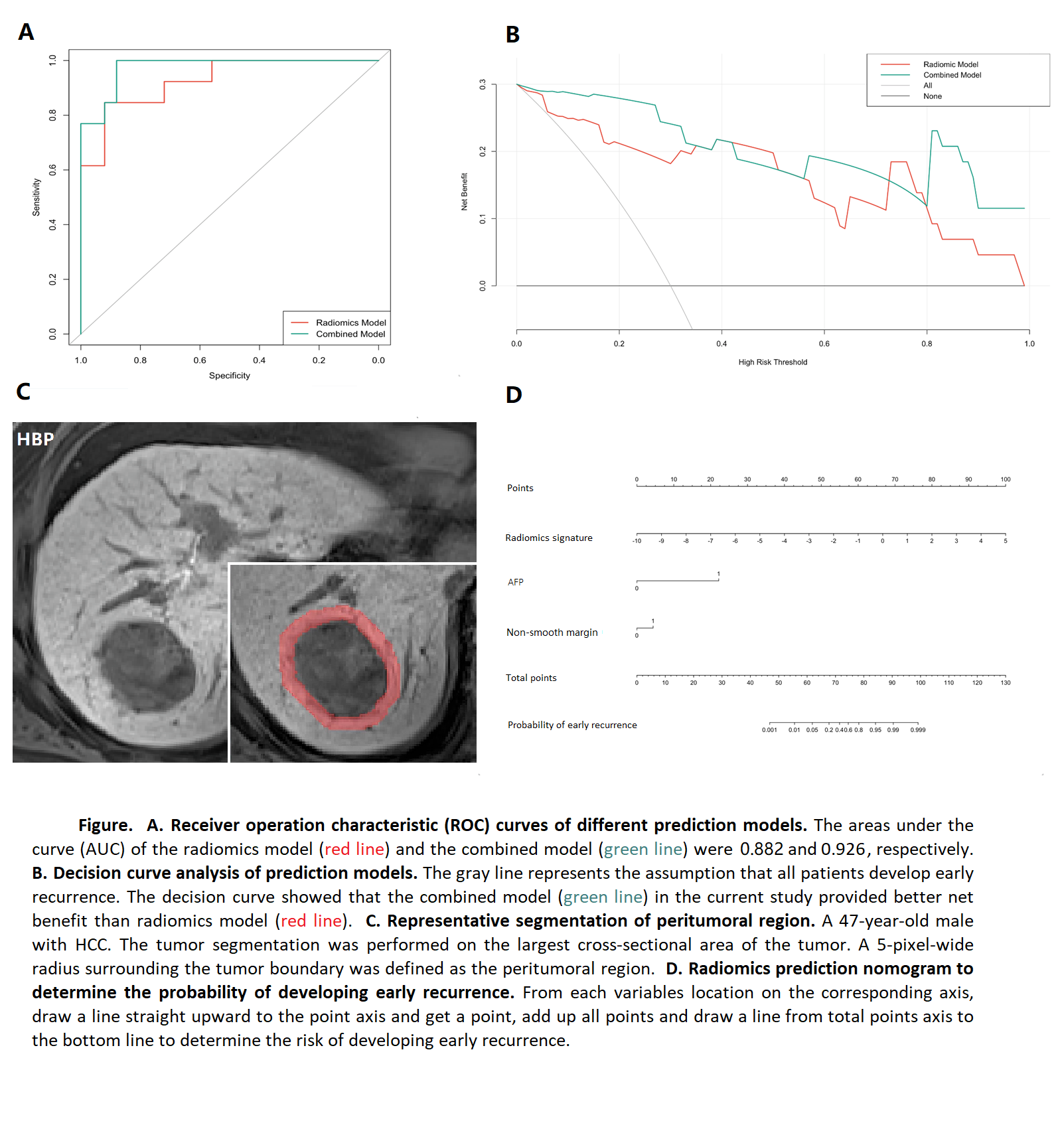
Figures