1151
Chronic hepatic encephalopathy in early developing brain, neurometabolic changes differ depending on the age of disease onset, in vivo longitudinal 1H MRS study1CIBM, EPFL, Lausanne, Switzerland, 2Service of Biomedicine, Neurometabolic Unit, Lausanne University Hospital, Lausanne, Switzerland, 3Swiss Center for Liver Disease in Children, University Hospitals Geneva, Geneva, Switzerland
Synopsis
Chronic hepatic encephalopathy (CHE) due to chronic liver disease (CLD) causes irreversible cognitive deficits in children. We aimed to study possible differences in neurometabolic changes in rat model of CLD&CHE depending on the age of the disease onset (post-natal day 15 vs 21), using in-vivo 1H-MRS. We showed differences in Gln, tCho, Lac, Asc, GSH and neurotransmitter concentrations between p15 and p21 pups during the evolution of the disease. These differences suggest that age of disease onset and its coincidence with neurodevelopmental processes play an important role and may result in different vulnerability to the disease depending on the age.
Introduction:
Chronic hepatic encephalopathy (CHE) is a serious neuropsychiatric disorder due to chronic liver disease (CLD) in adults and children. It is known that children are more affected by CLD than adult patients, with long-lasting cognitive deficits after liver transplantation1-4. Previously we showed that neurometabolic changes due to CHE were more severe and appeared earlier in young compared to adult rats after bile duct ligation (BDL)5. This finding led us to hypothesize that the effect of CLD on the brain may depend on the neurodevelopmental window at the time of the CLD onset. Therefore, using in vivo longitudinal 1H-MRS, we aimed to study whether CLD affects brain metabolism differently when the disease is acquired at two different moments of brain development in rats, at post-natal day 15 (p15) and p21, something never studied to date.Methods:
In this study, BDL surgery was performed on 7 male Wistar rats at p15. 8 animals were sham operated at matched age to take into account brain development. Changes in p15 BDL-rats were compared to rats from the previous study, operated at p21 (12 BDLs, 9 shams)5. Based on previous reports, rat brain maturation at p15 and p21 is approximately equivalent to 4 and 9 months in humans6. Blood sampling and in vivo 1H-MRS in hippocampus (VOI=2x2.8x2mm3) were performed at week 2,4 and 6 after BDL surgery. All MR experiments were performed on a 9.4T system (Varian/Magnex Scientific) using home-built 14mm diameter quadrature 1H-surface coil as a transceiver and ultra-short-echo time SPECIAL spectroscopy sequence (TE/TR=2.8/4000ms,160averages)7. First&second order shims were adjusted using FASTMAP (linewidth=9-11Hz)8. Concentrations of metabolites were calculated by LCModel using water as reference. Water content in the rat brain was shown to do not change significantly beyond p28, stabilizing at 80% 9 (first MRS scan in our study was at p29 = week2 in p15 BDL-rats). For statistics:ANOVA with Bonferroni correction.Results&Discussion:
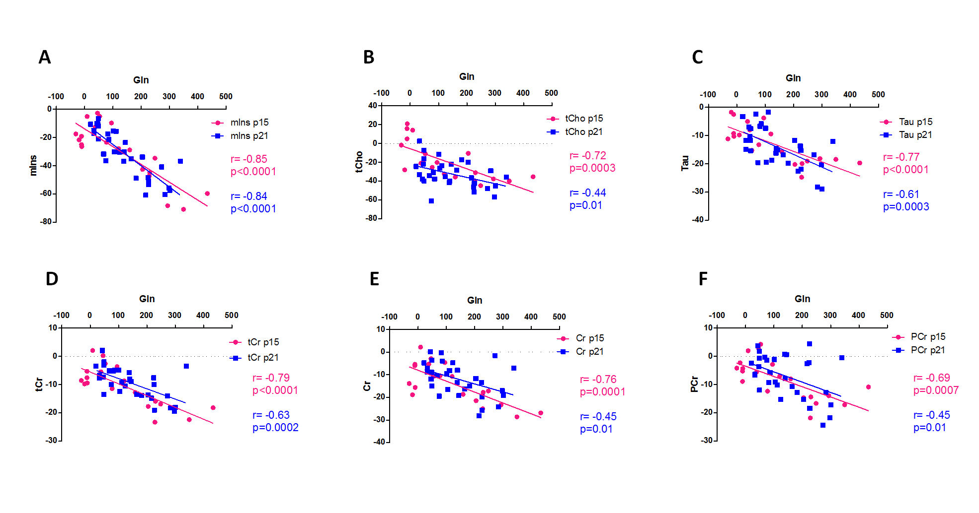
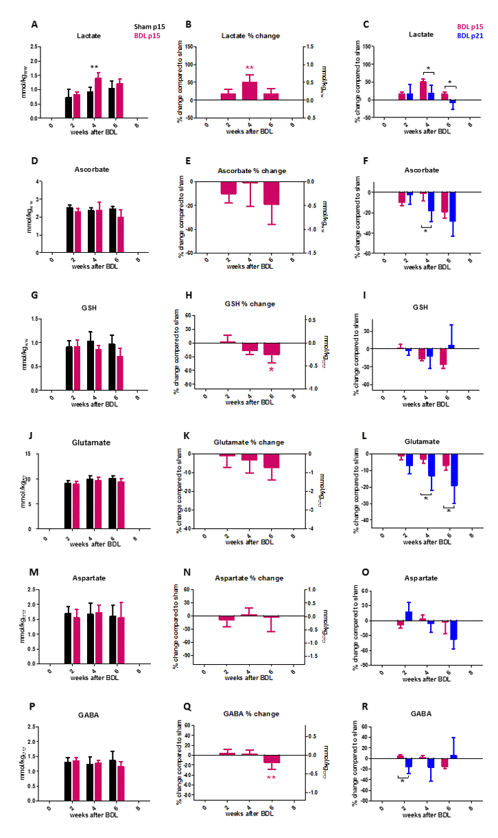
Both p15 and p21 showed increased plasma bilirubin and ammonium from week2 post-BDL (signs of CLD), and there was no difference between p15 and p21 BDL-rats. Both groups showed increased brain Gln due to ammonia detoxification (known change in CHE), visible already from the spectra (Fig1). Interestingly, p15 BDL rats did not display an increase in brain Gln at week2 (Fig2C), contrary to p21 animals, despite the same levels of plasma ammonium at week2. This may be due to immature glutamine synthetase (GS) activity which becomes fully active at around p2610. This could lead to higher levels of ammonia in the brains of p15 rats and greater toxicity. From week4 after BDL, Gln increase in p15 “caught up” with p21 and at week 6 the increase was greater in p15 than p21 (Fig2C). The osmoregulatory response to compensate for Gln increase, was very similar in p15 and p21 rats both in terms of % decrease of brain organic osmolytes (Fig3) and in terms of the correlation between this decrease and the increase in Gln (Fig4). Only tCho showed a delayed decrease in p15 BDL-rats. This difference might suggest difference in phospholipid metabolism between these 2 groups during the 2 weeks that follow BDL surgery. There was no difference in decrease of Cr and PCr in p15 and p21 BDL rats (Fig3). However lactate increased significantly more in p15 BDL-rats. Antioxidant response differed as well (Fig5): ascorbate decrease was significantly stronger in p21 BDL-rats while p15 rats showed a significant decrease only 6 weeks after BDL. GSH had a tendency to diminish to a greater extent in p15 BDL-rats, suggesting that neuronal and glial cells could be affected differently in p15 and p21 BDL rats. This is also supported by the fact that all measurable neurotransmitters(Glu,Asp,GABA) were very little affected in p15 BDL-rats compared to p21(Fig5J-R). This difference between p15 and p21 could be also related with the maturation of amino-acid enzymatic apparatus of the cell that stabilizes only after p20-p2610-11.Conclusions:
We showed, for the first time, differences in brain metabolic changes between p15 and p21 BDL-rats. These differences suggest that in this model rats may be more vulnerable to the neurological consequences of CLD according to age of disease onset, probably owing to coincident neurodevelopmental processes. Gln, tCho and Asc alteration appeared later while Lac and GSH changed earlier in the evolution of the disease. Interestingly, concentrations of neurotransmitters were very little affected in p15 BDL-rats compared to p21. Whether these differences in neurometabolic changes between p15 and p21 translate also into different neurological problems and long-term effect, especially after liver transplantation, requires further studies.Acknowledgements
Funding support was provided by the SNSF project no 310030_173222/1, the EU: FP7-PEOPLE-2012-ITN project 316679 TRANSACT and by the CIBM (UNIL, UNIGE, HUG, CHUV, EPFL, as well as the Leenaards and Jeantet Foundations), the CHUV and the HUG.References
[1] Robertson CMT. Neurocognitive outcomes at kindergarten entry after liver transplantation at <3 yr of age. Pediatr Transplant 2013; 17:621–630
[2] Gilmour SM. School Outcomes in Children Registered in the Studies for Pediatric Liver Transplant (SPLIT) Consortium. Liver Transplant 2010; 16:1041–1048.
[3] Ng V. Development and validation of the pediatric liver transplantation quality of life: A disease-specific quality of life measure for pediatric liver transplant recipients. J Pediatr 2014; 165:547–555.e7
[4] Sorensen LG. Longitudinal study of cognitive and academic outcomes after pediatric liver transplantation. J Pediatr 2014; 165:65–72.e2.
[5] Rackayova et al. Proc. Intl. Soc. Mag. Reson. Med. 22 (2014) 0808.
[6] Workman AD, Charvet CJ, Clancy B, et al (2013) Modeling Transformations of Neurodevelopmental Sequences across Mammalian Species. J Neurosci 33
[7] Mlynárik V, et al. Localized short- echo-time proton MR spectroscopy with full signal-intensity acquisition. Magn Reson Med 2006;56:965–970
[8] Gruetter R, Tkác I. Field mapping without reference scan using asymmetric echo-planar techniques. Magn Reson Med 2000;43(2):319–323.
[9] Souza S. W. De, Dobbing J. (1971) Cerebral Water Rat Edema and in Developing Cation Brain : I. Normal Content Postmortem in Developing Changes. Exp. Neurol. 32, 431–438.
[10] Bayer SM, McMurray WC (1967) The Metabolism of Amino Acids in Developing Rat Brain. J Neurochem 14:695–706
[11] Agrawal HC, Davison AN, Kaczmarek LK (1971) Subcellular distribution of taurine and cysteinesulphinate decarboxylase in developing rat brain. Biochem J 122:759–763.)
Figures
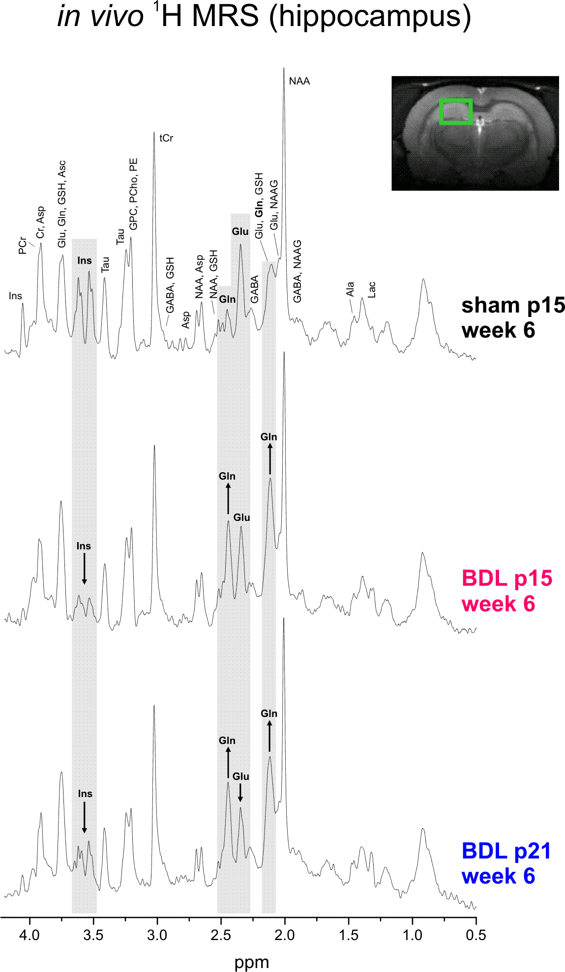
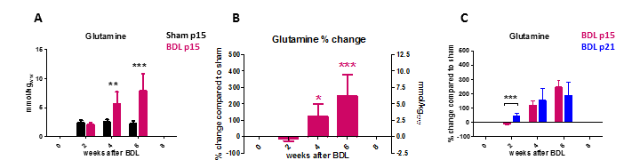
Fig2: P15 showed significant increase in brain Gln only at week4(A), delayed compared to p21(C). At week2, increase in Gln was significantly stronger in p21 BDL compared to p15 but from week4 there was no difference in Gln increase between p15 and p21. At week6, p15 rats reached even higher Gln increase than p21 BDL.
This applies to Fig2,3,5:
(First column)Absolute concentration in mmol/kgww in p15 BDL and sham. (Second column)Percental change in p15 BDL compared to sham at corresponding age. (pink* is compared to change at week 2)
(Third column)Comparison between percental change in p15 BDL and p21 BDL.
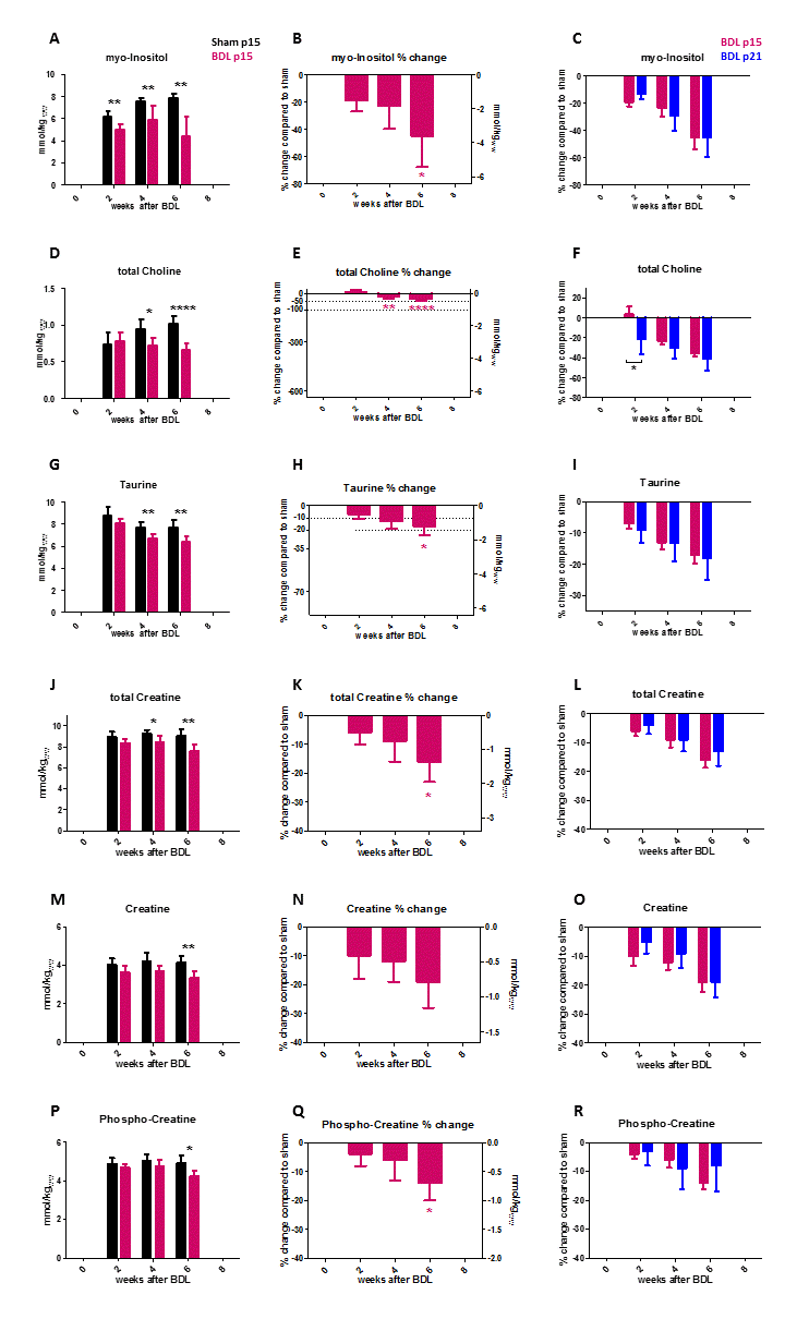
Fig3: There was no difference between p15 and p21 in the decrease in metabolites playing role in osmoregulatory answer (but also phospholipid and energy metabolism). Only tCho showed delayed decrease in p15 BDL rats, similary to Gln. Decrease of tCho in p15 was significant only from week 4 but reached the same decrease at week 6 than p21 BDL rats. Changes observable in shams: increase in myo-Inositol and tCho and decrease in Tau (A,D,G) are in agreement with previously published (Rackayova et al, Proc. Intl. Soc. Mag. Reson. Med. 26 (2018), 5428)