1145
Longitudinal multimodal MRI monitoring of a combination therapy using hydrogen enriched water and minocycline in ischemic stroke1Radiology, Stony Brook University, Stony brook, NY, United States, 2Neurology, Stony Brook University, Stony brook, NY, United States
Synopsis
Hydrogen or minocycline individually has been shown to be neuroprotective in experimental ischemic stroke. This study evaluated the efficacy of combined hydrogen enriched water with minocycline in an ischemic stroke rat stroke model using longitudinal MRI and behavioral tests using a double-blinded design. We found that the combination therapy reduced lesion volumes, white-matter damage, and behavioral deficits, compared to individual or vehicle treatment alone. These findings suggest that combination of hydrogen water with minocycline has positive therapeutic effects in ischemic stroke.
INTRODUCTION
Stroke is a leading cause of death and long-term disabilities 1. Neuroprotective agent capable of reducing brain damage after stroke has remained elusive (Ginsberg, 2008; Tymianski, 2014). Molecular hydrogen 2 is a known antioxidant agent with ideal redox profile and ability to pass though cell membranes to access all cellular compartments. Minocycline 3 is a widely used antibiotic and is known to inhibit the activation of matrix metallo-proteinase-9 (MMP-9) and poly(ADP-ribose) polymerase (PARP), which contribute to the pathogenesis of ischemic brain tissue damage. Both hydrogen and minocycline have excellent safety profiles, have been previously demonstrated individually to reduce infarction in animal models of stroke 4, 5. We postulate that the combined use of hydrogen and minocycline may have additive or even synergistic protective effects against ischemic brain damage, reducing resultant injury and hence aiding recovery after stroke. Thus, in this study, we tested the efficacy of the combined treatment of hydrogen enriched water (H2W) and minocycline (M) in an experimental ischemic stroke model.METHODS
Male Sprague-Dawley rats (250-350g) underwent 60-mins middle-cerebral-artery occlusion (MCAO) under 2.0% isoflurane. Three groups of studies were performed in a double blinded experimental design: vehicle (oral water and intraperitoneal saline, n=6), ii) H2W (n=6), and iii) H2W+M (oral H2W and intraperitoneal M, n=6) were administered immediately after reperfusion, and again 1 day and 2 days following stroke onset. H2W dose is 10ml/kg with saturation of 1.60ppm at atmospheric pressure, and minocycline dose is 20mg/kg.
Diffusion-weighted images, continuous arterial spin labeling, and T2 MRI were acquired at 30mins (during occlusion and prior to treatment) and 90mins (after reperfusion), 1, 4 and 7 days after stroke at 7T. Initial lesion volumes were defined by the core tissue volumes using threshold mean ADC of normal hemisphere minus 3x standard deviation 6. Infarct volumes were derived from T2 maps using threshold of mean T2 value of normal hemisphere plus 2x standard deviation 6. Edema correction was applied. Two regions of interest (ROIs) were generated to separate initial core and mismatch area, based on 30mins ADC and CBF data. CBF value was quantified on these two areas across time and normalized to CBF value of the contralateral corresponding area. The FA value of the corpus callosum (CC) on ipsilateral and contralateral hemisphere to stroke was calculated based on FA maps at each time point. The Garcia neurological scores were measured at Day 1, 4 and 7 after MCAO. Unpaired two-tailed t-tests were used in this study.
RESULTS
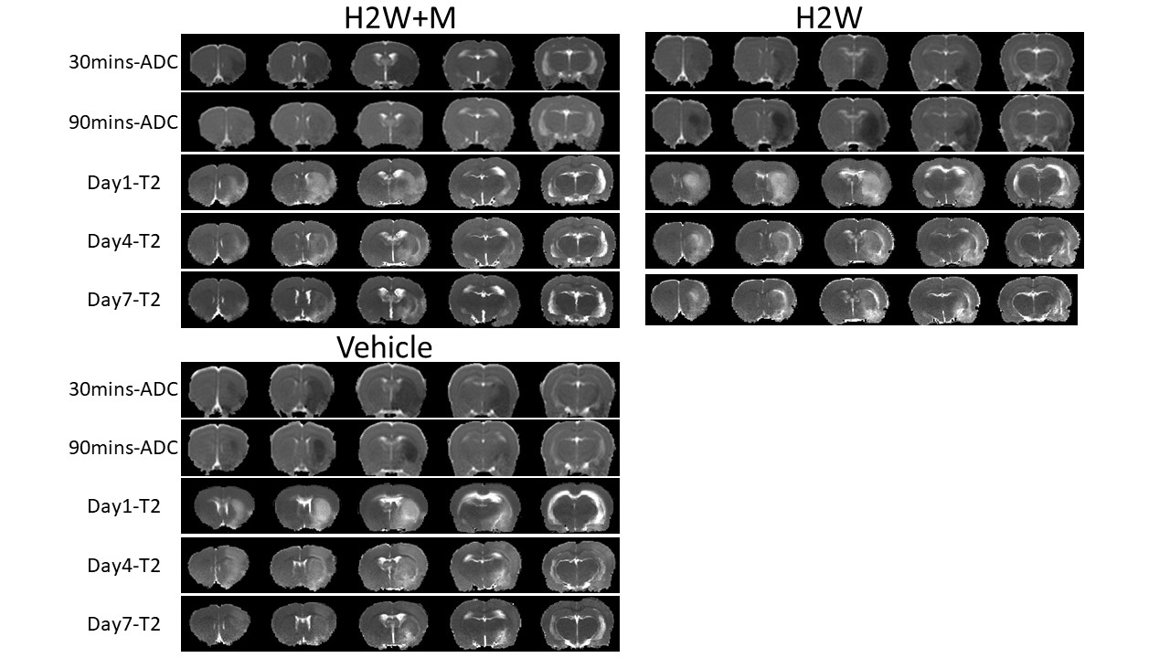
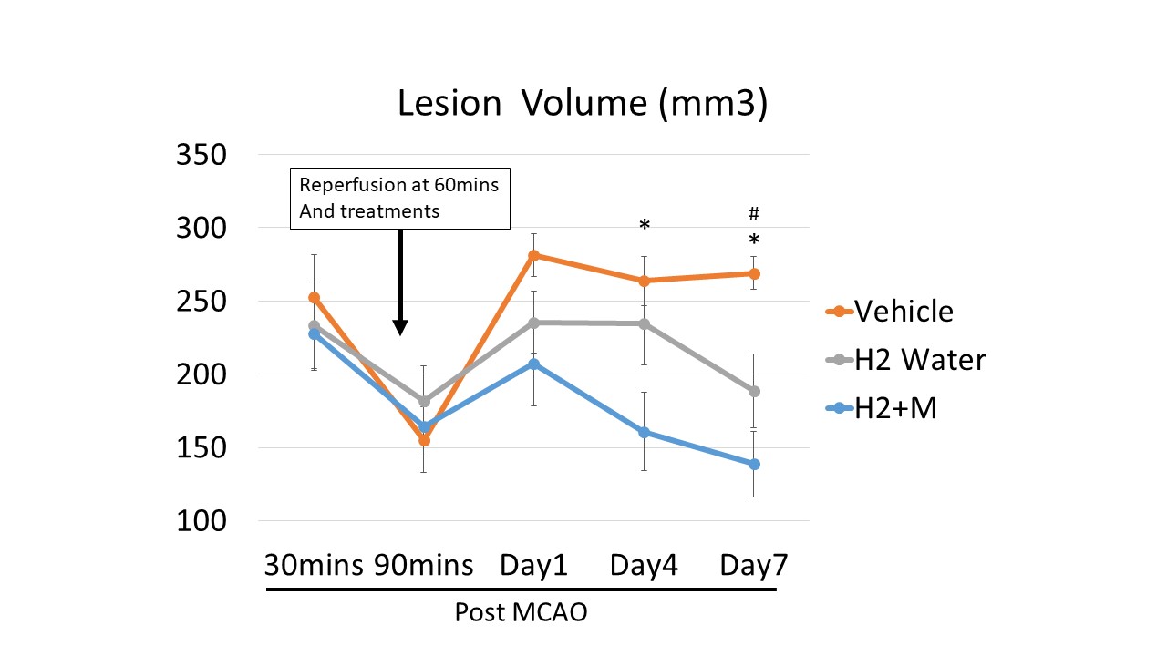
Figure 1 shows representative ADC and T2 map images from the vehicle, H2W and H2W+M groups at 30mins, 90mins, Day1, Day4 and Day7 after MCAO. Lesion volumes by MRI are shown in Figure 2. The initial lesion volumes at 30mins and 90mins of all groups were not statistically different from each other as expected (253±28, 233±29 versus 227±25mm3, P>0.05). The lesion volume at Day4 of the H2W+M group was smaller than that of vehicle group (161±26 versus 264±16mm3, p<0.05). At Day7, the lesion volumes of both H2W+M and H2W groups were also smaller than the vehicle group (139±22 and 189±25 versus 269±11mm3, p<0.05).
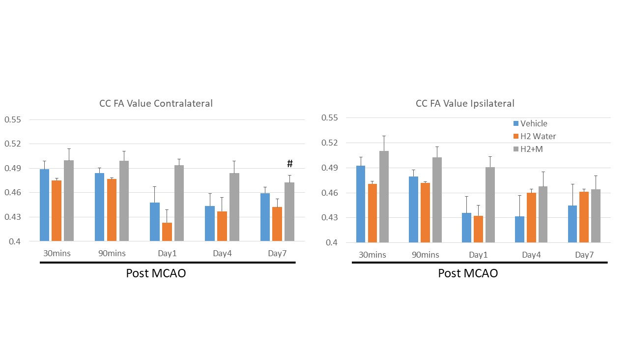
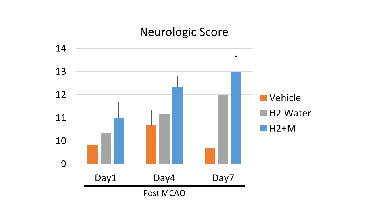
The CC FA value were consistently higher in the combination treatment group compared to the vehicle and H2W only groups, albeit not statistically significant (Figure 3). Normalized CBF of mismatch and core regions are shown in Figure 4. There were trends of increased CBF in the core region in the two treatment groups, and significantly in mismatch region of combination therapy group at Day7 (p<0.05). Neurologic behavioral data (Figure 5) showed that the combined treatment group generally did better than vehicle and H2W alone, reaching statistical significance at Day7 when compared to the vehicle group (p<0.05).
DISCUSION AND CONCLUSION
Both hydrogen and minocycline have been demonstrated individually to be neuroprotective in animal stroke models, based on different and potentially additive or synergistic mechanisms. Hydrogen likely ameliorates toxic free radical species generated by several processes, including excitotoxicity and tissue reperfusion, whereas minocycline likely ameliorates inflammation, blood-brain barrier breakdown, and zinc toxicity [7,8]. These effects could account for the lesion reduction and behavioral improvement observed in our study.
In conclusion, combined treatment of hydrogen-enriched water and minocycline was effective in reducing lesion volume and behavioral deficits compared to vehicle treatment in a stroke model. Future studies will evaluate different occlusion durations, different doses, and longer-term recovery with additional behavioral measures. Given that both hydrogen and minocycline have excellent safety profiles, additional supportive experimental data could readily lead to a clinical trial in ischemic stroke using non-invasive MRI as a key readout.
Acknowledgements
No acknowledgement found.References
1. Benjamin EJ, Virani SS, Callaway CW, Chamberlain AM, Chang AR, Cheng S, et al. Heart disease and stroke statistics-2018 update: A report from the american heart association. Circulation. 2018;137:e67-e492
2. Ren J, Chen YI, Mackey AM, Liu PK. Imaging rhodopsin degeneration in vivo in a new model of ocular ischemia in living mice. FASEB J. 2016;30:612-623
3. Yang Y, Salayandia VM, Thompson JF, Yang LY, Estrada EY, Yang Y. Attenuation of acute stroke injury in rat brain by minocycline promotes blood–brain barrier remodeling and alternative microglia/macrophage activation during recovery. J Neuroinflammation. 2015;12:26
4. Moller AR. Minocycline: A novel stroke therapy. Journal of Neurology & Stroke. 2015;2
5. Iketani M, Ohsawa I. Molecular hydrogen as a neuroprotective agent. 2016:1-8
6. Jiang Z, Watts LT, Huang S, Shen Q, Rodriguez P, Chen C, et al. The effects of methylene blue on autophagy and apoptosis in mri-defined normal tissue, ischemic penumbra and ischemic core. PLoS One. 2015;10:e0131929
Figures