1143
Computer-aided classification of intervertebral disc degeneration based on fractal dimension1The affiliated hospital of Chinese traditional medical university, Xian Yang, China, 2Siemens Healthcare, Scientific marketing, Xi'an, China
Synopsis
Magnetic resonance imaging (MRI) is considered to be the best imaging method to evaluate the IVD degeneration. However, grading systems for degeneration severity evaluation are based on qualitative descriptions of disc image, which impairs the detection of small changes in intervertebral discs. In order to obtain a tool for objective and continuous grading of IVD degeneration. Fractal method was used to analyze the spatial distribution of intervertebral disc signals. We found that fractal dimension value displayed the strongest association with the clinical grading of disc degeneration severity, suggesting that IVD fractal analysis is a suitable tool for objective and continuous IVD degeneration classification.
Introdunction
Magnetic resonance imaging (MRI) is considered to be the best imaging method to evaluate the IVD degeneration, which is very important for their diagnosis and grading1. The Pfirrmann classification system focuses on the signal intensity of nucleus pulposus or the structural morphology in sagittal T2-weighted MR images. The qualitative classification of IVD degeneration was affected by the subjective influence of the observers, and the grading of degeneration was classified as a non-continuous grades, which impairs the detection of small changes in intervertebral discs, especially the small changes after treatment2. Fractal dimension is an index of important texture feature, which has a strong correlation with observers’subjective assessment of image roughness or irregularity, may offer sensitive imaging biomarkers for IVD degeneration classification. This study was to explore the feasibility of fractal analysis to decode IVD heterogeneity with purpose to obtain a tool for objective and continuous grading of IVD degeneration.Methods
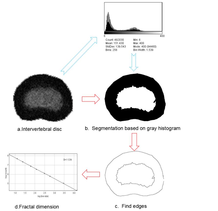
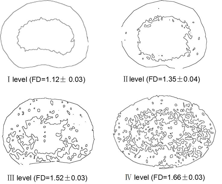
A 3 Tesla scanner was used, and conventional T2-weighted MR images were obtained, and a total of 180 lumbar discs were analyzed. All the intervertebral discs collected were classified according to Pfirrmann classification system. Severely degenerated intervertebral disks with collapsed disk space were excluded from the assessment because the new classification system was designed to classify relatively early degenerative disks and we wanted to avoid possible volume averaging by the endplate in the slice. Fractal analysis was performed using ImageJ (National Institutes of Health, Bethesda, MD) program with box-counting method(Fig1). Mann–Whitney U test was performed to examine the statistical significance between FD value and Pfirrmann grade.Results
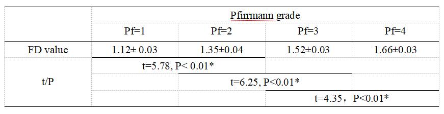
Fractal dimension value displayed the strongest association to the clinical grading of disc degeneration severity (P<0.01). The fractal dimension (FD) of Pfirrmann 1 IVDs, Pfirrmann 2 IVDs, Pfirrmann 3 IVDs and Pfirrmann 4 IVDs were 1.12± 0.03, 1.35±0.04,1.52±0.03 and 1.66±0.03, respectively(Fig2, Fig4). There was significant difference in the fractal dimension between Pfirrmann groups (P < 0.01). Statistical analysis indicates that FD value can effectively separate between stages of degeneration.Conclusion and discussion
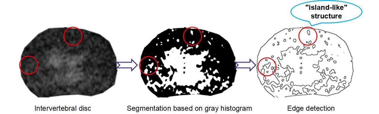
The Fractal dimension value associated well with IVD degeneration, suggesting that IVDs fractal analysis is a suitable tool for objective and continuous IVD degeneration classification. The increase of fractal dimension of intervertebral disc is mainly caused by two factors: First, in degeneration process, the distinction between nucleus pulposus and annulus fibrosus becomes irregular; Second, the "island-like" irregular structure is detected in annulus fibrosus of discs, which increases the complexity of intervertebral discs(Fig2,Fig3). The mechanism of loss of distinction between nucleus pulposus and annulus fibrosus is considered to be an increased number of AF fissures are developed that may result in leakage of NP into the AF. The "island-like" structure were found to be consistent with High Intensity Zones in the annulus. The HIZ has the potential to be a good indicator for discogenic low back pain3. In this study, the fractal method has been used to quantitatively and continuously detect these pathological changes in the process of degeneration. Compared with the conventional grading system, it has higher accuracy and objectivity. It may be a clinical tool for characterization of regional IVD degeneration effects.Acknowledgements
No acknowledgement found.References
[1] An H, Anderson P, Haughton V, et al. Introduction: disc degeneration:summary. Spine.2004;29:2677 –8
[2] Pfirrmann CW, Metzdorf A, Zanetti M, Hodler J, Boos N. Magnetic resonance classification of lumbar intervertebral disc degeneration. Spine.2001; 26(17):1873–1878.
[3] Peng B, Hou S, Wu W, Zhang C, Yang Y (2006) The pathogenesis and clinical significance of a high-intensity zone (HIZ) of lumbar intervertebral disc on MR imaging in the patient with discogenic low back pain. Eur Spine J. 2006;15(5):583-587.
Figures