1139
Variational Networks for Accelerating Biexponential 3D-T1rho Mapping of Knee Cartilage1School of Medicine, New York University, New York, NY, United States, 2Institute of Computer Graphics and Vision, Graz University of Technology, Graz, Austria
Synopsis
Quantitative T1ρ imaging using biexponential models usually requires multiple spin-lock times, which makes the acquisition time demanding. Recently, Compressed Sensing (CS) has demonstrated significant reduction in data acquisition time in MRI applications in general, and T1ρ relaxation mapping of knee cartilage in particular. However, biexponential T1ρ mapping error using CS is much higher than that of monoexponential T1ρ mapping error for the same acceleration factor. One possible approach to reduce artifacts, improving image and mapping quality, is to learn a Variational Network (VN) for image reconstruction. Here, we compare a VN, trained with real knee cartilage images, against the best CS approaches known for biexponential T1ρ mapping. Our results show that the VN produced biexponential maps superior to CS, with lower T1ρ mapping error.
Introduction:
Biexponential 3D-T1ρ mapping of knee cartilage can provide more information related to macromolecules such as proteoglycan and collagen1. T1ρ mapping can be used for early detection of knee osteoarthritis2. Several T1ρ-weighed images with different spin-lock times (TSLs) are required to estimate biexponential T1ρ parameters, i.e. short and long components and their corresponding fractions, which significantly increases the scan time. Compressed Sensing (CS) has been successfully used for accelerating biexponential T1ρ mapping of knee cartilage3. However, biexponential T1ρ mapping errors are nearly 3 times higher than its monoexponential counterpart4. Recently, learned Variational Networks (VN) has been utilized for high-quality knee joint image reconstructions, being superior to some CS reconstructions5. In this work, we evaluate the performance of VN against CS for accelerating biexponential 3D-T1ρ mapping of knee cartilage.Methods:
Multiple 3D-T1ρ-weighted knee joint datasets were acquired using a modified 3D-Cartesian turbo-Flash sequence on a 3T clinical scanner (Prisma, Siemens Healthcare, Germany) with a 15-channel Tx/Rx knee coil. Imaging sequence parameters were: TR/TE=1500ms/4ms, flip angle=8°, matrix size=256×128×64, spin-lock frequency=500Hz, slice thickness=2mm, FOV=120mm2, and receiver bandwidth=515Hz/pixel. Seven fully sampled knee datasets (n=7, age=28.5±8.5 years) were acquired in sagittal plane (healthy volunteers) with 10 TSLs including 2/4/6/8/10/15/25/35/45/55ms, total acquisition time of 32:05(min:sec).
The CS reconstruction3 is posed as: $$x=\arg\min_x ||m-Ax||_2^2+λR(x).$$
Where $$$x$$$ represents one reconstructed 2D image or a series of images (different TSLs, 2D+time reconstructions), $$$A=SFC$$$, where $$$C$$$ denotes coil sensitivities, $$$F$$$ represents the spatial FFT, $$$S$$$ is the sampling mask and $$$λ$$$ is the regularization parameter. The following regularization functions are considered: the $$$l_1$$$-norm, as $$$R(x)=||Tx||_1$$$, where $$$T$$$ is the a spatial finite difference (SFD) or spatiotemporal finite differences (STFD); the low rank plus sparse (L+S) reconstruction, where $$$ x=l+s$$$ is a decomposition on a sparse part $$$s$$$ and a low rank $$$l$$$ part, where $$$R(x)$$$ is set as $$$λ_l ||l||_*+λ_s ||Ts||_1$$$, and $$$T$$$ is spatial finite difference. Except for the SFD, the other two regularizations were among the best methods for biexponential T1ρ mapping3. The CS problem is solved with 150 iteration of FISTA-FGP6. The VN is given by the algorithm5:
$$x_{t+1} = x_t – (λ_t A^*(m-A x_t) + \sum_{i=1}^N K_{t,i}^*Φ'_t(K_{t,i} x_t) ), 1 ≤ t ≤ 10$$
Where $$$t$$$ represents iteration index (or layer). The VN parameters, i.e. convolutional filters $$$K_{t,i}$$$ (N=24, size=11x11), activation functions $$$Φ'_t$$$, and step-sizes $$$λ_t$$$, are learned from data. The sampling pattern follows 2D Poisson Disk, and it is the same for all datasets and TSLs with the same acceleration factor (AF), central area of k-space is used to estimate coil sensitivities with ESPIRiT7. Data from 3 volunteers (3x256slices x10TSL=7680 images) were used to train the VN parameters and the $$$λ$$$ in the CS methods. Data from another 4 volunteers were used for testing. After reconstruction, curve fitting is performed using the biexponential model:
$$|x(t,n)|=a_s(n)exp\left(\frac{-t}{τ_s(n)} \right)+a_l(n)exp\left(\frac{-t}{τ_l(n)}\right)+b,$$
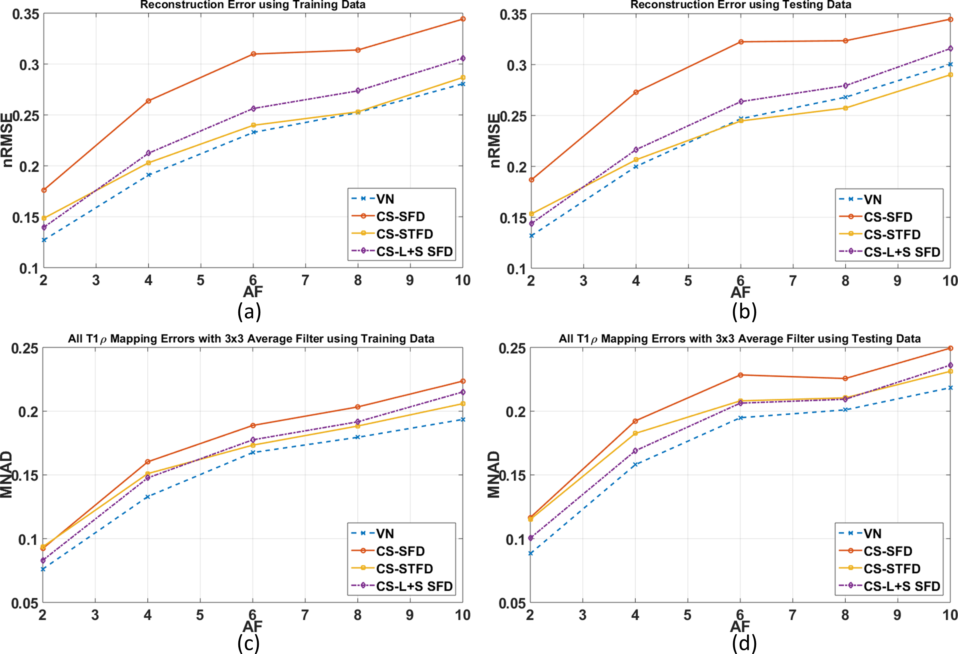
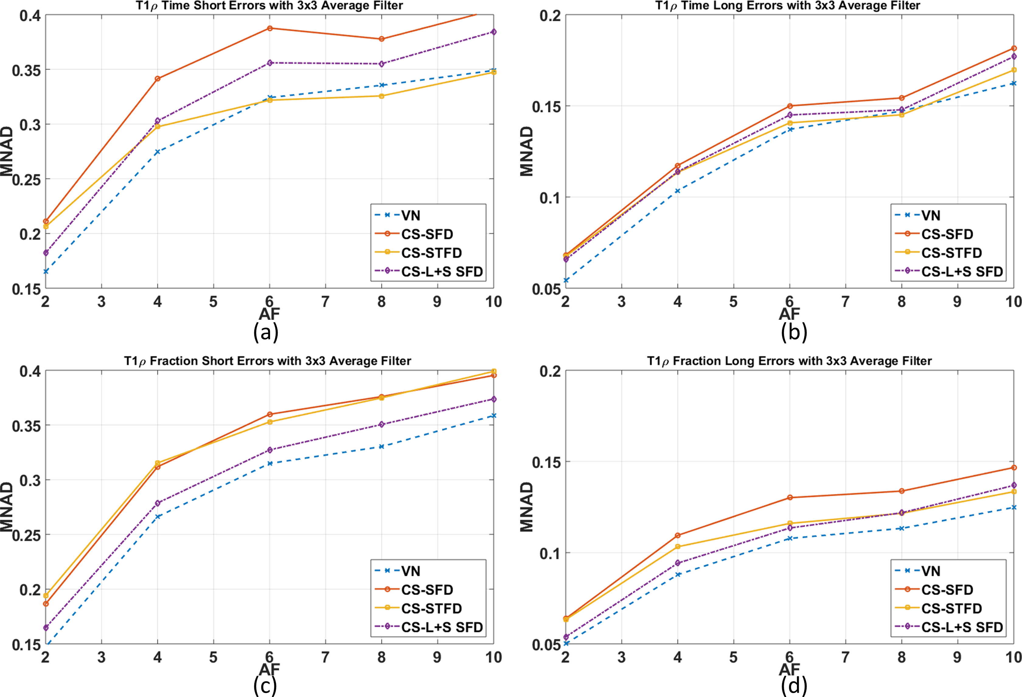
where $$$a_s(n)$$$ and $$$a_l(n)$$$ are magnitudes of short and long components, while $$$τ_s(n)$$$ and $$$τ_l(n)$$$ are T1ρ relaxation times of short and long exponentials. Non-linear least squares is utilized as cost function, where the minimization is done utilizing conjugate gradient Steihaug’s trust-region (CGSTR)8. A 3x3 voxel averaging is utilized prior to the fitting process. Monoexpontial fitting is done first, and used as initial solution for biexponential fitting. The fitting process occurs only in specific ROIs of the 3D image related to the knee cartilage (medial, lateral and patellar cartilage). We compare the reconstructions and estimated T1ρ relaxation times/fractions with the reference data using normalized root mean square error (nRMSE) and median of the normalized absolute deviation (MNAD) respectively3.
Results and Discussion:
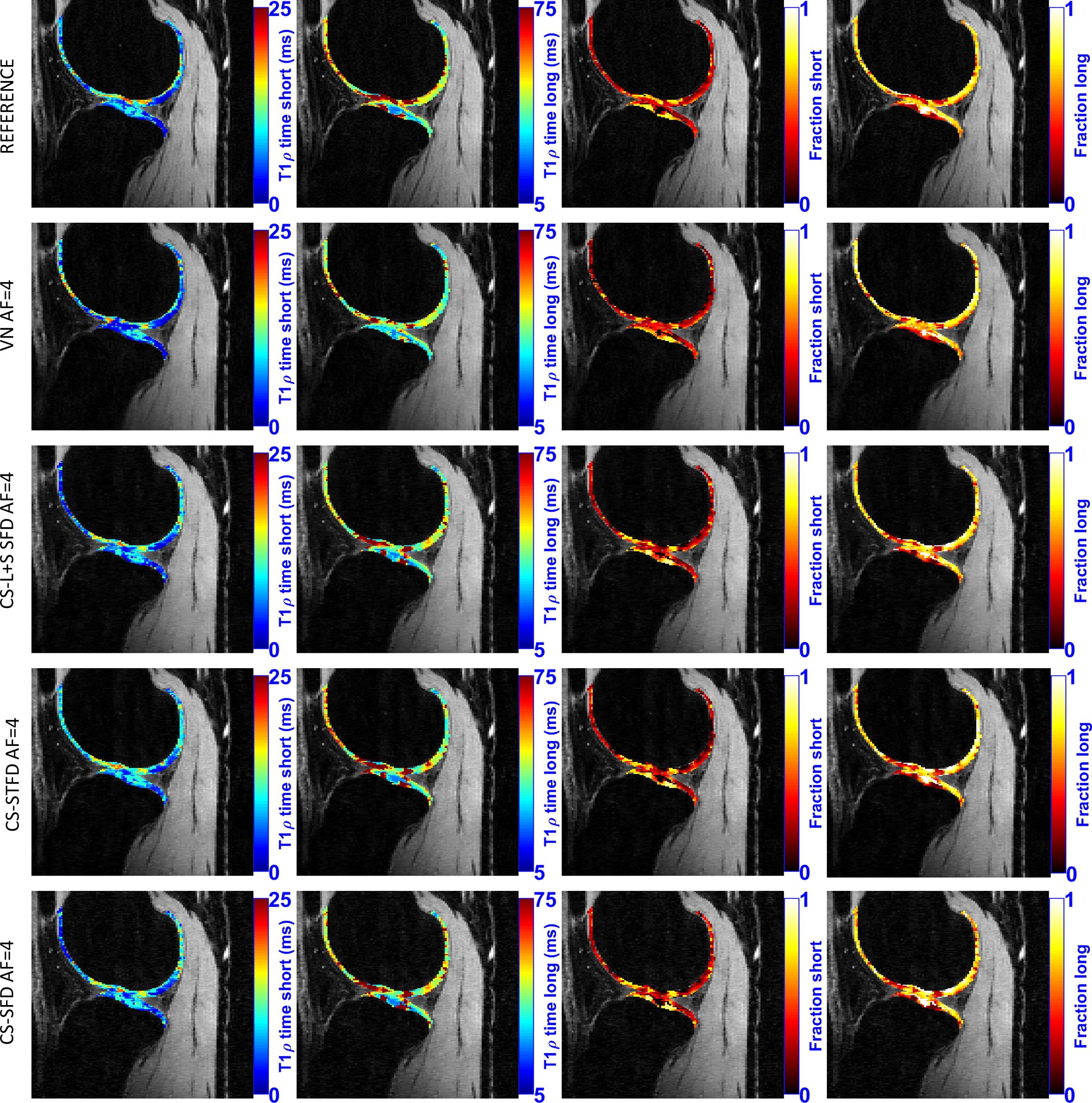
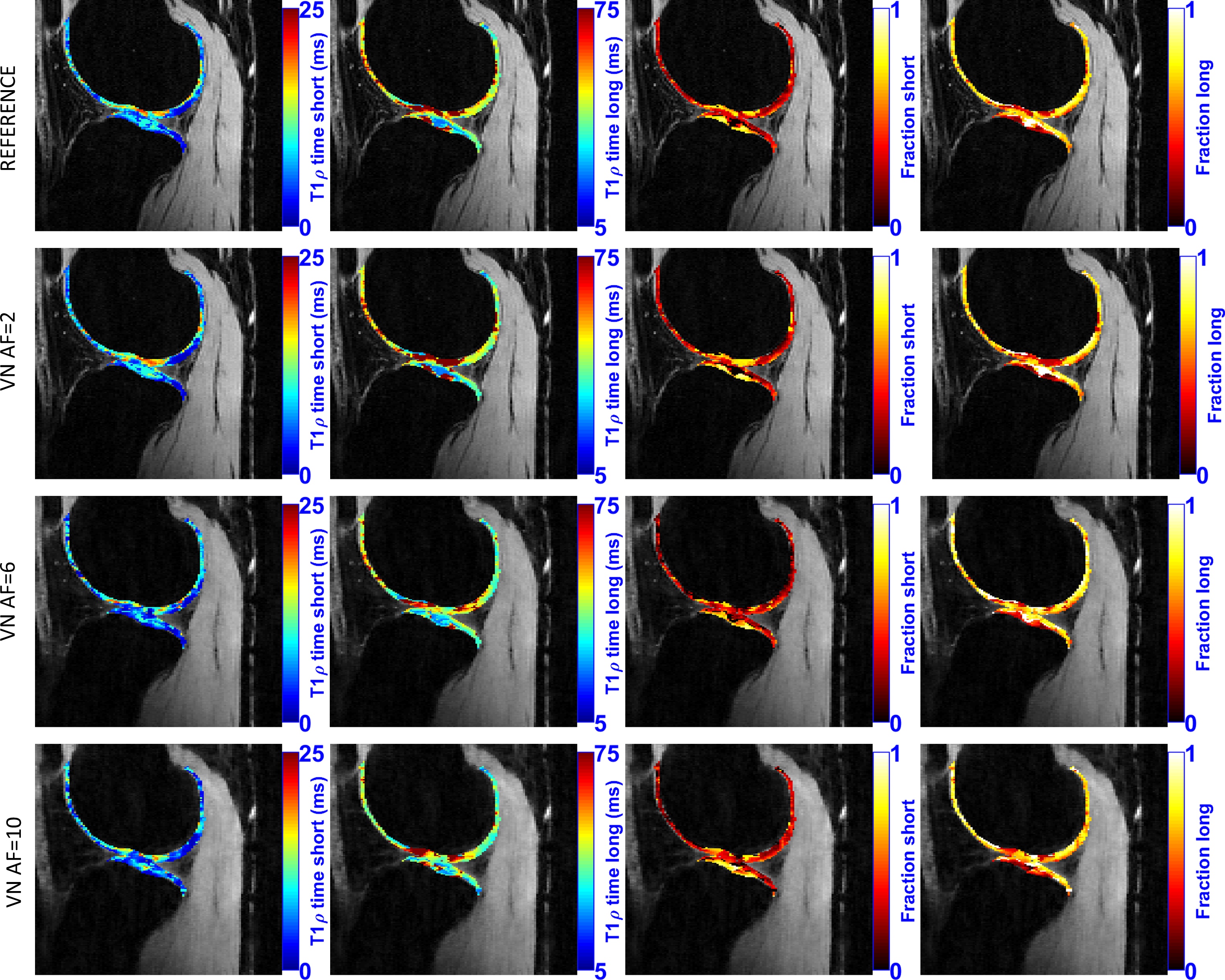
The VN performed better than CS methods for biexponential T1ρ mapping of knee cartilage in terms of nRMSE and MNAD for training and testing datasets (Figure 1). The superior quality of the VN comes from the use of better spatial information (24 convolutional filters 11x11) learned from the training data. Comparing the error in individual components of the biexponential model (Figure 2), we observed that VN improved the results in nearly of all components. Some visual T1ρ results are shown in Figures 3 and 4. VN requires a long training time (~48h), however, reconstruction time is much lower (1.5sec @NVIDIA-M40-GPU-12GB) compared to CS methods (225sec @ intel-i7-1.6GHz-48GB parallel-cluster).Conclusion:
VN is a promising approach to accelerate biexponential T1ρ mapping of knee cartilage. The VN performed better than the CS techniques with faster reconstruction time and lower T1ρ mapping errors, showing great robustness on this quantitative problem. Our experiments show that the VN performed well even with the diverse contrast and SNR produced by acquisition with different TSL, a result that was not yet obvious from previous research9. This is also the first time that this reconstruction approach is validated on biexponential mapping.Acknowledgements
This work was supported in part by NIH grants R01-AR060238, R01-AR067156, R01-AR068966, and NIH R01-EB024532 and was performed under the rubric of the Center for Advanced Imaging Innovation and Research (CAI2R, www.cai2r.net) an NIBIB Biomedical Technology Resource Center (NIH P41 EB017183).References
1. Sharafi A, Xia D, Chang G, Regatte RR. Biexponential T1ρ relaxation mapping of human knee cartilage in vivo at 3 T. NMR Biomed. 2017;30:e3760. doi: 10.1002/nbm.3760.
2. MacKay JW, Low SBL, Smith TO, Toms AP, McCaskie AW, Gilbert FJ. Systematic review and meta-analysis of the reliability and discriminative validity of cartilage compositional MRI in knee osteoarthritis. Osteoarthr. Cartil. 2018. doi: 10.1016/j.joca.2017.11.018.
3. Zibetti MVW, Sharafi A, Otazo R, Regatte RR. Compressed sensing acceleration of biexponential 3D-T1ρ relaxation mapping of knee cartilage. Magn. Reson. Med. 2018. doi: 10.1002/mrm.27416.
4. Zibetti MVW, Sharafi A, Otazo R, Regatte RR. Accelerating 3D-T 1ρ mapping of cartilage using compressed sensing with different sparse and low rank models. Magn. Reson. Med. 2018;80:1475–1491. doi: 10.1002/mrm.27138.
5. Hammernik K, Klatzer T, Kobler E, Recht MP, Sodickson DK, Pock T, Knoll F. Learning a variational network for reconstruction of accelerated MRI data. Magn. Reson. Med. [Internet] 2018;79:3055–3071. doi: 10.1002/mrm.26977.
6. Beck A, Teboulle M. Fast gradient-based algorithms for constrained total variation image denoising and deblurring problems. IEEE Trans. Image Process. 2009;18:2419–2434. doi: 10.1109/TIP.2009.2028250.
7. Uecker M, Lai P, Murphy MJ, Virtue P, Elad M, Pauly JM, Vasanawala SS, Lustig M. ESPIRiT-an eigenvalue approach to autocalibrating parallel MRI: Where SENSE meets GRAPPA. Magn. Reson. Med. 2014;71:990–1001. doi: 10.1002/mrm.24751.
8. Steihaug T. The conjugate gradient method and trust regions in large scale optimization. SIAM J. Numer. Anal. 1983;20:626–637. doi: 10.1137/0720042.
9. Knoll F, Hammernik K, Kobler E, Pock T, Recht MP, Sodickson DK. Assessment of the generalization of learned image reconstruction and the potential for transfer learning. Magn. Reson. Med. [Internet] 2018:1–13. doi: 10.1002/mrm.2735
Figures