1136
Fully automatic segmentation of wrist cartilage on MR images by convolutional neural network1The International Research Center Nanophotonics and Metamaterials, University of Information Technology Mechanics and Optics, Saint-Petersburg, Russian Federation, 2Federal Almazov North-West Medical Research Center, Saint-Petersburg, Russian Federation, 3APHM, Service de Radiologie, Hôpital de la Conception, Marseille, France, 4CRMBM, Aix-Marseille Universite, Marseille, France
Synopsis
A fully automatic, wrist cartilage segmentation method on magnetic resonance images is developed and validated. The method is based on convolutional neural networks (CNN). Cartilage segmentations obtained with the CNN showed a substantial agreement with manual segmentations for the full 3D wrist images and a good agreement for central coronal slices. The proposed method provided cartilage masks having a high concordance with manually obtained ones.
INTRODUCTION
A gold standard of the articular cartilage degradation
assessment in patients with osteo- and rheumatoid arthritis is based on
radiographic measurements of the joint space narrowing. This method is indirect
and implies multiple measurements of distances between articular surfaces.
Magnetic resonance imaging (MRI) allows the direct measurement of cartilage
thickness. Manual and automatic tissue
segmentation techniques can be used for cartilage extraction from 3D MR images to
evaluate total cartilage volume of a studied joint1,2. However, for such a
complex joint as the wrist, the manual segmentation procedure
becomes time consuming, and no automatic methods have been proposed for this
purpose so far. A novel, deep learning-based method has shown a great potential
for tissue segmentation in musculoskeletal MRI3,4. In this study, we develop and validate an
automatic procedure of wrist cartilage segmentation from 3D VIBE MR images
based on a convolutional neural network (CNN).
METHODS
The study was conducted on 20 3D VIBE wrist images (acquired at 1.5T Magnetom Espree, Siemens) of 8 healthy volunteers and 3 patients suffered osteoarthritis. 3D images were converted into 1760 coronal slices, from which on 341 the cartilage was manually segmented by an experienced radiologist. The data were splitted into 4 datasets for the CNN training, development, test and validation (fig.1). The CNN input was a 28x28 pixels patch surrounding the pixel which had to be classified. The network architecture was constructed using Keras neural network library and had eight layers (2DConvolution, 2DMaxpooling, 4 2DConvolution, Flatten and Dense) and a two-class output. All the 3D data of subjects involved into the test phase were unseen by the CNN during the training phase. Dice coefficient (DSC) was calculated for the test data set. In order to validate the performance of CNN, a comparison with manual segmentation procedure results was held. A medial coronal slice of every 3D image (20 in total – validation dataset, also unseen by CNN) was segmented manually by 3 observers (twice by two of them, and once by one), and by the trained CNN. The manual segmentations made by the first observer in the first session were considered as a ground truth (GT). For the manual procedure, the intra- and interobserver concordance indices (CI) were calculated. The CNN-assessed segmentations were compared with GT through DSC and CI calculation.
RESULTS
The results of comparative
analysis are summarized in Table 1. The trained CNN successfully segmented
cartilage pixels as illustrated in Figure 2. One session of 20 slices manual
segmentation of validation dataset made by a single observer took averagely
about one hour, while the CNN processed the same dataset in 5 minutes.
DISCUSSION
The obtained here DSC value (0.73) is slightly lower than for the CNN-assisted knee cartilage segmentations (0.80-0.82)3,4 . Taking into account a more complex structure of wrist joint and very limited training data, the obtained value can be considered as satisfactory. For the validation dataset the DSC reached 0.81, as it contained only medial coronal slices (fig.2 (a)) with high percentage of cartilage and higher contrast of cartilage pixels relatively to the surrounding tissues. In noisy images with low contrast of cartilage, the subcutaneous fat was occasionally considered by the CNN as cartilage (circled areas on fig. 2(b)). However, it still achieves higher concordance with the GT than the observers. Interestingly, the results obtained for both manual and CNN assisted segmentation showed similar CIs for healthy volunteers and patients. Images of patients contained pathological changes such as bone marrow edema (fig. 2(b)), cortex erosion (fig. 2(c)), ganglion cyst (fig. 2(d)). A vessel in a capitate bone of the second patient (fig. 2(d)) was not considered by CNN as cartilage. These results demonstrate a capability of the developed CNN to distinguish cartilage from other anatomical structures and possible joint abnormalities. The lack of input data for CNN training was a limitation of this study.
CONCLUSION:
We have proposed a fast and fully automatic CNN-based tool for cartilage segmentation in wrist 3D MR images. Using this tool, the measurement of cartilage volume for the purpose of cartilage degradation study may be performed with higher accuracy than manually. Data augmentation and 3D approach will be used in the future to increase quality of the CNN performance quality.Acknowledgements
This work was financially supported by the Government of the Russian Federation through the ITMO Fellowship and Professorship Program. This work was supported by the Russian Science Foundation (Project No. 18-79-10167).References
1 Zink JV, Souteyrand P, Guis S et al. Standardized quantitative measurements of wrist cartilage in healthy humans using 3T magnetic resonance imaging. World Journal of Orthopedics 2015; 6(8): 641-648.
2 Peterfy CG, Van Dijke CF, Janzen DL et al. Quantification of articular cartilage in the knee with pulsed saturation transfer subtraction and fat-suppressed MR imaging: optimization and validation. Radiology 1994;192(2):485-91.
3 Prasoon A, Petersen K, Igel C et al. Deep feature learning for knee cartilage segmentation using a triplanar convolutional neural network. In: Med Image Comput Comput Assist Interv 2013;16(Pt 2):246-53.
4 Zhou Z, Zhao G, Kijowski R et al. Deep convolutional neural network for segmentation of knee joint anatomy. Magn Reson Med 2018;00:1–12.
Figures
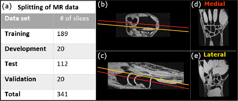
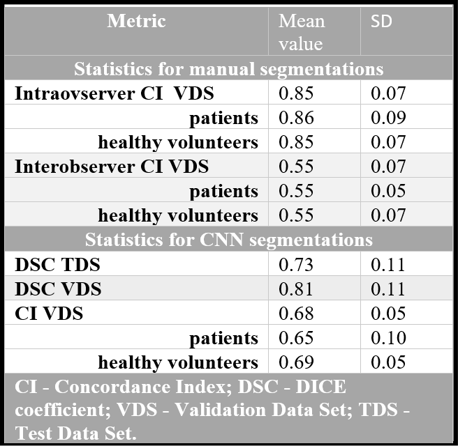
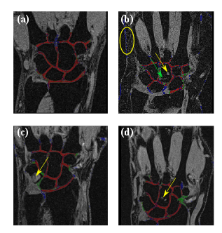
Figure 2. Illustrations of CNN performance: (a) - a medial slice from the validation dataset with good performance (DSC = 0.82); (b) - a medial slice with the subcutaneous fat (circled in yellow) considered by CNN as a cartilage; (b,c,d) - CNN performance on the images of unseen patients from the validation dataset. The yellow arrows point to the high signal intensity lesions. The green arrow (f) points to the vessel. The developed CNN assigned correctly these structures as non-cartilage. Red color corresponds to the pixels correctly segmented as the cartilage; green – incorrectly assigned to the background; blue – incorrectly assigned to the cartilage.