1134
Imaging Post-Transplant Allogeneic Rats with Acquired Immune Tolerance Using Hyperpolarized [1-13C] Pyruvate MRI1Radiology, University of Pennsylvania, Philadelphia, PA, United States, 2Surgery, University of Pennsylvania, Philadelphia, PA, United States
Synopsis
Post-transplant, lungs are clinically monitored using regular radiography and/or CT scans to detect rejection. We previously demonstrated that lactate-to-pyruvate ratio derived via hyperpolarized (HP) [1-13C]-pyruvate MRI is an earlier predictor of lung rejection than microCT in an orthotopic rat lung transplantation model. In this study, we imaged transplanted allogeneic lungs in a recipient rats with acquired immune tolerance to resolve the signal contribution of ischemia-reperfusion injury from that of tissue rejection. Transplanted rat lungs with acquired immune tolerance exhibit similar behavior to that of syngeneic lung transplants before abolishing their acquired tolerance.
Introduction
Post-transplant, lungs are clinically monitored using regular radiography and/or CT scans to detect rejection. We previously demonstrated that lactate-to-pyruvate ratio derived via hyperpolarized (HP) [1-13C]-pyruvate MRI is an earlier predictor of lung rejection than microCT in an orthotopic rat lung transplantation model1. In this study, we imaged transplanted allogeneic lungs in recipient rats with acquired immune tolerance to resolve the signal contribution of ischemia-reperfusion injury from that of tissue rejection.Methods
Left lungs were transplanted in syngeneic rats (inbred Lewis(L) to Lewis), allogeneic rats (Wistar-Furth(WF) to inbred L), and allogenic rats with acquired immune tolerance (inbred WFxL to inbred L). In the latter cohort, immune tolerance was acquired via injection of neonatal stem cells from WFxL into neonatal inbred Lewis rats. Orthotopic transplant surgery was performed as previously described, with a single-suture bronchial anastomosis technique and a proximal cuffing approach for vascular anastomoses2. Total surgery duration was about 3.5h, with approximate ischemia time of 110 minutes. Animals were monitored post-surgery to assess recovery. HP [1-13C]-pyruvate MR imaging was performed on days 3, 7 and 14 (as well as day 28 for the immune tolerant cohort). Animals were imaged while supine in a 4.7T magnet (Varian Inc.). HP [1-13C]-pyruvate (28.6mg, 15mM OX063, 1.5mM Dotarem Gd) was polarized using a HyperSense DNP polarizer, and ~1.2mL (4mL/kg, 80mM) of HP agent was injected via the tail vein over 6s. HP [1-13C]-pyruvate chemical shift imaging (CSI) was performed using a 2D slice selective phase-encoded FID-CSI sequence (TR/TE= 35.7/0.35ms, α= 9°, FOV= 45x45x10mm3), as previously reported3. Spectra were reconstructed, processed and analyzed using custom MATLAB scripts. A subset of animals were sacrificed and their lungs fixed for histological processing.Results
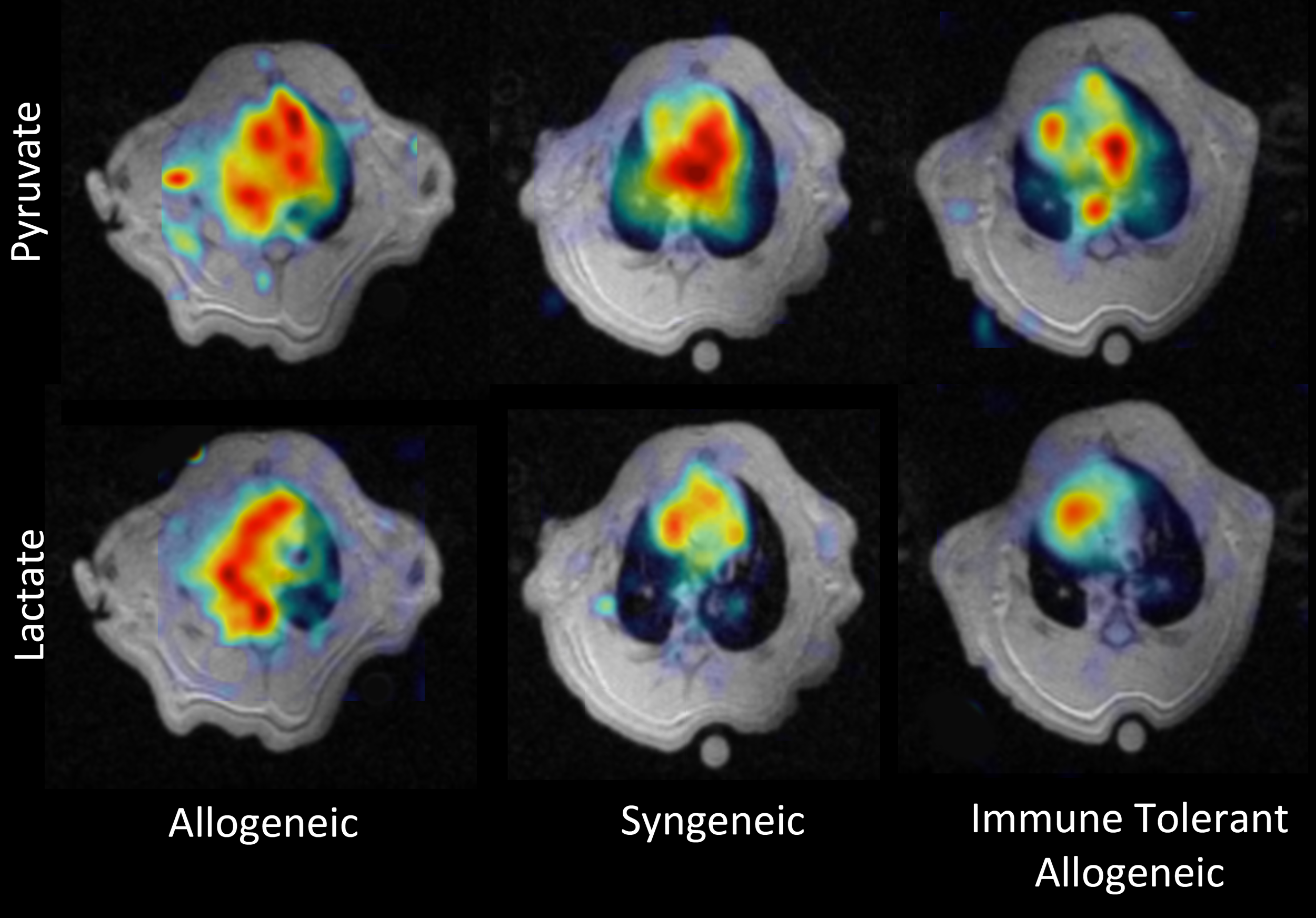
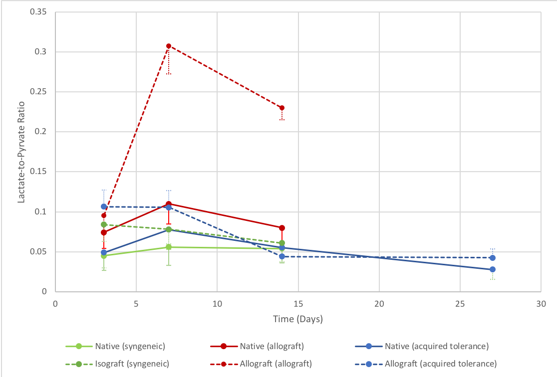
Figure 1 shows normalized pyruvate and lactate colormaps for all three cohorts on day 7. The representative allogeneic rat lung shows a much higher lactate signal in the allograft (left lung). In comparison, the other cohorts show minimal differences between the native and transplanted (left) lungs, as reflected quantitatively in figure 2. On day 3, the average lactate-to-pyruvate ratios of all three transplanted lungs (dashed lines) range between 0.08 to 0.11 compared to a range of 0.05 to 0.07 for the native lungs (solid lines). By day 7, the rejected allograft (red line) has a lactate-to-pyruvate ratio of 0.3±.04, a 2.7-fold increase compared to the other cohorts, which remained below 0.11. Most importantly, the immune tolerant allograft (blue line) follows a similar trajectory to the syngeneic graft (green line)—showing no signs of rejection. We see a similar trend on day 14: lactate-to-pyruvate remains elevated in the allograft (0.23±.02), whereas the other transplanted and native lungs remain below 0.08. While the immune tolerant cohort was also imaged on day 28 to assess the presence of chronic rejection, the lungs remained viable at this timepoint, as shown by their low lactate-to-pyruvate (below 0.04).Discussion
Findings in both allogeneic and syngeneic cohorts mirror those in our previous study [1]. Pyruvate signal can be used as a marker for perfusion, whereas, based on immunohistological findings from [1], lactate signal corresponds to the activity of neutrophils (post-surgery ischemia-reperfusion), CD4+ and CD8+ cells (the latter two drive the rejection process). As seen in figure 2, the general trend is a slight increase in lactate-to-pyruvate ratio in all non-rejected lungs on day 7 (compared to day 3). This is most likely because of hyperperfusion following surgery, which subsides by day 7. In contrast, the greater than 2.7-fold increase in lactate-to-pyruvate in the allograft results from increased CD4+ and CD8+ infiltration and activity as the host rejects the allograft. The most important finding of this study is that the immune tolerant cohort shows no allograft rejection. Indeed, as expected, our results indicate that the immune tolerant cohort has a similar response to that of the syngeneic cohort. However, future studies will allow us to separate the respective contributions of signal derived from post-surgical inflammation and rejection by abolishing this cohort’s tolerance via the injection of naive Lewis T-cells (that have no tolerance for WFxL cells) and then imaging the rejection process in the absence of any contributing inflammatory signal.Conclusions
Transplanted rat lungs with acquired immune tolerance exhibit similar behavior to that of syngeneic lung transplants. The next steps of this study will be to abolish the acquired tolerance so as only to image the signal derived from the tissue rejection process, allowing us to differentiate between the signals emanating from ischemia-reperfusion injury and rejection.Acknowledgements
No acknowledgement found.References
[1] Siddiqui, et al. HP [1-13C] Pyruvate-derived metabolic biomarkers are an early predictor of lung rejection in the rat lung transplantation model. ISMRM, Paris, 2018.
[2] Habertheuer, et al. Innovate, simplified orthotopic lung transplantation in rats. Journal of Surgical Research, 2013.
[3] Pourfathi, et al. In-vivo Assessment of Lung Injury Using Hyperpolarized Carbon-13 MRI in a Two-hit Model of Acid Aspiration and VILI. ISMRM, Singapore, 2016.
Figures