1131
A fluorine-19 MR characterization of ABL-101 - a novel tracer with high biocompatibility and MR sensitivityEmeline Darçot1, Roberto Colotti1, David Brennan2,3, Graeme A Deuchar3, Celestine Santosh2,3, and Ruud B van Heeswijk1,4
1Radiology, Lausanne University Hospital (CHUV), Lausanne, Switzerland, 2Institute of Neurological Sciences, Queen Elizabeth University Hospital, Glasgow, United Kingdom, 3Aurum Biosciences Ltd, Glasgow, United Kingdom, 4Center for BioMedical Imaging (CIBM), Lausanne and Geneva, Switzerland
Synopsis
A major challenge that slows down the translation of fluorine-19 (19F) MRI for inflammation monitoring and cell tracking into clinical practice is the need for perfluorocarbons (PFCs) that have good biocompatibility while also being suitable for 19F MRI. We therefore characterized ABL-101, a perfluoro(t-butylcyclohexane) emulsion, as a 19F MRI tracer. ABL-101 had T2/T1 ratios and detection limits similar to PFCs developed specifically for MRI. This combined with the short clearance half-life of this intravenous emulsion make ABL-101 a very promising candidate as a tracer in future clinical trials that use 19F MRI.
Introduction
While fluorine-19 (19F) MRI for inflammation monitoring or cell tracking has recently been increasingly used[1], one of the main challenges that prevent its translation into clinical practice is the availability of perfluorocarbons (PFCs) with good biocompatibility that are also suitable for 19F MRI. Among the PFCs that have been investigated for 19F MRI, perfluorooctyl-bromide (PFOB) and perfluorodecalin (PFD) have been shown to have short clearance half-lives, have been used in several clinical trials and thus fulfill the biocompatibility requirement[2]. Perfluoropolyether (PFPE) and perfluoro-15-crown-5 ether (PFCE) fulfill the MRI suitability requirement with favorable MRI properties, but have only partially known or low biocompatibilities[3]. ABL-101 (previously known as Oxycyte) is currently under clinical development for intravenous use by Aurum Biosciences Ltd (Glasgow, UK), and contains the PFC perfluoro(t-butylcyclohexane). ABL-101 was initially developed to improve oxygen delivery following brain injury. It has been investigated as oxygen carrier[4-6], but has not yet been used for 19F MRI. The goal of this study was therefore to characterize ABL-101 for its use as a 19F MRI tracer.Methods
Two sets of two ABL-101 phantoms were made for NMR at 9.4T and 14.1T (AV4 400MHz and AV3HD 600MHz, Bruker), and for MRI at 3T (Prisma, Siemens Healthcare). Both sets were made with undiluted ABL-101 (PFC concentration=1.20M, 19F concentration=24M) and diluted ABL-101 in agar gel (2%w/v in distilled water; PFC concentration=300mM). T1 and T2 relaxation times of the CF3 resonance were measured at 3T, 9.4T, and 14.1T, as well as at 24°C and 37°C. Bloch equation simulations were performed in Matlab (MathWorks, Natick, Massachusetts, USA) to determine optimal repetition times (TR) and echo train lengths (ETL) for a turbo spin echo (TSE) pulse sequence with and without longitudinal magnetization restoration (LMR), and the optimized flip angle of a balanced steady-state free precession (bSSFP) pulse sequence was calculated[7] for 24°C and 37°C, at 3T and for both the CF3 singlet and the CF-CF2 multiplet. A dilution series of ABL-101 and agar gel was created to calculate the detection limit (lowest detectable concentration at 10min scan time in 1mm3) with TSE. Finally, three mice received tail vein injections of 3ml/kg body weight of ABL-101 and were scanned once a week to determine the ABL-101 clearance half-life in the liver and spleen.Results
The CF3 group of ABL-101 had a well-separated uncoupled resonance (>50ppm to the CF2 multiplet) that was used for imaging in this study (Figure 1). All relaxation times decreased with the magnetic field strength (Table 1). The T2/T1 ratios at 3T were very similar at 0.23 and 0.24 at 24°C and 37°C respectively, while the optimal bSSFP flip angle varied little, from 51° to 52°, respectively. The LMR had a higher impact on the optimal TSE parameters (LMR off: TR=1880ms, ETL=20 vs. LMR on: TR=920, ETL=10 at 24°C, respectively; Figure 2). The ABL-101 detection limit was 15.9 mM 19F atoms/10min/1mm3 (Figure 3). A large 19F MR signal was observed in the spleen and liver of all mice at day 1 after injection, and the clearance half-lives of ABL-101 in the mouse spleen and liver were 6.85±0.45 days and 3.20±0.35 days, respectively (Figure 4).Discussion
The spectral profile of ABL-101 enabled regular imaging without the need of additional methods that compensate for multiple resonances[8,9]. At 3T, the T1/T2 ratios of ABL-101 were similar to those of other PFCs that have a similar spectral profile: the ABL-101 T2/T1 ratio was very close to that of PFPE and PFOB at 37°C[10], which indicates that the obtainable SNR per unit of time and per 19F atom should at least be similar. This was confirmed with the detection limit, which was lower than that of PFOB, PFPE or PFCE with TSE[10]. The clearance half-life of ABL-101 was shorter than those determined with MRI for other PFCs such as PFCE, PFOB, and PFD, (250 days, 12 days, and 9 days, respectively[3]), although this might be due to the lower injected dose in our study: prior studies with PFOB with lower doses found similar clearance half-lives of 3-8 days[2,11].Conclusion
The characteristics of ABL-101 as a 19F MRI tracer are similar to those of PFCs developed specifically for MRI. Simultaneously, the biocompatibility of this emulsion (acceptable tissue clearance half-life) is similar to other PFCs that have been used in large doses in clinical trials. Overall, ABL-101 is thus a very promising candidate for future clinical development in trials investigating the use of 19F MRI for cell tracking or in vivo monitoring of inflammation.Acknowledgements
An ABL-101 sample was generously donated by Aurum Biosciences (Glasgow, Scotland). We would like to thank Aurélien Bornet, PhD and Emilie Baudat, PhD for performing the high-field NMR experiments, Laurent Lecomte for veterinary assistance with the mouse study, Matthias Stuber, PhD for insightful discussions on the study design, and Katarzyna Pierzchala, PhD for her help with the construction of the ABL-101 phantoms. This study was supported by grants from the Swiss National Science Foundation (SNSF, number PZ00P3-154719 and 32003B-182615).References
[1] J Ruiz-Cabello et al., NMR in Biomedicine, 2010; [2] JG Riess et al. Chem Rev, 2001; [3] C Jacoby et al., NMR in Biomedicine, 2014 [4] GA Deuchar et al. Theranostics, 2018; [5] Z Zhou et al. Neurosurgery, 2008; [6] A Haque et al. Lung, 2016; [7] K Scheffler et al. Eur Radiol, 2003; [8] MJ Goette et al. Magn Reson Med 2015; [9] RB van Heeswijk et al. Magn Reson Med 2018; [10] R Colotti et al. Magn Res Med, 2016; [11] RM Mitten et al. Biomater Artif Cells Artif Organs, 1988.Figures
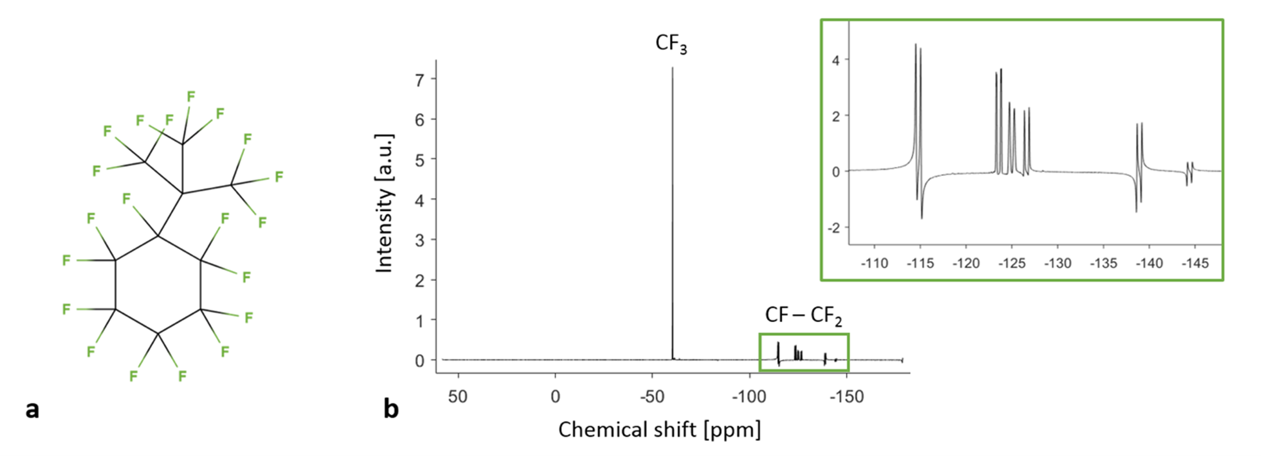
Figure 1. ABL-101 structure and spectrum. a.
Perfluoro(t-butylcyclohexane), the main perfluorocarbon in ABL-101, is composed of 5 inequivalent CF2
groups, 1 CF group and 3 equivalent CF3 groups. b. The ABL-101 spectrum has one main resonance that corresponds to
the CF3 groups, and a multiplet of several smaller resonances (green
inset) that corresponds to the J-coupled CF-CF2 groups. This
spectrum was obtained at 14.1T.
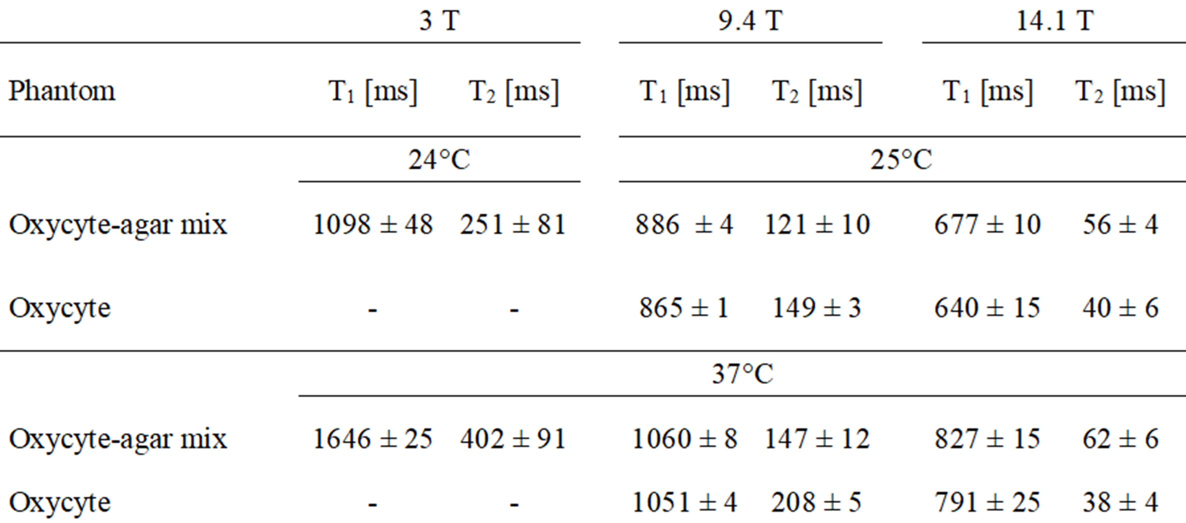
Table 1. Fluorine-19 relaxation times of the ABL-101
CF3 resonance. The T1 and T2
relaxation times were measured at temperatures of 25°C and 37°C, and at
magnetic field strengths of 3T, 9.4T and 14.1T. Pure ABL-101 had a PFC
concentration of 1.2M (19F concentration 24M), while the ABL-101-agar
mix had a PFC concentration of 0.30M (19F concentration 6.0M).
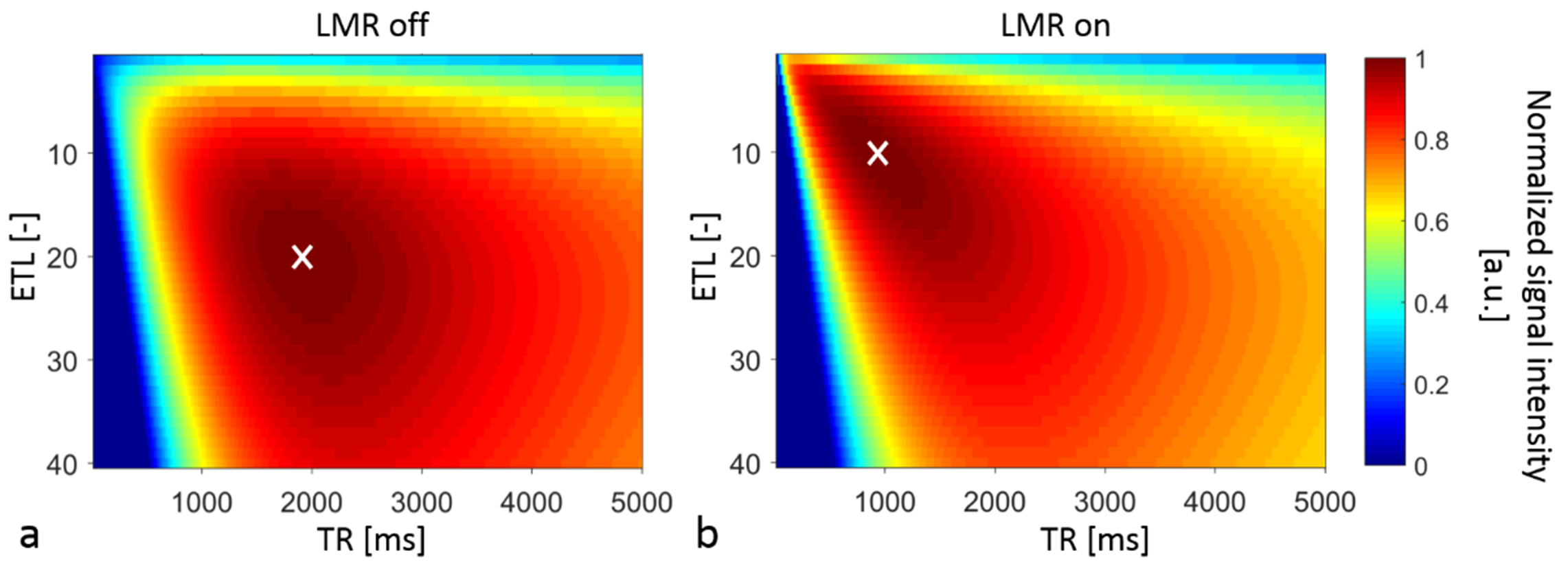
Figure 2. TSE parameter optimization for the ABL-101 CF3
resonance at 3T and 24°C. a. Map of the normalized signal intensity
with the longitudinal magnetization restoration (LMR) off or b. on. With BW = 130 Hz/px and TE =
13ms for both simulations, the optimized parameters were ETL = 20, TR = 1880 ms
and ETL = 10, TR = 920ms for LMR off and on, respectively (white crosses).
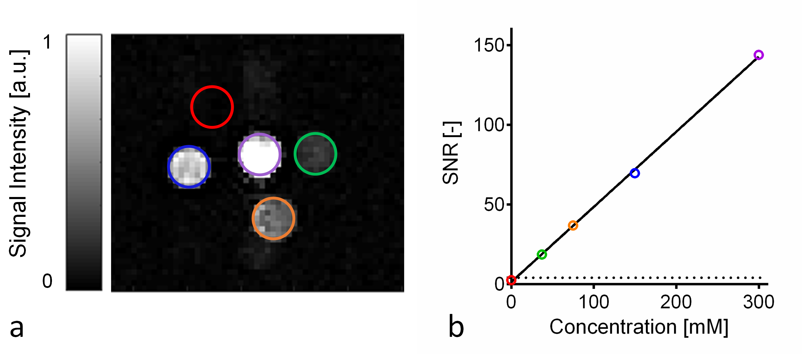
Figure 3. Detection limit of ABL-101 at 3T. a.
19F image of the ABL-101 phantom: four tubes are visible; the
colored ROI of the fifth tube without ABL-101 contains just noise (red ring). b. Fit of the SNR in the tubes as a
function of the corresponding ABL-101 concentration. The dotted line
indicates the Rose limit below which signal can be confused with noise (SNR =
4). A linear fit resulted in the equation (solid line), which provides a
detection limit of 15.9 mM of 19F atoms for an acquisition time of 10
minutes and spatial resolution 1mm3.
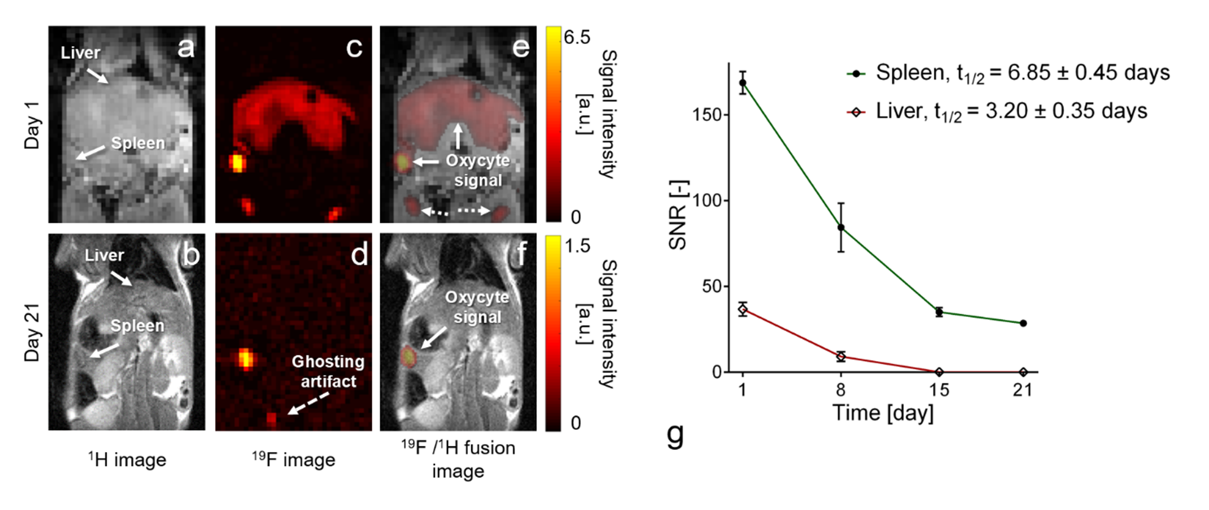
Figure 4. Detection of ABL-101 signal in mouse liver and spleen at 3T and
related clearance half-lives. a, b. 1H
anatomical images, c, d. 19F
images and e, f. 1H/19F
fused images, one and 21 days after injection, respectively. The 19F
signal (white arrows) observed at day 1 in liver has completely been cleared at
day 21. 19F signal was observed in potential lymph nodes (dotted
arrows) and as a small ghosting artifact from a reference tube (dashed arrow). g. SNR measurements from day one to 21
after injection (n=3 animals, dosage=3mL/kg).