1126
Novel Fluorine-18 Labeled Fe3O4@Al(OH)3 Nanoparticles as Dual Contrast Agents for Simultaneous PET/MRI1Biomedical MRI, Department of Imaging and Pathology, KU Leuven, Leuven, Belgium, 2MoSAIC, Department of Imaging and Pathology, KU Leuven, Leuven, Belgium, 3NANOMAG Group, Department of Applied Physics, Research Technological Institute, Universidade de Santiago de Compostela, Santiago de Compostela, Spain, 4Radiopharmaceutical Research, Department of Pharmaceutical and Pharmacological Sciences, KU Leuven, Leuven, Belgium, 5Nanohealth and Optical Imaging Group, Department of Imaging and Pathology, KU Leuven, Leuven, Belgium, 6Nuclear Medicine and Molecular Imaging, Department of Imaging and Pathology, KU Leuven, Leuven, Belgium
Synopsis
The emergence of (pre)clinical PET/MRI scanners is accompanied by the development of novel contrast agents. Aluminum hydroxide magnetic nanoparticles (Fe3O4@Al(OH)3 NPs) can overcome limitations of iron oxide NPs after labeling with Na18F, allowing visualization with PET (high sensitivity, low background) and MRI (high resolution, longitudinal NP follow-up). In this study, labeling of novel Fe3O4@Al(OH)3 nanostructures with Na18F was rapid. In vivo, radiolabeled NPs accumulated in liver of healthy mice, while cells labeled with radiolabeled NPs were also seen in lungs. Our results indicate the potential of these novel nanostructures for cell tracking with high sensitivity, specificity and resolution using PET/MRI.
Purpose
New contrast agents have been developed for simultaneous positron emission tomography/magnetic resonance imaging (PET/MRI) – an imaging approach combining the advantages of both modalities. However, radiolabeling of the contrast agents is often time-consuming1,2. Iron oxide (Fe304) nanoparticles (NPs) have demonstrated great potential as negative contrast agents for MRI, e.g. for cell tracking applications3. By embedding an aluminum hydroxide (Al(OH)3) coating shell with Fe3O4 NPs, direct and fast labeling of these nanostructures with 18F--labeled sodium fluoride (Na18F) becomes possible, allowing thus visualization of the particles with both PET and MRI4,5.
Materials and Methods
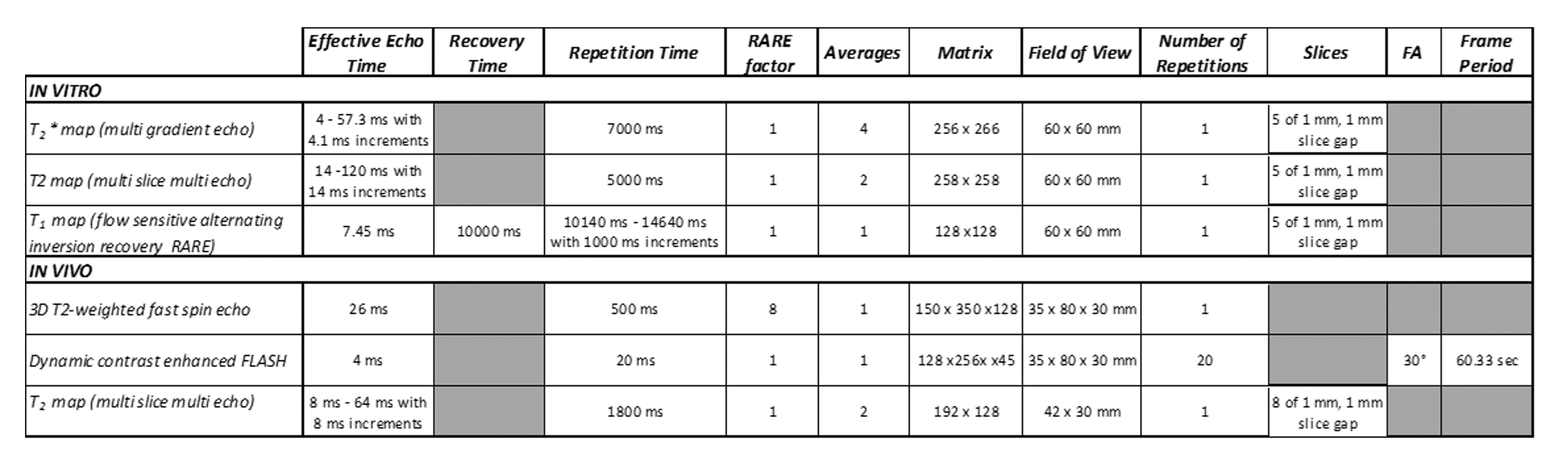
Nanostructures of Fe3O4 NPs embedded within Al(OH)3 coating shell (Fe3O4@Al(OH)3 nanostructures) were synthesized by forced chemical hydrolysis of pre-prepared magnetic particles. NPs were coated with polyacrylic acid to increase stability, followed by magnetic separation and re-dispersion in Milli-Q water. Labeling efficiency was evaluated after addition of Na18F to NPs (11.85µg iron in Milli-Q) while shaking. Afterwards NPs were centrifuged and resuspended in different media to test labeling stability. At different time points samples were taken for instant thin layer chromatography (iTLC, analysis using γ-counter). Serial dilutions of NPs and different numbers of mouse mesenchymal stem cells (mMSCs) labeled with radiolabeled NPs were resuspended in equal amounts saline/2% agar. Afterwards, a 1h static PET and T2*, T2 and T1 maps were simultaneously acquired to evaluate their in vitro contrast properties. For in vivo biodistribution studies, Fe3O4@Al(OH)3 NPs or 100,000 mMSCs labeled with NPs were injected via the tail vein of healthy 6-7-week-old C57BL/6 mice. To this end, NPs (11.85µg iron) were labeled with 3 MBq Na18F (0.9±0.2 MBq upon injection) or mMSCs were labeled with radiolabeled NPs (labeling NPs with 10 MBq Na18F; 1h mMSC labeling with NPs containing 0.38mM iron in saline; upon injection: 0.25±0.08 MBq). Before and after injection of NPs/cells, a whole-body 3D T2-weighted scan and parametric T2 map were acquired. A static 1h PET scan and dynamic contrast-enhanced (DCE)-MRI were acquired simultaneously with injection of NPs/cells. All data were acquired on a BioSpec 70/30 MRI equipped with a 3-ring SiPM based PET insert (Bruker BioSpin - scan parameters: Table 1). PET and DCE-MRI scans were analyzed in PMOD v3.9 and 3D Slicer, respectively. Other analyses were performed in ImageJ. After scanning, animals were sacrificed, and liver and lungs were isolated and analyzed using the γ-counter.
Results
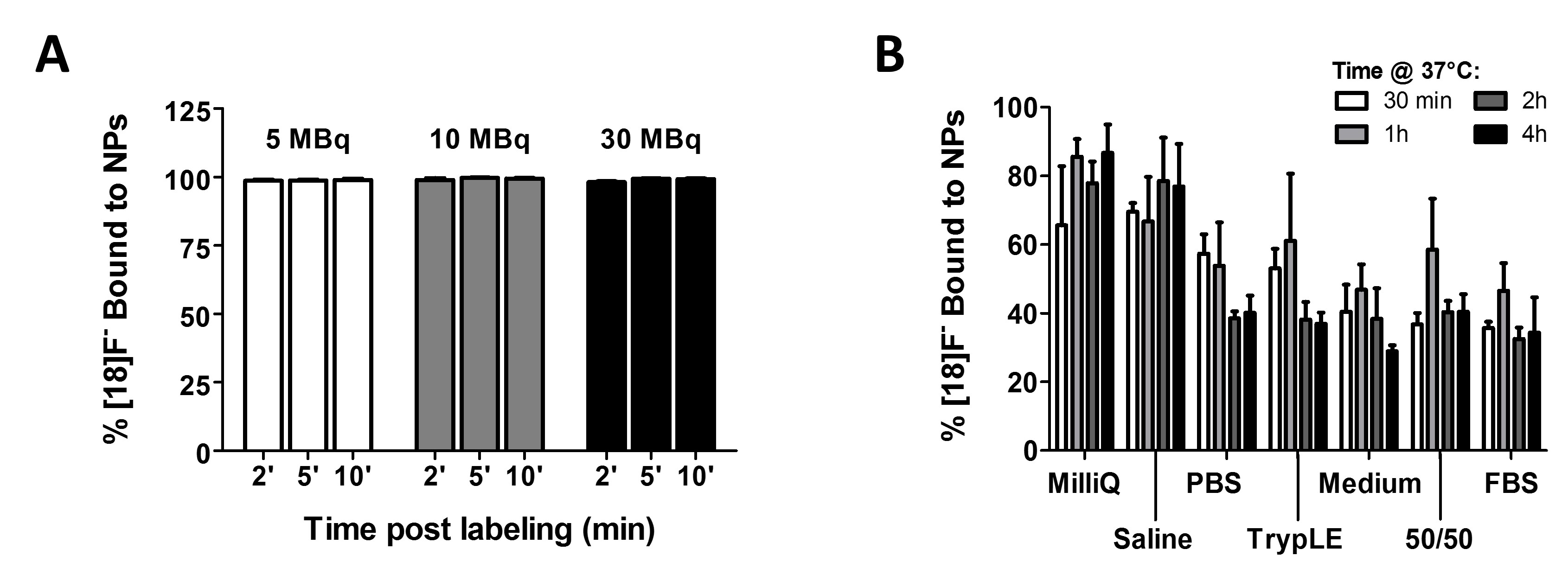
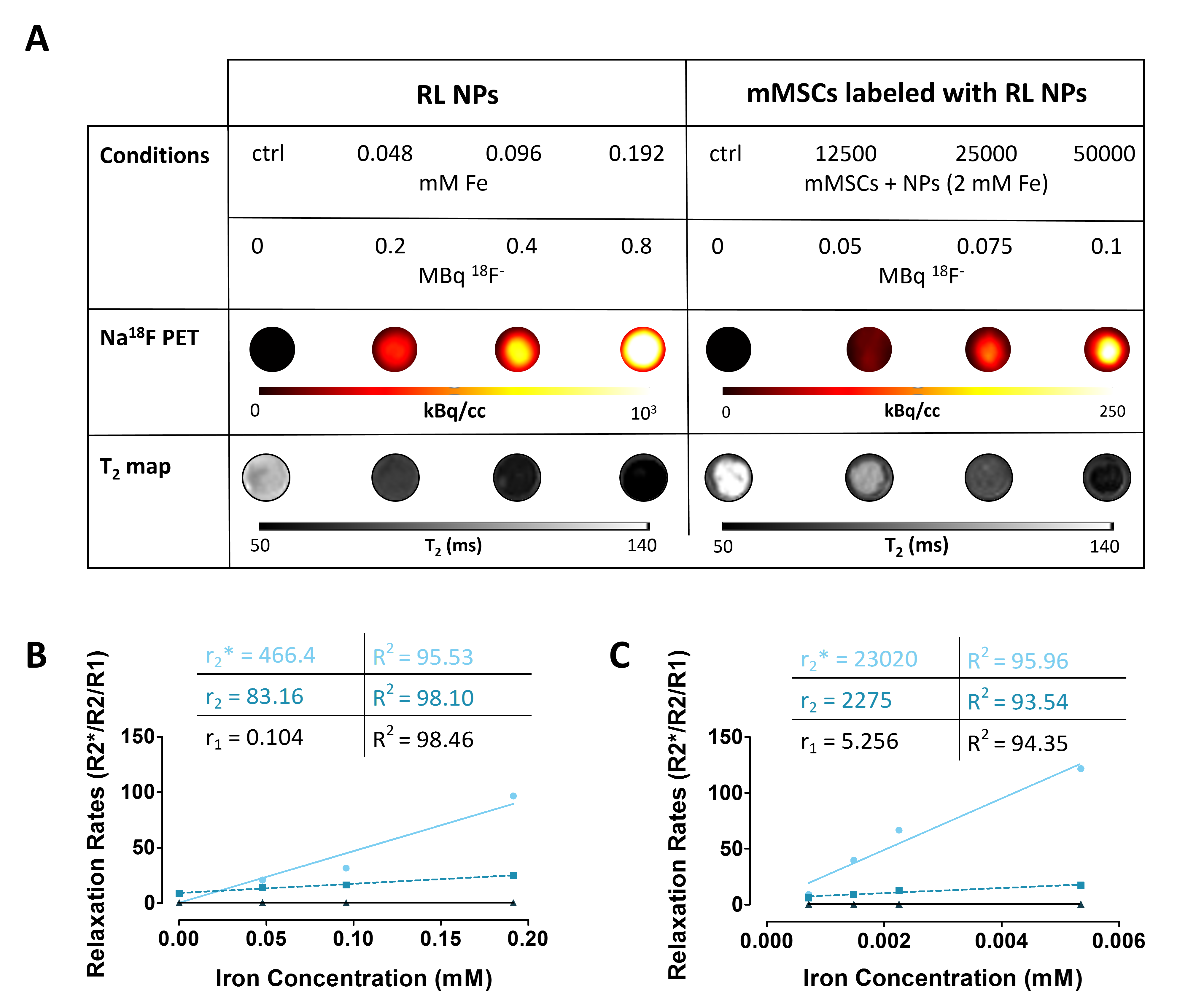
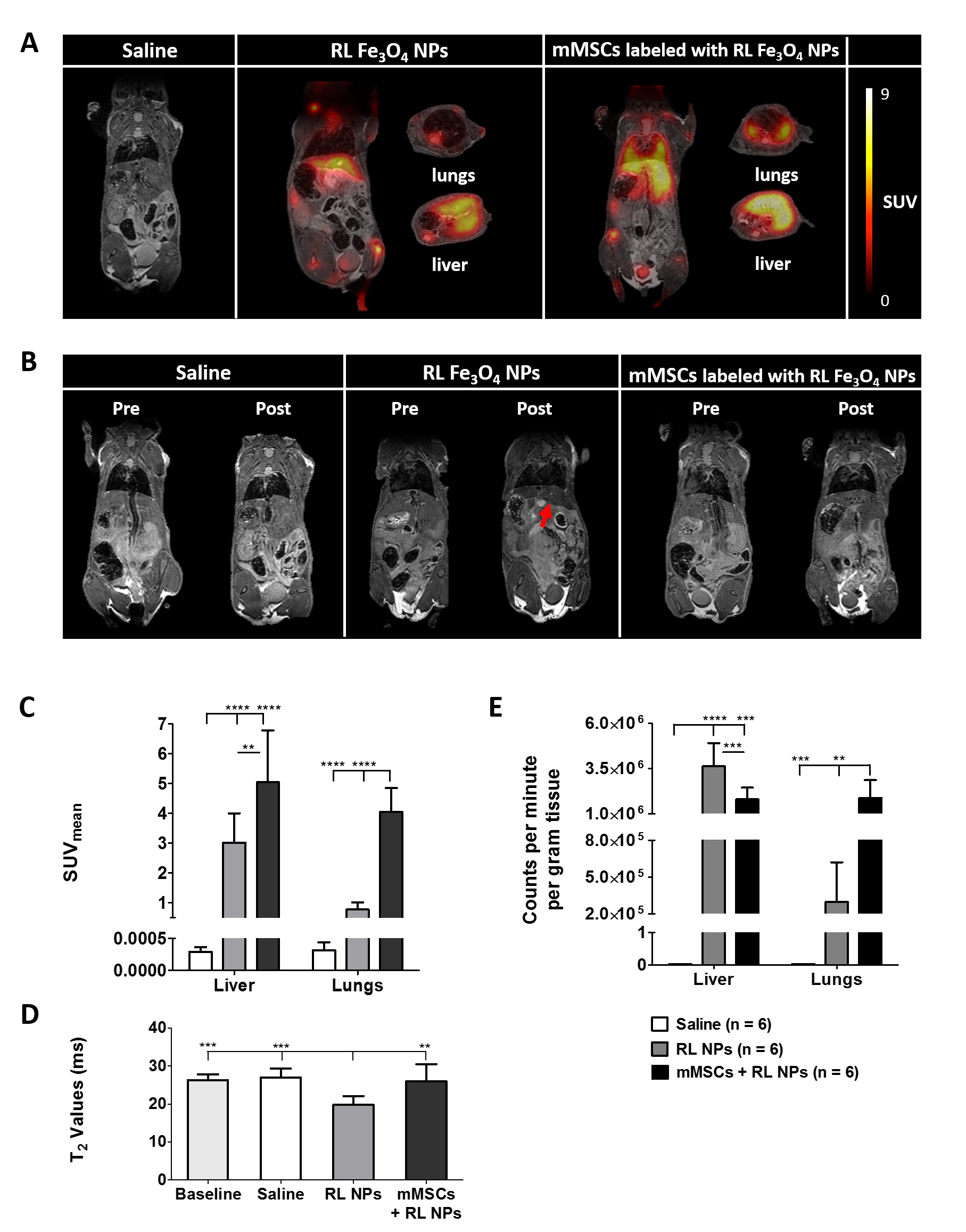
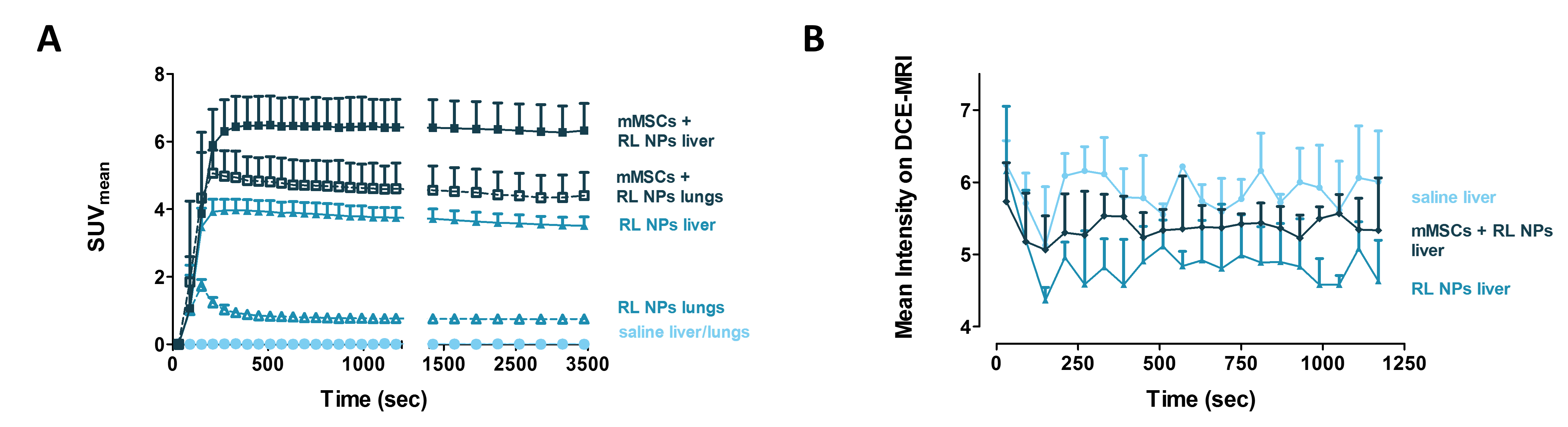
Labeling of Fe3O4@Al(OH)3 NPs in Milli-Q is fast (>97% Na18F bound after 2’ - Fig. 1A). Stability of the radiotracer bound to the NPs after 4h was the highest in Milli-Q (86.6% Na18F bound) and saline (76.9%), while it was less constant in other conditions (Fig. 1B). In vitro, we were able to visualize small amounts of radiolabeled Fe3O4@Al(OH)3 NPs and radiolabeled mMSCs on both PET and MRI (Fig. 2). After their intravenous injection, most radiolabeled NPs were visualized in the liver with both PET (increased standardized uptake values; SUV) and MRI (decreased T2 values). Conversely, an increased lung SUV value was visualized after injection of radiolabeled mMSCs when compared to injection of radiolabeled NPs. Moreover, increased SUV values in bone and spleen were measured (Fig. 3A-D). In vivo findings were confirmed by ex vivo γ-counter measurements (Fig. 3E). Furthermore, dynamic reconstruction of Na18F PET and DCE-MRI scans yielded time-activity curves showing rapid accumulation of the NPs and mMSCs in the liver, which stayed stable over time. While SUV increase was transient after NPs injection, it was stable after mMSCs injection (Fig. 4).
Discussion and Conclusion
In conclusion, we were able to visualize radiolabeled NPs and mMSCs labeled with these NPs in vitro and in vivo with simultaneous PET/MRI. The high potential of (cell) imaging using PET/MRI contrast agents/tracers is based on 1) detection of cells/NPs also in organs with a hypointense MR background, e.g. lungs, 2) limited background signal in PET, and 3) the combined advantages of both imaging techniques (i.e. high resolution, anatomical information, good soft tissue and long lasting contrast for MRI, high sensitivity and specificity for PET)5–7. Combining both techniques provides additional information on the stability of radiolabeling or cell labeling in vivo based on the distribution and stability of contrast (e.g. liver= free NPs; lungs or targeted site= labeled cells; spleen= clearance of NPs/cells, bone= defluorination of NPs). Each individual imaging technique would not be able to provide a complete picture of the whole-body distribution of NPs/cells over time in vivo. Future PET/MRI studies could include targeted tumor therapy, providing in-depth information on the active targeting mechanism of the NPs and passive targeting mechanism of stem cells towards tumors, as has been shown, but not elucidated using other techniques8,9.
Acknowledgements
This work was funded by the European Horizon 2020 ‘PANA’ project under grant agreement 686009 and the KU Leuven program financing ‘In Vivo Molecular Imaging Research’ (IMIR).References
1. Chen, F. et al. In Vivo Integrity and Biological Fate of Chelator-Free Zirconium-89-Labeled Mesoporous Silica Nanoparticles. ACS Nano 9, 7950–9 (2015).
2. Bouziotis, P. et al. 68 Ga-radiolabeled AGuIX nanoparticles as dual-modality imaging agents for PET/MRI-guided radiation therapy. Nanomedicine 12, 1561–1574 (2017).
3. Himmelreich, U. & Dresselaers, T. Cell labeling and tracking for experimental models using Magnetic Resonance Imaging. Methods 48, 112–124 (2009).
4. McBride, W. J., Sharkey, R. M. & Goldenberg, D. M. Radiofluorination using aluminum-fluoride (Al18F). EJNMMI Res. 3, 36 (2013).
5. Jauregui-Osoro, M. et al. Biocompatible inorganic nanoparticles for [18F]-fluoride binding with applications in PET imaging. Dalton Trans. 40, 6226–37 (2011).
6. Massoud, T. F. & Gambhir, S. S. Molecular imaging in living subjects: seeing fundamental biological processes in a new light. Genes Dev. 17, 545–80 (2003).
7. Sauter, A. W., Wehrl, H. F., Kolb, A., Judenhofer, M. S. & Pichler, B. J. Combined PET/MRI: one step further in multimodality imaging. Trends Mol. Med. 16, 508–515 (2010).
8. Leibacher, J. & Henschler, R. Biodistribution, migration and homing of systemically applied mesenchymal stem/stromal cells. Stem Cell Res. Ther. 7, 7 (2016).
9. Krueger, T. E. G., Thorek, D. L. J., Denmeade, S. R., Isaacs, J. T. & Brennen, W. N. Concise Review: Mesenchymal Stem Cell-Based Drug Delivery: The Good, the Bad, the Ugly, and the Promise. Stem Cells Transl. Med. 7, 651–663 (2018).
Figures