1110
First-in-human trial of Gd-based theranostic nanoparticles: MRI assessment of uptake and biodistribution in patients with multiple brain metastases1NHTherAguix, Crolles, France, 2CHU Grenoble Alpes, Grenoble, France, 3GIN, Inserm, Université de Grenoble, Grenoble, France, 4Institut des Sciences Moléculaires, CNRS, Université de Bordeaux, Bordeaux, France, 5Institut Lumière Matière, CNRS, Université de Lyon, Villeurbanne, France, 6Neurospin, CEA, Gif-sur-Yvette, France
Synopsis
We report here the main MRI findings of a dose escalation phase 1 clinical trial with intravenous administration of theranostic AGuIX nanoparticles, conducted in 15 patients with brain metastases with the objective of evaluating the efficacy of the nanoparticles as contrast agent and radiosensitizer for brain metastases. The nanoparticles were found to enhance four different histological types of brain metastases up to one week after nanoparticle administration. Quantitative measurements of nanoparticle concentration in all types of brain metastases were obtained two hours after administration to patient - and incidentally two hours before the first session of whole brain radiotherapy.
Introduction
The use of radiosensitizers is an effective approach to increase the curative efficacy of radiotherapy and to limit some undesirable side effects1. Among nanoscale size radiosentitizer, the theranostic nanoparticles, combining both diagnostic and radiosensitizing properties on the same nano-object, represent an elegant solution to achieve these objectives2. We present here the main MRI findings obtained in a first-in-human phase 1 clinical trial with intravenous administration of Gd-based nanoparticles, in patients with multiple brain metastases from four types of primary tumors (NSCLC, colon, melanoma, breast cancer).Materials and Methods
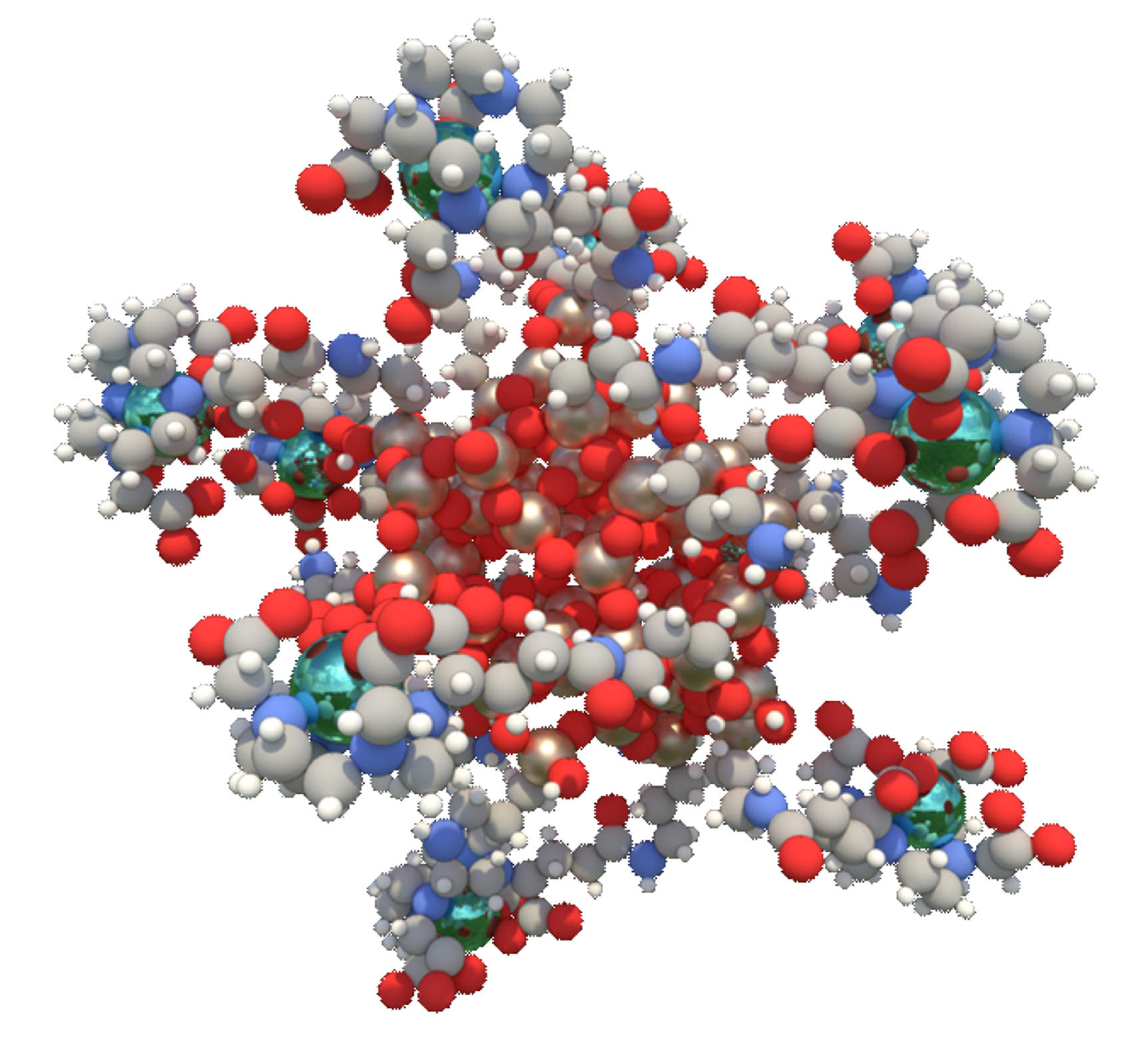
This study is part of the prospective dose escalation phase I-b clinical trial Nano-Rad (NCT02820454) to evaluate the safety and potential of the radio-sensitizing nanoparticle AGuIX® (NH TherAguix, France) in combination with whole brain radiotherapy for the treatment of brain metastases. Patients (N=15) with multiple brain metastasis ineligible for local treatment by surgery or stereotactic radiation were recruited. The AGuIX theranostic agent is composed of a polysiloxane network surrounded by DOTA cyclic ligands covalently grafted to the matrix3. Their hydrodynamic diameter is 3 ± 0.1 nm and their mass is 8.5 ± 1 kDa. On average each nanoparticle presents on its surface 10 DOTA ligands chelating gadolinium ion (Fig. 1). The longitudinal relaxivity r1 at 3 Tesla is equal to 8.6 mM-1.s-1 per Gd3+. At D1, the patients were administered intravenously with solution of AGuIX nanoparticles at different doses of 15, 30, 50, 75 or 100 mg/kg body weight. Two hours post administration, the patients followed a MRI session including a 3D T1-weighted inversion recovery gradient echo sequence and a 3D FLASH sequence with multiple flip angles. The latter sequence was used to compute T1 maps and AGuIX concentration based on changes in T1 values following administration of the nanoparticles. The MRI acquisitions were performed at 3 Tesla (Philips Achieva) using a 32-channel head coil. The patients then underwent a whole brain radiation therapy (30 Gy delivered in 10 sessions). Similar MRI sessions were performed at D8, D28 and D100, the last two sessions including injection of Dotarem® (Guerbet, France) contrast agent.Results
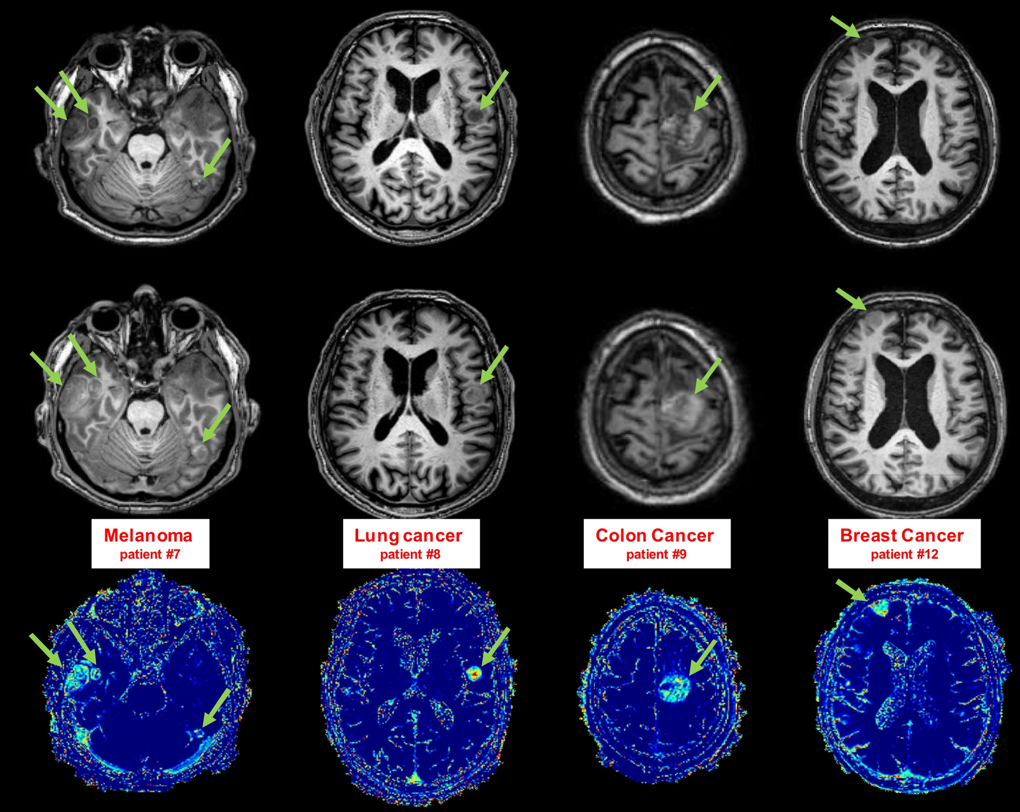
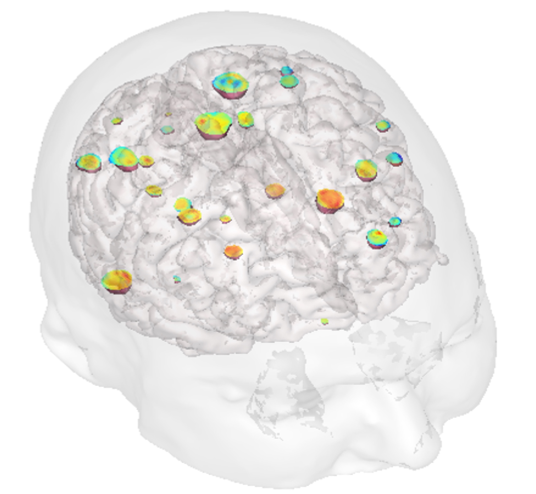
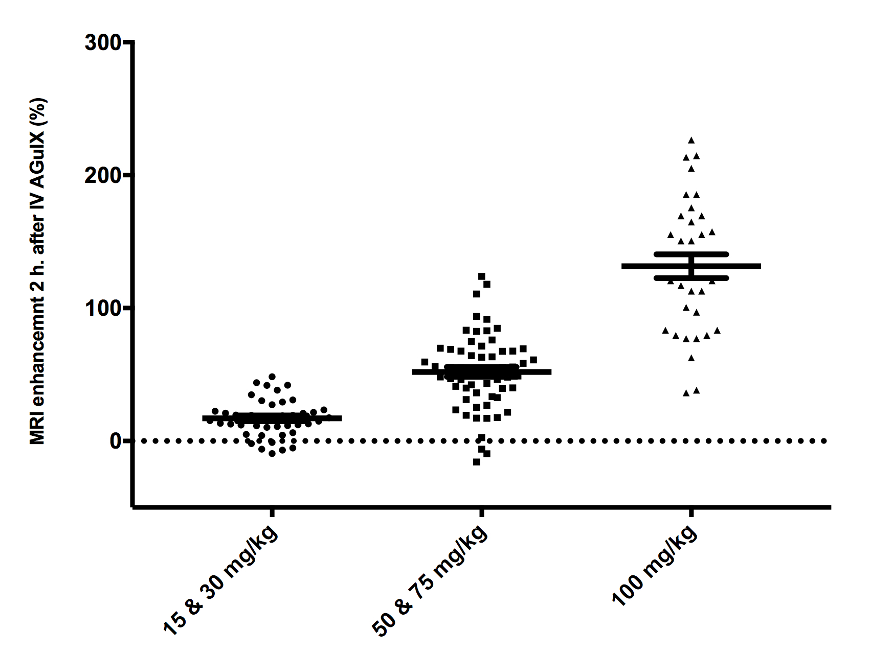
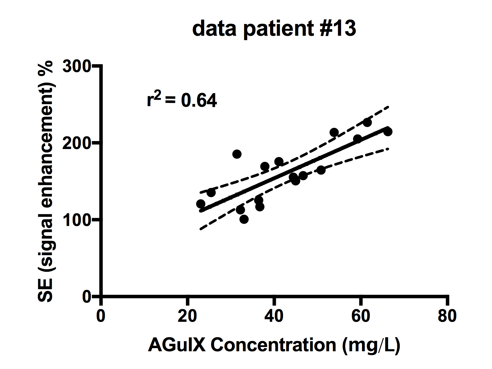
Two hours after AGuIX injection, MRI signal enhancements (SE) were observed for all histological types of brain metastases, all patients and all doses administered. Tumor enhancements are exemplified in Fig. 2 and Fig 3 for each type of primary tumour. The SE were found to increase with the administered dose of AGuIX nanoparticles (Fig. 4). The mean AGuIX concentration in metastases of the patients administered with the highest dose were measured to range between 17.3 and 48.7 mg/L. The correlation between MRI enhancement and nanoparticle concentration was assessed for patients with the largest administered dose. The correlation is exemplified in Fig. 5 with MRI data from a patient with NSCLC metastases. A positive correlation between the two MRI parameters was observed with a relationship close to linearity in the range of values measured. No significant MRI enhancement and no T1 variations were observed in any of healthy brain regions. For patient administered with the largest dose, persistence of MRI enhancement was noticed in metastases at D8, one week after administration of nanoparticles.Discussion and conclusions
The largest dose of
nanoparticle administered to the patients corresponds to the amount of Gd3+
injected in one dose of clinically-used contrast agent such as Dotarem. The SE
was measured to be similar for AGuIX and Dotarem administration. Importantly,
these initial results show that nanoparticle uptake and signal enhancement are
present in all the different histological types of metastases investigated. The
computed concentration of nanoparticles in the patients injected with the
highest dose is of the same order of magnitude as the nanoparticle
concentration obtained previously in animal models4,5. At the
highest AGuIX dose, all observable metastases (diameter above 1 cm) were
contrast-enhanced up to 8 days after the nanoparticle were administered,
illustrating the accumulation and delayed clearance of nanoparticles from the
metastasis, as previously observed in preclinical studies6. The
linear relationship observed between the signal enhancement and the
nanoparticle concentration makes it possible to consider the SE, in the
investigated range, as a simple and robust index for measuring nanoparticle
concentration in metastases.
In summary, the preliminary results of this clinical trial demonstrate that intravenous injection of AGUIX is effective for enhancing different histological types of brain metastases in patients, with good tolerance to intravenous injection of AGuIX nanoparticle up to the highest 100 mg/kg dose. All these findings and observations make it possible to confidently envision and implement a second translation step to the clinic of these theranostic nanoparticles within the framework of a phase 2 clinical trial.
Acknowledgements
The authors acknowledge the Centre Hospitalier Universitaire (CHU) of Grenoble for sponsoring and supporting the clinical trial Nano-Rad and the company NH TherAguix for providing AGuIX nanoparticles. The authors are grateful to Yohan Pietras (CHU Grenoble) for planning of MRI sessions and patient's scanning. The MRI facility IRMaGe is partly funded by the French program ‘Investissement d’Avenir’ run by the French National Research Agency, grant ‘Infrastructure d’avenir en Biologie Sante’ [ANR-11-INBS-0006].
References
1. S.L. Liauw, P.P. Connell, R.R. Weichselbaum. New paradigms and future challenges in radiation oncology: an update of biological targets and technology. Sci Transl Med. 5, 173sr2 (2013).
2. G. Le Duc et al. Toward an image-guided microbeam radiation therapy using gadolinium-based nanoparticles. ACS Nano. 5, 9566-9574 (2011).
3. F. Lux et al. Ultrasmall rigid particles as multimodal probes for medical applications. Angew Chem 50(51), 12299-12303 (2011).
4. C. Verry et al. MRI-guided clinical 6-MV radiosensitization of glioma using a unique gadolinium-based nanoparticles injection. Nanomedicine 11, 2405-2417 (2016).
5. A. Bianchi et al. Targeting and in vivo imaging of non-small-cell lung cancer using nebulized multimodal contrast agents. Proc Natl Acad Sci U S A. 111 9247-9252 (2014).
6. S. Kotb et al. Gadolinium-Based Nanoparticles and Radiation Therapy for Multiple Brain Melanoma Metastases: Proof of Concept before Phase I Trial. Theranostics 6(3):418-427 (2016).
Figures