1102
Deep Learning-enabled Diffusion Tensor MR Fingerprinting1Computer Science, Technical University of Munich, Garching, Germany, 2GE Healthcare, Munich, Germany, 3Cardiff University Brain Research Imaging Centre (CUBRIC), Cardiff University School of Psychology, Cardiff, United Kingdom, 4Division of Psychological Medicine and Clinical Neurosciences, Cardiff University School of Medicine, Cardiff, United Kingdom, 5Imago7 Foundation, Pisa, Italy, 6Department of Neuroradiology, Klinikum rechts der Isar, Munich, Germany, 7Department of Physics, Technical University of Munich, Garching, Germany
Synopsis
MR Fingerprinting enables the quantification of multiple tissue properties from a single, time-efficient scan. Here we present a novel Diffusion Tensor MR Fingerprinting acquisition scheme that is simultaneously sensitive to T1, T2 and the full diffusion tensor. We circumvent the long-standing issue of phase errors in diffusion encoding and expensive dictionary matching by using a neural network architecture capable of learning the non-linear relation between fingerprints and multiparametric maps, robustly mitigating motion, undersampling and phase artifacts. As such, our framework enables the simultaneous quantification of relaxation parameters together with the diffusion tensor from a single, highly accelerated acquisition.
Introduction
MR Fingerprinting (MRF) provides a framework for simultaneous quantification of multiple tissue parameters from a single, time-efficient acquisition1. Recently, attempts have been made to develop diffusion-weighted MRF techniques. However, the transient nature of MRF signals makes them highly susceptible to severe motion artifacts, especially when considering the full diffusion tensor (DT). Susceptibility to motion, together with the exponential scaling of the dictionary with the dimensionality of the parameter space, pose a significant challenge and limit existing diffusion-weighted MRF applications to the estimation of the apparent diffusion coefficient2-5. Here we present a novel Diffusion Tensor MRF (DT-MRF) acquisition, where diffusion-encoding gradients in all three spatial dimensions encode the full DT. Inspired by image quality transfer ideas6,7, we take advantage of neural networks to recover information from images corrupted from phase errors due to motion and spatial undersampling artefacts to reliably reconstruct T1 and T2 maps together with the DT maps from a single acquisition.Methods
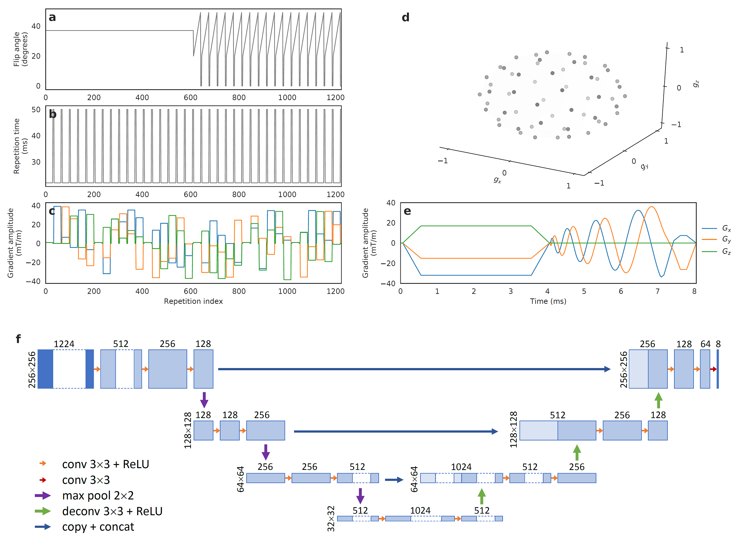
Building on diffusion-weighted steady-state free precession literature8, we propose a diffusion-sensitive MRF-type acquisition as follows (Figure 1a-e). Diffusion-encoding directions are varied randomly every 34 repetitions, with non-diffusion-weighted unbalanced gradients added every six directions. In total, 30 diffusion directions, chosen based on the electrostatic repulsion algorithm9, are acquired. An initial inversion pulse is followed by a train of constant flip angles with repeating variable flip angle ramps in the latter part of the sequence to increase T1 and T2 sensitivity. Repetition times are set constant during diffusion-encoding with longer waiting periods in-between directions. During each repetition, one arm of an undersampled spiral interleave is acquired. Image time-series are obtained using sliding-window reconstruction.
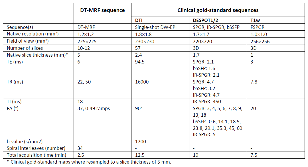
In an IRB-approved study, data from eleven patients with Multiple Sclerosis (MS) and nine healthy subjects were acquired on a 3T HDx MRI system (GE Healthcare, Milwaukee, WI) using an eight channel receive-only head RF coil, after obtaining written informed consent. The protocol (see Table 1) included diffusion tensor imaging (DTI), DESPOT1/2 sequences and a high resolution T1w acquisition for co-registering all modalities. In addition to these clinical sequences, 8-12 axial slices, covering the middle portion of the brain were acquired with our DT-MRF sequence. We obtained T1 and T2 maps from DESPOT1/2 and calculated the DT from the DTI dataset. These clinical gold-standard maps constitute the ground-truth for the deep learning approach.
Bypassing conventional dictionary matching10,11, we propose a modified UNET architecture to learn a non-linear function between the temporal evolution of the DT-MRF magnitude images, and the quantitative relaxation and DT maps, scaled from 0 to 1, as output (Figure 1f). The model was implemented using TensorFlow and trained for 650 epochs using L2 loss with ADAM optimizer, batch size of 5, learning rate of 1e-4 and dropout rate of 0.5. Aiming at an efficient and robust reconstruction method, we trained our model on healthy subjects and MS patients. We calculated mean diffusivity (MD), axial diffusivity (AD), radial diffusivity (RD) and fractional anisotropy (FA) metrics from the predicted and gold standard DT12.
Results
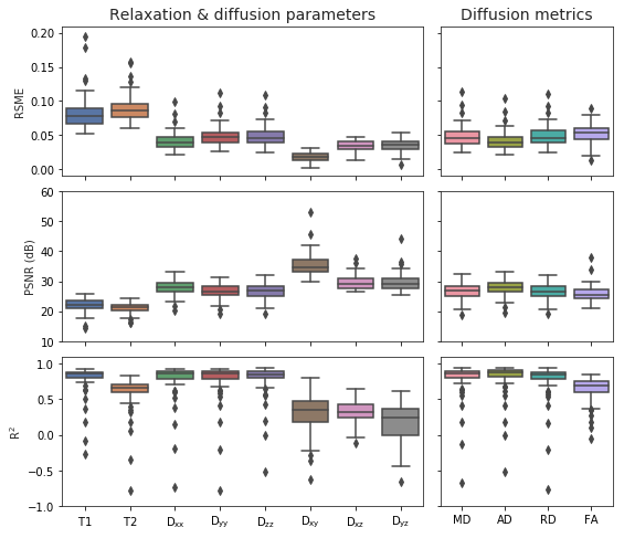
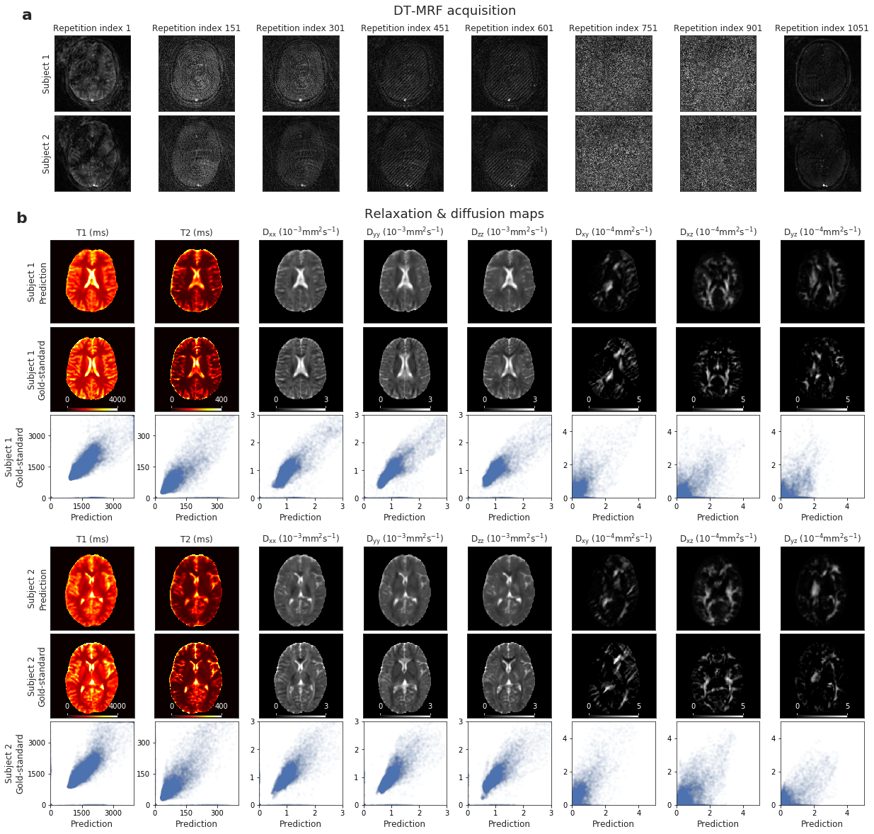
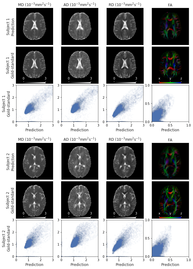
Predicted maps correlate highly with the DESPOT1/2 and DTI gold-standard, respectively (Figure 2). The quantification errors for the diffusion metrics also indicate good agreement with the gold-standard. Figure 3 shows estimated maps for T1, T2 and the DT elements together with the reference for two representative test datasets. High consistency between the predicted maps and traditional, gold-standard methods can be visually observed. The corresponding diffusion metrics confirm this good correspondence and demonstrate that directional information in the predicted DT is preserved (Figure 4).Discussion
The proposed DT-MRF scheme simultaneously encodes T1, T2 and the DT with a highly accelerated acquisition (12s/slice). In combination with the deep learning-based reconstruction, this enables fast parameter inference, providing artifact-free quantitative relaxation and DT maps whilst making expensive postprocessing pipelines of conventional multi-modality imaging redundant. Despite achieving high correlation with respect to gold-standard DTI, the network does not fully recover the characteristic fiber structure of high anisotropy areas but still captures directional information. We expect that further improvements in the diffusion-encoding and neural network reconstruction will be able to even further improve its predictive quality and is hence subject of future work.Conclusion
We propose a novel DT-MRF acquisition and reconstruction framework. Taking advantage of deep learning architectures, we reconstruct multiparametric outputs from spatiotemporal MRF data corrupted by motion, phase errors and undersampling artifacts. We provide a proof-of-concept for simultaneous quantification of T1 and T2 relaxation maps and DT maps with a MRF-type acquisition scheme that goes beyond ADC mapping alone. Additionally, we show that circumventing conventional dictionary matching paves the way to innovative higher-dimensional MRF applications.Acknowledgements
No acknowledgement found.References
1. Ma D, Gulani V, Seiberlich N, et. al: Magnetic Resonance Fingerprinting. Nature 495(7440), 187-192 (2013)
2. Cohen O, Rosen MS. Simultaneous Diffusion, PD, T1, and T2 Mapping with Optimized MR Fingerprinting EPI. Proc Intl Soc Mag Reson Med (2018)
3. Rieger B, Akçakaya M, Schad LR, Weingärtner S. Simultaneous quantification of T1, T2 and Apparent Diffusion Coefficient using Magnetic Resonance Fingerprinting based on Echo Planar Imaging. Proc Intl Soc Mag Reson Med (2018)
4. Jiang Y, Hamilton JI, Lo WC, et. al. Simultaneous T1, T2 and Diffusion Quantification using Multiple Contrast Prepared Magnetic Resonance Fingerprinting. Proc Intl Soc Mag Reson Med (2017)
5. Jiang Y, Hamilton JI, Wright KL, et. al. Simultaneous Quantification of T1, T2 and Diffusion with Diffusion-weighted drive-equilibrium prepared Magnetic Resonance Fingerprinting. Proc Intl Soc Mag Reson Med (2016)
6. Alexander DC, Zikic D, Ghosh A, et. al. Image quality transfer and applications in diffusion MRI. NeuroImage 152(2017), 283-298 (2017)
7. Tanno R, Worrall DE, Ghosh A, et. al. Bayesian Image Quality Transfer with CNNs: Exploring Uncertainty in dMRI Super-Resolution. MICCAI (2017)
8. McNab JA, Miller, KL. Steady-state diffusion-weighted imaging: theory, acquisition and analysis. NMR in biomedicine 23(7), 781-793 (2010)
9. Jones DK, Horsfield MA, Simmons A. Optimal strategies for measuring diffusion in anisotropic systems by magnetic resonance imaging. Magn Reson Med 42(3), 515-525 (1999)
10. Gómez PA, Molina-Romero M, Ulas C, Buonincontri G, Sperl J, Jones DK, Menzel MI, Menze BH. Simultaneous parameter mapping, modality synthesis, and anatomical labeling of the brain with MR fingerprinting. MICCAI (2016)
11. Gómez PA, Buonincontri G, Molina-Romero M, Sperl J, Menzel MI, Menze BH. Accelerated parameter mapping with compressed sensing: an alternative to MR Fingerprinting. Proc Intl Soc Mag Reson Med (2017)
12. Basser PJ, Mattiello J, LeBihan D. Estimation of the effective self-diffusion tensor from the NMR spin echo. J Magn Reson B 103(3), 247-254 (1994)
Figures