1098
Relax-MANTIS: REference-free LAtent map-eXtracting MANTIS for efficient MR parametric mapping with unsupervised deep learning1Medical Physics, University of Wisconsin-Madison, Madison, WI, United States, 2Radiology, University of Wisconsin-Madison, Madison, WI, United States, 3Biomedical Engineering, University of Wisconsin-Madison, Madison, WI, United States
Synopsis
The purpose of this work was to develop and evaluate a novel deep learning-based framework termed Reference-free Latent map-eXtracting MANTIS (Relax-MANTIS) for efficient MR parameter mapping. Our approach incorporated end-to-end CNN mapping, the concept of cyclic loss to enforce data fidelity and without the need of explicit training references. Our results demonstrated that the proposed framework produced accurate and robust T1 mapping in knee and low-SNR lung UTE MRI. The good quantitative agreement to the reference method suggests that Relax-MANTIS allows potentially accelerated quantitative mapping without modifications of scan protocol and sequence for high-resolution knee and whole lung T1 quantification.
INTRODUCTION
While several deep learning methods1,2 have focused on highly efficient image reconstruction for conventional MRI, applications for MR parameter mapping have been limited. A recent deep learning-based reconstruction framework, Model-Augmented Neural neTwork with Incoherent Sampling (MANTIS)3, demonstrated a great capability of reconstructing quantitative maps directly from undersampled images using supervised learning. In line with MANTIS, here we developed a variant of MANTIS for efficient parameter mapping using unsupervised learning. In our new method termed Reference-free Latent map-eXtracting MANTIS (Relax-MANTIS), we aimed to obtain quantitative maps directly using the efficient image-to-parameter Convolutional Neural Network (CNN) mapping with the incorporation of an MR signal model. We demonstrated our method on variable flip angle (VFA) T1 mapping in both knee and low-SNR ultrashort echo time (UTE) lung MRI.METHODS
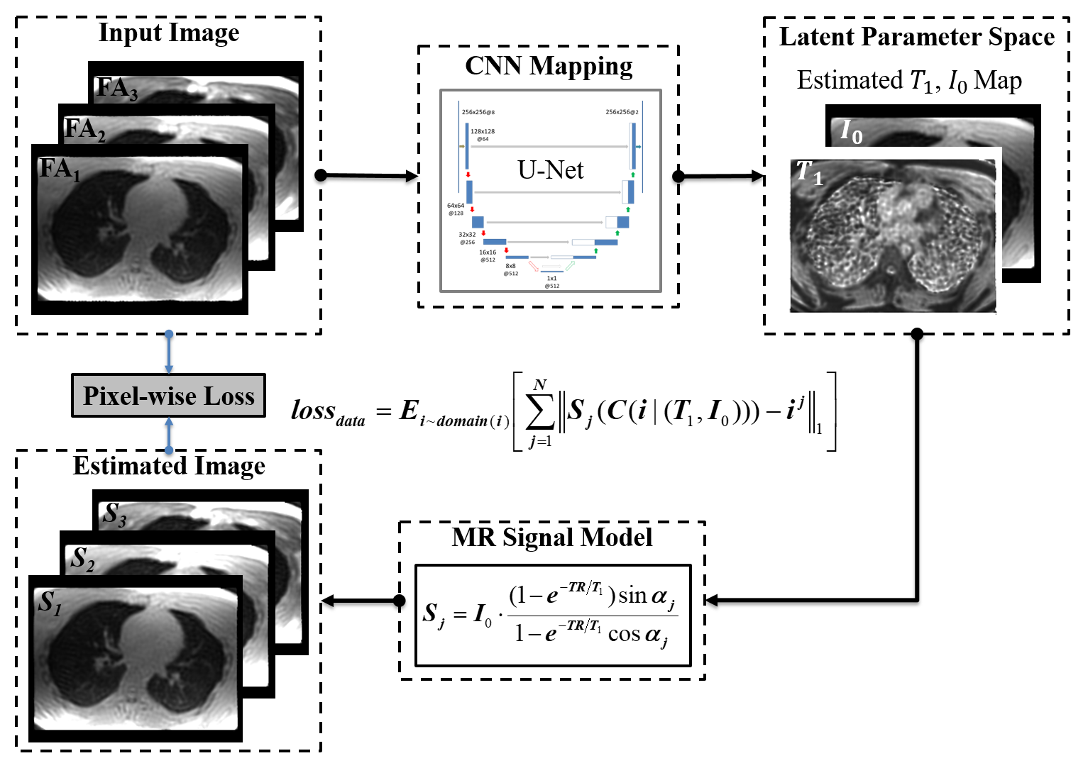
(a) Quantitative Imaging: The Relax-MANTIS framework (Figure 1) performs a learning process to estimate latent parameter maps, for instance, T1 and proton density images $$$ I_{0}$$$, using CNN mapping and the known MR signal model for the parameters of interest. Like MANTIS, the CNN mapping was implemented for converting the VFA image data directly to the T1 and $$$ I_{0}$$$ maps. However, Relax-MANTIS relaxes the requirement for the use of reference quantitative maps as supervised learning, but ensures that the extracted parameter maps from end-to-end CNN mapping produce estimated images matching the VFA images (i.e. model-augmented data consistency). The training objective is to learn parameters and using the generator CNN, $$$C(i|T_{1},I_{0})$$$: $$\hat{T_{1}},\hat{I_{0}}=argmin_{T_{1},I_{0}}(E_{i\sim domain(i)}[\sum_{j=1}^N\parallel S_{j}(C(i|T_{1},I_{0}))-i^{j} \parallel_{1}])$$ where $$$i$$$ are input VFA images, $$$i^{j}$$$ is the input image at the $$$j^{th}$$$ FA, $$$N$$$ is the total number of FAs, $$$S_{j}(\cdot)$$$ is the MR signal model, denotes the $$$l_{1}$$$ norm, $$$E$$$ is the expectation operator.
(b) Network Implementation: We used U-Net as convolutional encoder/decoder for performing the end-to-end CNN mapping4. The network was trained on an Nvidia GeForce GTX 1080Ti card using adaptive gradient descent optimization with a learning rate of 0.0001 for 300 epochs.
(c) Evaluation: Two image datasets were used as follows. 1) 34 spoiled gradient echo VFA healthy knee data sets (30/4 for training and evaluation) at 3.0T (GE Discovery MR750): 16cm FOV, 3 FAs = 3°, 7° and 18°, TR/TE = 4.9/2.3 ms, 3mm thickness, 256×256×32 matrix. 2) 12 low-SNR (10.9±2.9) 3D radial UTE VFA5 data sets of the healthy lungs (9/3 for training and evaluation) with full chest coverage at 1.5T (GE SignHDx): 32cm FOV, 5 FAs = 2°, 4°, 6°, 10° and 14°, TR/TE=2.86/0.08 ms, ~30,000 projections per FA, and total scan time=~14 minutes. Due to the inherently low proton density in lung, the two additional FAs (6°and 14°) were acquired to solve for T1 using standard non-negative least-squares fitting (NNLS)6. All UTE images were reconstructed at 1.25mm isotropic resolution (320×356×320 matrix) and registered to the UTE at 10° FA for each subject. For both knee and lung data, parameter maps were also calculated using standard NNLS and the widely used maximum likelihood variable projection method (VPM) with a grid search estimator7,8.
RESULTS
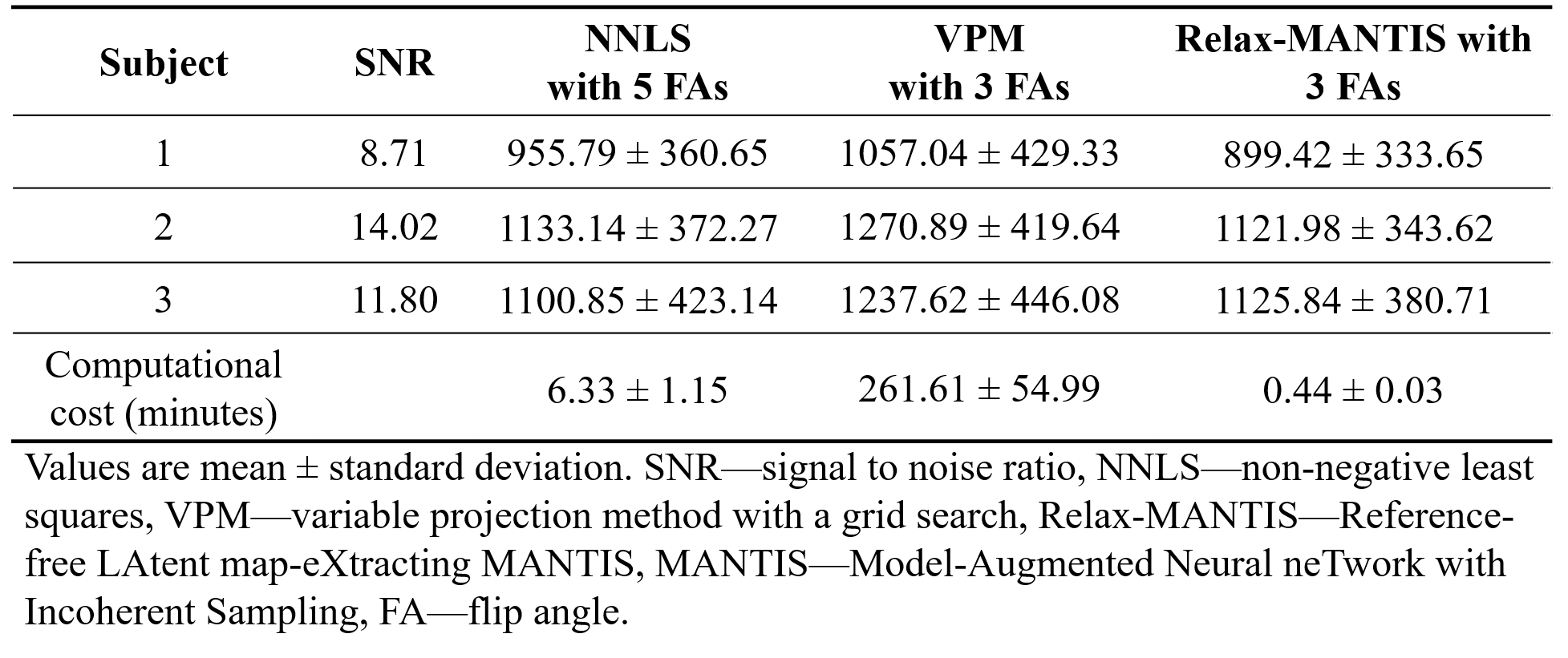
Examples of the estimated MR parameter maps from knee VFA data suggest qualitatively similar voxelwise T1 maps estimated using VPM and Relax-MANTIS referring to the standard NNLS (Figure 2), and $$$ I_{0}$$$estimated by Relax-MANTIS is notably more homogeneous as a result of de-noising functionality of CNN. For the lung VFA example with SNR=8.7, the parameter maps from Relax-MANTIS with only 3 FAs qualitatively agree better with the standard NNLS with 5 FAs (Figure 3), which is further confirmed by comparing the whole lung T1 histogram distributions. The T1 statistics on the three lung UTE testing data in Table 1 suggests that Relax-MANTIS with 3FAs yielded closer average whole lung T1 with smaller standard deviation relative to the measurements using standard NNLS with 5 FAs.DISCUSSION
The direct image-to-parameter transformation enabled by the model-augmented CNN mapping with unsupervised learning provides a promising approach for efficient and robust MR parameter mapping. The proposed framework produced accurate solutions for parameters in both knee and low-SNR lung imaging applications. Relax-MANTIS provides good quantitative agreement with the standard NNLS while used only 3 FAs, resulting in a 40% scan time reduction for a robust whole lung T1 quantification. Further scan-time reduction may be achieved by using MANTIS with reduced k-space acquisition. The proposed Relax-MANTIS framework can potentially be extended to support other quantitative imaging, such as B0/B1 field estimation with incorporation of the corresponding MR signal models.CONCLUSION
With a combination of efficient end-to-end CNN mapping and model-based data fidelity reinforcement, the Relax-MANTIS framework allows an efficient and robust latent-parameter extraction with high apparent parametric image quality.Acknowledgements
No acknowledgement found.References
[1] Hammernik K, Klatzer T, Kobler E, Recht MP, Sodickson DK, Pock T, et al. Learning a variational network for reconstruction of accelerated MRI data. Magn Reson Med 2018;79:3055–71. doi:10.1002/mrm.26977.
[2] Mardani M, Gong E, Cheng JY, Vasanawala S, Zaharchuk G, Alley M, et al. Deep Generative Adversarial Networks for Compressed Sensing Automates MRI. IEEE Trans Med Imaging 2017;PP:1. doi:10.1109/TMI.2018.2858752.
[3] Liu F, Feng L, Kijowski R. MANTIS: Model-Augmented Neural neTwork with Incoherent k-space Sampling for Efficient Estimation of MR Parameters. ISMRM Work OnMachine Learn Part II, Cap Hilton, Washington, DC, USA 2018.
[4] Ronneberger O, Fischer P, Brox T. U-Net: Convolutional Networks for Biomedical Image Segmentation 2015:1–8. doi:10.1007/978-3-319-24574-4_28.
[5] Bell LC, Johnson KM, Fain SB, Kruger SJ, Nagle SK. T1 mapping of the lungs using DESPOT1 approach with 3D radial UTE acquisition. Abtract# 4234 20th ISMRM Meet Melbourne, Aust 2012.
[6] Chang L-C, Koay CG, Basser PJ, Pierpaoli C. Linear least-squares method for unbiased estimation of T1 from SPGR signals. Magn Reson Med 2008;60:496–501. doi:10.1002/mrm.21669.
[7] Nataraj G, Nielsen J-F, Scott C, Fessler JA. Dictionary-Free MRI PERK: Parameter Estimation via Regression with Kernels. IEEE Trans Med Imaging 2018;37:2103–14. doi:10.1109/TMI.2018.2817547.
[8] Golub G, Pereyra V. Separable Nonlinear Least Squares: the Variable Projection Method and its Applications. Inverse Probl 2003;19:R1–26.
Figures