1073
Dual-Imaging Modality Approach to Evaluate Cerebral Hemodynamics in Growth-Restricted Fetuses: Oxygenation and Perfusion1Department of Biomedical Engineering, Wayne State University, Detroit, MI, United States, 2Department of Radiology, Wayne State University, Detroit, MI, United States, 3Department of Obstetrics and Gynecology, Wayne State University, Detroit, MI, United States, 4Institute for Biomedical Research, Waterloo, ON, Canada, 5Perinatology Research Branch, NICHD/NIH/DHHS, Bethesda, MD, United States
Synopsis
Cerebral blood perfusion has shown to be a sensitive biomarker of brain sparing at early stages of fetal growth restriction (FGR) compared to conventional Doppler estimates. Blood perfusion along with cerebral blood oxygenation may provide a holistic view of the fetal brain metabolism during FGR. Fractional moving blood volume, an ultrasound-based; and susceptibility weighted imaging, an MRI-based technique were used to estimate fetal cerebral blood perfusion and oxygenation, respectively. A significantly negative and positive correlation was found between cerebral blood perfusion and oxygenation, in normal growth and FGR fetuses, respectively. This dual modality based model will improve assessment of fetal well-being.
Introduction
A reduced middle cerebral artery pulsitility (MCA-PI) or resistance index (MCA-RI) is considered a sign of vasodilatation and manifestation of blood redistribution to the fetal brain1,2. Hemodynamic changes in the fetal brain might occur before the MCA-PI reduction; therefore, other blood flow estimates, such as cerebral blood perfusion that shows blood flow changes in small vessels, could be a more sensitive biomarker of “brain sparing” at early stages of fetal growth restriction (FGR)3. Studies in animal models4 and human adults5 suggest a subsequent increase in venous blood oxygenation (SvO2) as a consequence of increased blood flow; however, this is not studied in human fetuses. Hence, having the information of SvO2 along with perfusion would provide a comprehensive understanding of the fetal cerebral metabolic status in FGR fetuses and might contribute to the clinical management of these pregnancies. Fractional moving blood volume (FMBV), an Ultrasound (US)-based technique, has been found to be more sensitive in detecting cerebral blood flow (CBF) redistribution compared to Doppler indices in fetuses with growth restriction6.
Magnetic resonance imaging (MRI) can provide additional diagnostic information over US. However, contraindications for contrast agents and the low temporal and spatial resolutions limit the use of MRI in studying blood perfusion in the fetal brain. Nevertheless, SWI has been shown to estimate SvO2 in second and third trimester healthy human fetuses7. Therefore, we propose a dual-modal imaging approach to estimate the global CBF and SvO2 using FMBV and SWI-MRI techniques, respectively, in normal growth and FGR fetuses.
Materials and Methods
Normal growth (n=33) and FGR fetuses (n=10) from singleton pregnancies between 20 and 40 weeks of gestation were evaluated. All fetal MRI scans were carried out on a 3.0T Verio (Siemens Healthineers, Erlangen, Germany) with the parameters mentioned in Table 1. MRI-based SWI sequence was obtained in the straight section of the superior sagittal sinus and processed as explained previously7. US and Doppler studies were performed using General Electric Voluson E8 (GE Healthcare, Milwaukee, WI, USA) ultrasound systems and 2-5 MHz probes. Blood perfusion was estimated using Power Doppler ultrasound (PDU) and FMBV from the frontal lobe in a mid-sagittal plane of the fetal brain. The temporal resolution of the video was 5 frames/ sec. FMBV was estimated by normalizing PDU signals obtained from the mid-sagittal region of interest to eliminate the effect of depth and tissue interfaces8. The association between fetal brain SvO2 and FMBV was calculated for normal growth and FGR groups. The distribution of fetal cerebral SvO2 and FMBV was evaluated across gestational ages for both the cohorts.Results
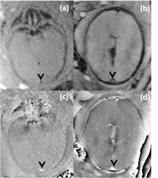
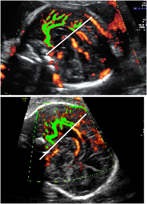
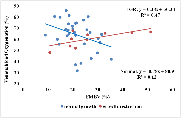
Figure 1 shows the representative images of magnitude and phase images of fetal brain of second and third trimester fetuses. Figure 2 shows the FMBV in the frontal lobe of the same fetal brains. In normal growth fetuses, SvO2 showed a mild decreasing trend (slope=-0.7±0.4; p=0.1), whereas FMBV showed a mild increasing trend (slope=0.2±0.2; p=0.23) with advancing gestation. A mild but significant negative association (slope=-0.78±0.3; p=0.04) between the SvO2 and FMBV estimates was found in normal growth fetuses (Figure 3). In FGR fetuses, the SvO2 values and trend were similar to normal growth fetuses, whereas FMBV values were higher and the trend across gestation was opposite compared to normal fetuses. Additionally, contrary to normal fetuses, the SvO2 and FMBV association was significantly positive in FGR fetuses (slope =0.38±0.12; p=0.02) (Figure 3).Discussion
The association between FMBV and SvO2 in normal growth fetuses is weak, suggesting that in normal circumstances the maintenance of values does not imply major changes in blood perfusion. In FGR fetuses, the redistribution of blood significantly increases blood perfusion to the brain to maintain normal oxygen saturation. Our results are in close agreement to the previous finding in growth restricted fetuses where a fetal blood sample was obtained by cordocentesis and blood gas values were correlated with Doppler velocimetry of the MCA9. The results showed significantly lower oxygen saturation in the umbilical cord and increased blood flow to the brain manifested a reduced PI/RI indices. Our study is the first in human fetuses applying two imaging modalities that have already validated for evaluation of oxygen saturation and blood perfusion, and provides evidence that combining MRI and US might improve the evaluation of hemodynamic changes in complicated fetuses.Conclusion
Combined MRI (SWI) and ultrasound (FMBV) techniques showed a significant association between cerebral blood oxygenation and blood perfusion in normal growth and FGR fetuses. This dual-imaging approach could contribute to the early detection of fetal "brain sparing” and brain oxygen saturation changes in high-risk pregnancies.Acknowledgements
No acknowledgement found.References
1. Figueroa‐Diesel H, et. al., Doppler changes in the main fetal brain arteries at different stages of hemodynamic adaptation in severe intrauterine growth restriction. Ultrasound in Obstetrics & Gynecology. 2007;30(3):297-302.
2. Turan O, et al.. Progression of Doppler abnormalities in intrauterine growth restriction. Ultrasound in Obstetrics & Gynecology. 2008;32(2):160-7.
3. Cruz-Martinez R, et al. Longitudinal brain perfusion changes in near-term small-for-gestational-age fetuses as measured by spectral Doppler indices or by fractional moving blood volume. American Journal of Obstetrics & Gynecology. 2010;203(1):42. e1-. e6
4. Shen Y, et al. In vivo measurement of tissue damage, oxygen saturation changes and blood flow changes after experimental traumatic brain injury in rats using susceptibility weighted imaging. Magnetic resonance imaging. 2007;25(2):219-27
5. Buch S, et al. Quantifying the changes in oxygen extraction fraction and cerebral activity caused by caffeine and acetazolamide. Journal of Cerebral Blood Flow & Metabolism. 2017;37(3):825-36
6. Hernandez‐Andrade E, et al. Changes in regional fetal cerebral blood flow perfusion in relation to hemodynamic deterioration in severely growth‐restricted fetuses. Ultrasound in Obstetrics & Gynecology. 2008;32(1):71-6.
7. Yadav BK, et al. Imaging putative foetal cerebral blood oxygenation using susceptibility weighted imaging (SWI). European radiology. 2017:1-7
8. Hernandez‐Andrade E, et al. Evaluation of fetal regional cerebral blood perfusion using power Doppler ultrasound and the estimation of fractional moving blood volume. Ultrasound in Obstetrics and Gynecology: The Official Journal of the International Society of Ultrasound in Obstetrics and Gynecology. 2007;29(5):556-61.
9. Vyas S, et al. Middle cerebral artery flow velocity waveforms in fetal hypoxaemia. BJOG: An International Journal of Obstetrics & Gynaecology. 1990;97(9):797-803
Figures