1063
Localized pure shift proton MRS for measurements on biological samples1Department of Electronic Science, Xiamen University, Xiamen, China
Synopsis
Localized proton magnetic resonance spectroscopy (MRS) provides a non-invasive tool for metabolic studies on biological systems by revealing valuable biochemical information, complementary to anatomical information delivered by MRI. However, due to limited proton chemical shift range and extensive J coupling splittings, spectral congestion is generally encountered in the resulting 1D spectra acquired by conventional proton MRS techniques, such as PRESS (Point resolved spectroscopy) and STEAM (STimulated Echo Acquisition Mode). In this abstract, we present a previously-unreported proton MAS approach to obtain localized 1D pure shift spectra with spectral simplification, potentially useful for studies on biological samples.
INTRODUCTION
Localized proton magnetic resonance spectroscopy (MRS) has proved to be a powerful and non-invasive technique for examining metabolite compositions and offering considerable biochemical information for metabolic studies on biological systems.1 Conventional localized proton MRS techniques based on single-voxel localization, such as STEAM (STimulated Echo Acquisition Mode) and PRESS (Point resolved spectroscopy), have been proposed for measurements on biological samples. In general, biological samples contain abundant metabolite compositions and this introduces numerous NMR resonances along with extensive J coupling splittings and inevitably yield spectral congestion in the resulting 1D spectra with limited proton chemical shift range. An NMR technique, “pure shift spectroscopy”, provides a feasible way for removing J coupling effects and enhancing spectral resolution with only chemical shifts present.2 In this abstract, a proton MRS approach based on the combination of pure shift module and single-voxel localization is introduced to acquire localized 1D pure shift spectra with spectral simplification for measurements on biological samples.METHODS
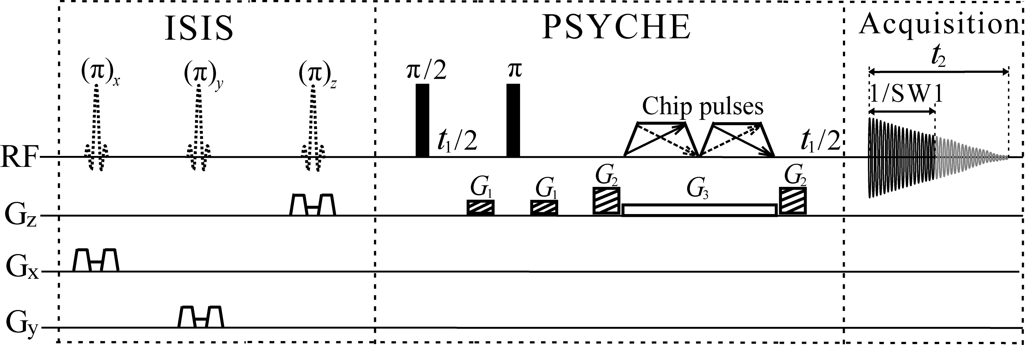
The proposed localized pure shift proton MRS sequence is shown in Fig. 1. This sequence is designed based on the concatenation of the pure shift module, PSYCHE (Pure Shift Yielded by Chirp Excitation)3, and the single-voxel localization module, ISIS (Image Selected In vivo Spectroscopy)4. The ISIS localization module, based on selective inversion of three orthogonal slices during the spin preparation period, is performed with eight different experiments for volume selection. The PSYCHE module, consisting of an indirect evolution period t1, two frequency-swept chirp pulses β matching with a simultaneous weak gradient G2 and coherence selection gradients G1 and G3, generates the desired pure shift signals. Acquired data in the t2 period contains the chunk data of 1/SW1 interval for each t1 increment, and a 1D data without J coupling effects is reconstructed after the concatenation of all chunked data as t1 increments. Finally, the localized 1D pure shift proton spectrum displayed in phase-sensitive mode is obtained after Fourier transform.
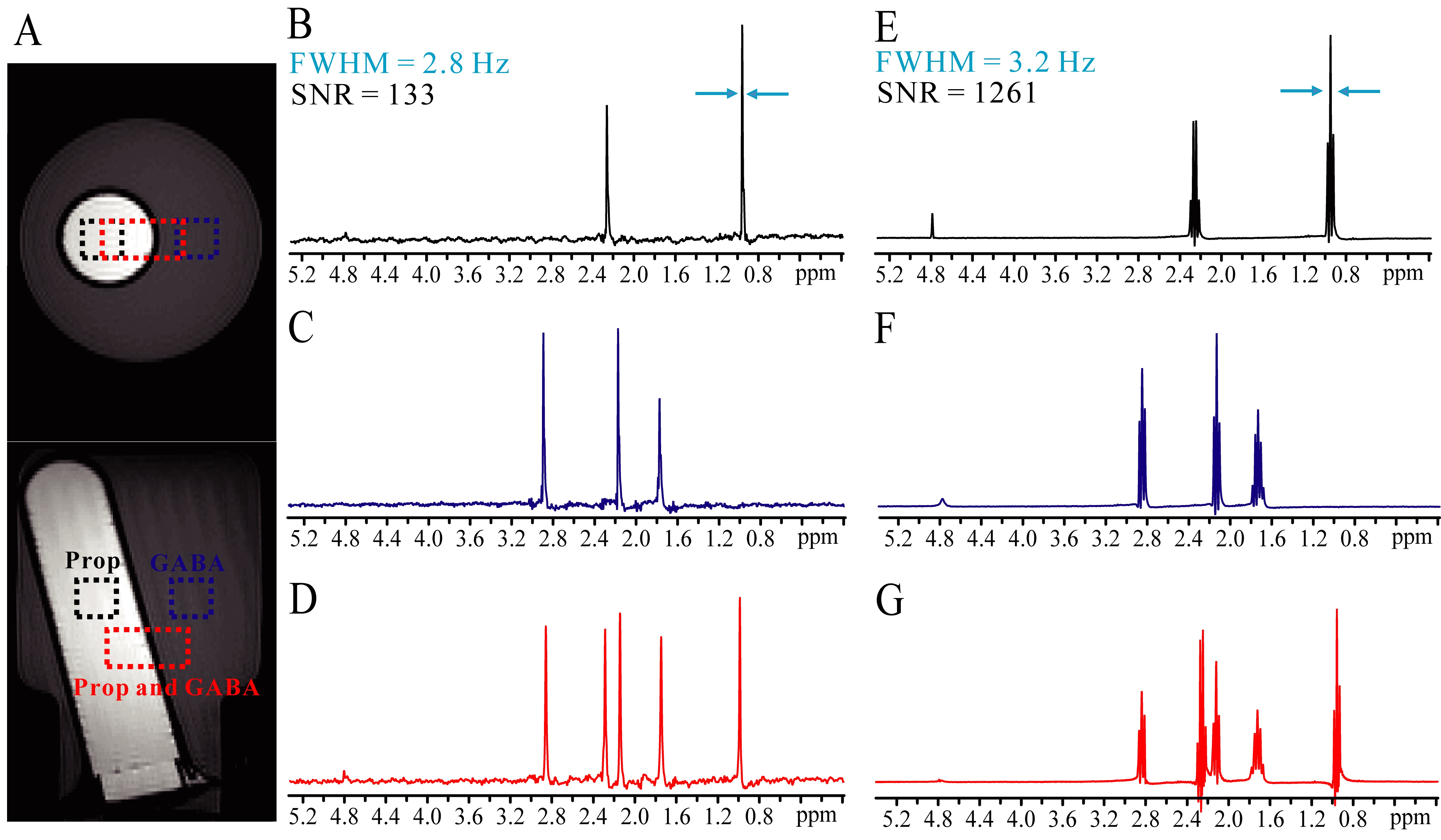
All experiments were performed on a Varian (Palo Alto, CA, USA) 7.0 T small animal MRI scanner at 293 K. A phantom built with two plastic bottles, filled with 1 M propionate (Prop) aqueous solution and 1 M γ-aminobutyric acid (GABA) aqueous solution, was used to demonstrate the performance of the localized pure shift MRS method. In addition, this method was also applied to in vitro pig brain tissue to show its applicability on biological samples.
RESULTS
The comparison results on the phantom sample acquired by the proposed pure shift MRS and the standard PRESS are shown in Fig. 2. Figure 2A is axial and coronal spin-echo images of the phantom sample, displaying three selected volumes, i.e. 5×5×5 mm3 for the Prop component (black dot frame), 5×5×5 mm3 for the GABA component (blue dot frame), and 5×10×5 mm3 for both two components (red dot frame). From localized 1D spectra obtained by the pure shift MRS approach (Fig. 2B ~ 2D), it can be seen that J coupling effect is suppressed and all multiplets are collapsed to singlets for each chemical shift size. In contrast, multiplet peaks are observed in 1D PRESS spectra (Fig. 2E ~ 2G).
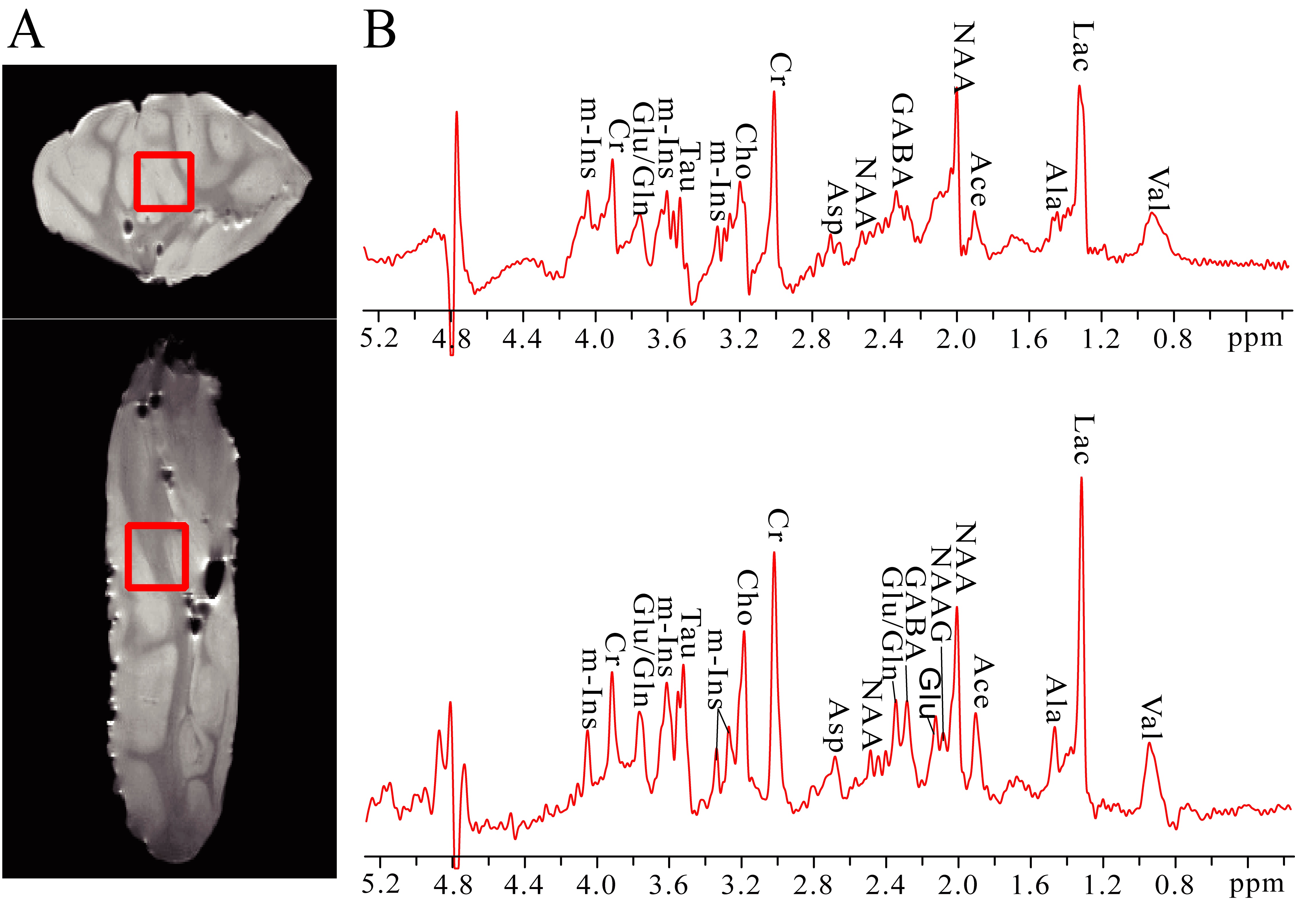
Experimental results on the in vitro pig brain tissue are shown in Fig. 3. The localized voxel with the size of 6×6×6 mm3 is marked in spin-echo images (Fig. 3A). Figure 3B shows the standard 1D PRESS spectrum acquired from the selected volume and most metabolites are assigned. The localized pure shift MRS spectrum from the same volume is given in Fig. 3C. The localized pure shift spectrum can yield better spectral resolution satisfactory for metabolite analyses.
DISCUSSION
The PSYCHE technique presents a promising manner for simplifying spectral information by removing J coupling splittings. Herein, we exploit the pure shift spectroscopy to proton MRS measurements on biological samples by combining the PSYCHE module with the ISIS localization module. Experiments on the phantom and the in vitro pig brain tissues shows the applicability of the proposed MRS approach on localized pure shift measurements. Due to the intrinsic signal penalty caused by the PSYCHE module, the sensitivity of localized pure shift MRS experiments is generally lower than that in conventional MRS experiments.CONCLUSION
Here, we present a previously-unreported MAS approach to obtain localized 1D pure shift proton spectra. Compared to conventional MRS approaches, the proposed MRS method can simplify spectral information by collapsing J coupling multiplets into singlets, thus potentially useful for metabolic studies on biological samples that contain abundant metabolite compositions.Acknowledgements
This work was partially supported by the NNSF of China under Grants 11675135 and 21327001.References
1. Michaelis T, Boretius S, Frahm J. Localized proton MRS of animal brain in vivo: Models of human disorders. Prog. Nucl. Magn. Reson. Spectrosc. 2009; 55(1):1-34.
2. Zangger K. Pure shift NMR. Prog. Nucl. Magn. Reson. Spectrosc. 2015; 86-87:1-20.
3. Foroozandeh M, Adams RW, Meharry NJ, et al. Ultrahigh-Resolution NMR Spectroscopy. Angew. Chem. Int. Edit. 2014; 53(27):6990-6992.
4. Ordidge RJ, Connelly A, Lohman JAB. Image-Selected in vivo Spectroscopy. A New Technique for Spatially Selective NMR Spectroscopy. J. Magn. Reson. 1986; 66:283-294.
Figures