1061
Double-Shot Semi-Adiabatic Localization for Ultra Short Echo Time Magnetic Resonance Spectroscopy1Biomedical Engineering, Columbia University, New York, NY, United States, 2Center for Magnetic Resonance Research and Department of Radiology, University of Minnesota, Minneapolis, MN, United States, 3GE Healthcare, Berlin, Germany, 4Radiology, Columbia University, New York, NY, United States
Synopsis
Modern magnetic resonance spectroscopic (MRS) pulse sequences which employ localization through adiabatic pulses suffer from long minimum echo times ($$$\ge$$$ 19 ms). Here we developed a novel MRS pulse sequence which allows for an echo time of 8 ms with adiabatic localization in two of the three spatial dimensions, at the cost of two-shot volume localization. The sequence is highly robust to B1 inhomogeneity and does not have any anomalous J-modulation since no slice-selective refocusing pulses are used. High spectral quality was obtained in phantom experiments, although strong lipid contamination was observed, consistent with previously reported two-shot localization methods.
Introduction
Typical MRS localization methods, PRESS1 and STEAM2, offer relatively short echo times (TE), however they both suffer from susceptibility to B1 inhomogeneity in addition to large chemical shift displacement errors (CDSE) due to the relatively low bandwidth of their amplitude-modulated pulses. To combat these issues, sequences with high bandwidth adiabatic pulses have been developed, such as LASER3 or sLASER4,5, at the cost of longer TE (e.g., min TE of sLASER of 24 ms for sLASER at 4T6 and longer for LASER). These echo times make J-difference editing for gamma-aminobutyric acid difficult. To reduce TE the sequence SPECIAL7 was developed which obtains spatial localization in one dimension through a pre-inversion pulse. Similarly, SPECIAL-sLASER was developed which replaced the amplitude-modulated 180° refocusing pulse with an adiabatic refocusing pulse pair, obtaining the benefits of adiabatic pulses at the cost of a minimum TE of 19 ms at 3T8.
Here a novel method was developed, referred to as DOuble Shot Semi-Adiabatic Localization (DOSSAL), which allows for adiabatic localization in two of the three spatial dimensions, minimum CDSE and anomalous J-modulation due to signal cancellation at the borders of the voxel9 at a TE of 8 ms on a clinical 3T scanner, thereby allowing improved estimation for coupled spin systems, macromolecules or additional time to increase the selectivity of editing pulses.
Methods
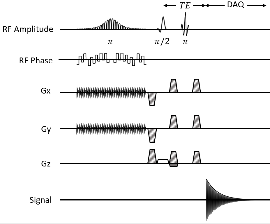
The DOSSAL sequence operates by employing a two-dimensional selective adiabatic inversion pulse10 followed by a slice-selective excitation pulse and a refocusing pulse (Figure 1). The inversion pulse consists of 30 spiral jinc subpulses with 5-turn spiral, and the subpulses are modulated by a hyperbolic secant envelope, providing adiabaticity, with a total duration of 40 ms. The excitation pulse was a minimum phase Shinnar-Le Roux11 (SLR) pulse with a duration of 1.80 ms and the refocusing pulse was a linear-phase SLR pulse with a duration of 5.00 ms. The 2D inversion pulse is toggled off every other TR and the acquired traces from the two cycles are subtracted to provide full three-dimensional localization. In order to reduce the subtraction artefact instead of turning the inversion pulse off every other TR, the positioning of the voxel was shifted to outside the subject’s head, as this method has been shown to reduce subtraction-derived lipid contamination8.
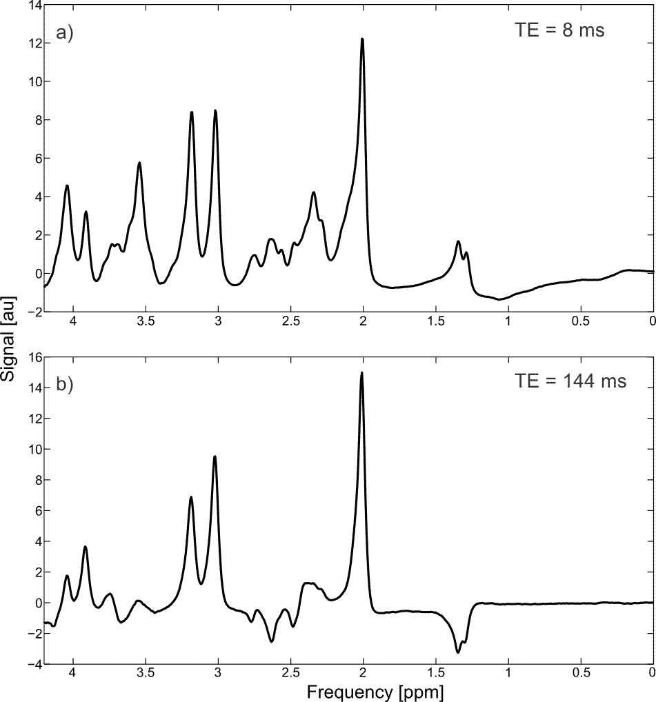
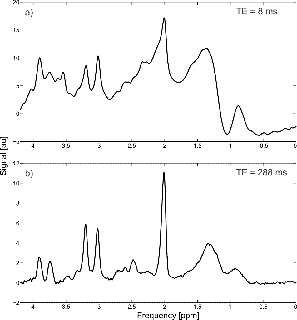
All experiments were performed on a MR750 3T MRI system (GE Healthcare, WI, USA) at the New York State Psychiatric Institute (NYSPI). Phantom experiments within GE’s “BRAINO” metabolite phantom were performed with the minimum achievable TE of 8 ms to demonstrate the sequence’s short-TE capabilities, in addition to an TE at 144 ms tailored to lactate detection. One healthy subject was scanned with the minimum TE in addition to a long TE (288 ms) experiment to reduce lipid contamination. All experiments had a TR of 1.5 s, 128 excitations and a cylindrical voxel with size of 5 x 5 x 2 cm3 (5 cm diameter in the inversion dimensions), and used VAPOR12 for water suppression, as well as outer-volume suppression (OVS) for sideband suppression.
Results and Discussion
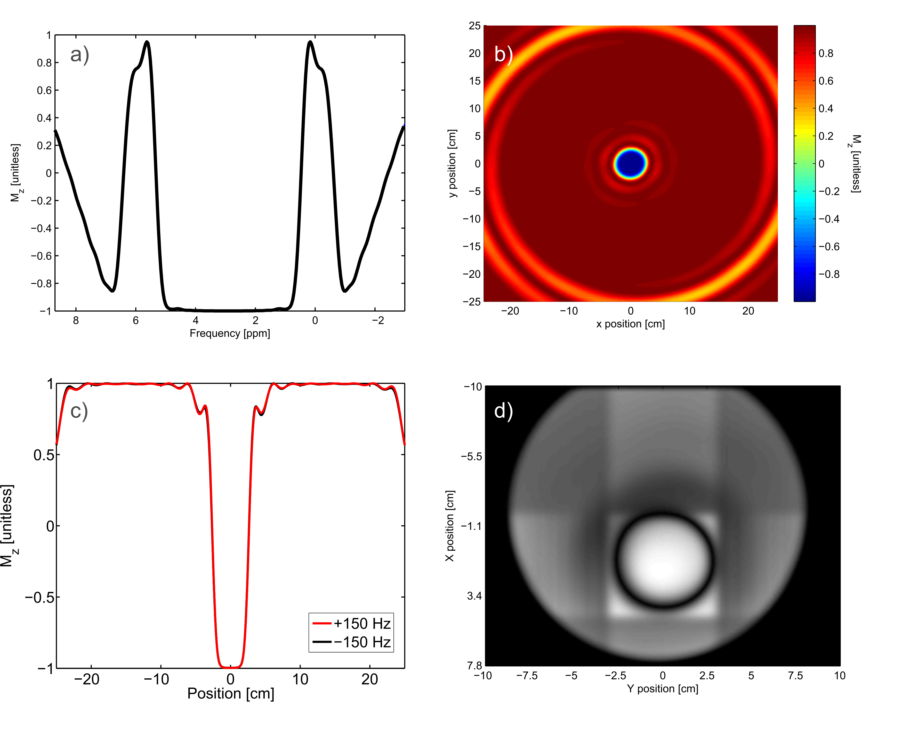
All spectral resonances upfield of water can be measured simultaneously (Figure 2a) at 3T. The inversion profile at the center frequency of the two-dimensional adiabatic RF pulse has little out-of-voxel signal (Figure 2b), aside from large side bands at about 25 cm distance radially from the center of the voxel, which was placed outside the subject’s head. Side bands can be observed closer to the voxel; however, they were further suppressed by the use of carefully placed OVS bands (Figure 2d). Due to the subpulses being of very short duration (and thus high bandwidth) there is virtually no CDSE (Figure 2c). High spectral quality was demonstrated in phantom experiments (Figure 3). Some baseline variation can be observed in the TE = 8 ms experiment at 0 to 1 ppm, which is likely due to this being the spectral region of the transition band of the pulse (as seen in Figure 2a). In vivo experiments demonstrated the functionality of the method. This proof-of-principle implementation, however, suffered from substantial lipid contamination at short TE (Figure 4a) that is largely reduced at longer TE due to the short lipid T2 relaxation time (Figure 4b). Additional methods, both experimental and data processing such as L2-based lipid suppression13, will be investigated to reduce the lipid contamination.Conclusion
A novel sequence was developed which provides full intensity signal, adiabatic localization in two dimensions, no anomalous J-modulation and CDSE at a TE of 8 ms, which could be used for improved estimation for coupled spin systems, macromolecules or highly selective editing sequences.Acknowledgements
Special thanks to the New York State Psychiatric Institute (NYSPI) and Dr. Feng Liu for their facilities and technical support. This research was supported by the National Multiple Sclerosis Society (NMSS, RG-5319).References
- Bottomley, P. A. Spatial Localization in NMR Spectroscopy in Vivo. Ann N Y Acad Sci 508, 333–348 (1987).
- Frahm, J. et al. Localized Proton NMR Spectroscopy in Different Regions of the Human Brain in Vivo Relaxation Times and Concentrations of Cerebral Metabolites. Magn Reson Med 63, 47–63 (1989).
- Garwood, M. & DelaBarre, L. The Return of the Frequency Sweep: Designing Adiabatic Pulses for Contemporary NMR. Jour Magn Reson 153, 155–177 (2001).
- Scheenen, T. W. J., Klomp, D. W. J., Wijnen, J. P. & Heerschap, A. Short echo time 1H-MRSI of the human brain at 3T with minimal chemical shift displacement errors using adiabatic refocusing pulses. Magn Reson Med 59, 1–6 (2008).
- Scheenen, T. W., Heerschap, A. & Klomp, D. W. Towards 1H-MRSI of the human brain at 7T with slice-selective adiabatic refocusing pulses. Magn Reson Mater Phy 21, 95–101 (2008).
- Oz, G. & Tkac, I. Short-echo, Single-shot, Full-indensity 1H MRS For Neurochemical Profiling at 4T: Validation in the Cerebellum andd Brainstem. Magn Reson Med 65, 1–19 (2011).
- Mlynárik, V., Gambarota, G., Frenkel, H. & Gruetter, R. Localized short-echo-time proton MR spectroscopy with full signal-intensity acquisition. Magn Reson Med 56, 965–970 (2006).
- Fuchs, A., Luttje, M., Boesiger, P. & Henning, A. SPECIAL semi-LASER with lipid artifact compensation for 1H MRS at 7 T. Magn Reson Med 69, 603–612 (2013).
- Lange, T., Dydak, U., Roberts, T. P. L., Bjeljac, M. & Boesiger, P. Pitfalls in Lactate Measurements at 3T. Am J Neuroradiol 27, 895–901 (2006).
- Jang, A., Wu, X., Auerbach, E. J. & Garwood, M. Designing 3D Selective Adiabatic Radiofrequency Pulses With Single and Parallel Transmission. Magn Reson Med 79, 701–710 (2018).
- Pauly, J., Le Roux, P., Nishimura, D. & Macovski, A. Parameter Relations for the Shinnar-Le Roux Selective Excitation Pulse Design Algorithm. IEEE Trans Med Imag 10, 53–65 (1991).
- Tkác, I., Starcuk, Z., Choi, I. Y. & Gruetter, R. In vivo 1H NMR spectroscopy of rat brain at 1 ms echo time. Magn. Reson. Med. 41, 649–56 (1999).
- Steel, A. et al. Metabolite-cycled density-weighted concentric rings k-space trajectory (DW-CRT) enables high-resolution 1 H magnetic resonance spectroscopic imaging at 3-Tesla. Sci. Rep. 8, 1–10 (2018).
Figures