1054
MR-Guided Pharmacological Intervention: Creating Causal Capabilities for fMRI1Radiology, University of Wisconsin - Madison, Madison, WI, United States, 2Medical Physics, University of Wisconsin - Madison, Madison, WI, United States, 3Biomedical Engineering, University of Wisconsin - Madison, Madison, WI, United States, 4Psychiatry, University of Wisconsin - Madison, Madison, WI, United States, 5Neuroscience, University of Wisconsin - Madison, Madison, WI, United States
Synopsis
Neuroscience is in need of precise interventional tools that alter local neural dynamics while monitoring whole brain network activity. We demonstrate methods to guide catheters to deliver and monitor pharmacologic alteration of a local brain region in anaesthetized Rhesus monkeys while monitoring changes in resting state functional connectivity MRI (rs-fcMRI) throughout all brain networks. Expected and unexpected alterations in rs-fcMRI after unilateral and bilateral infusions of inhibitory agents in the limbic system are provided. The approach shows promise for using the alterations to compute effective connectivity through fMRI.
Introduction:
Cognitive and behavioral functions are mediated by distributed networks of neurons involving multiple cortical and subcortical brain regions. Neuroscientists rely heavily on repeated insertions of electrophysiological probes in pre-clinical models to perform causal studies of these regions at the circuit level. At the larger network level, functional magnetic resonance imaging (fMRI) has served as the cornerstone of brain mapping tools for the past twenty years. However, the spatial and temporal resolution limits of fMRI provide primarily correlative information on brain connectivity. Determining how one region causally modulates and mediates activity in other regions remains difficult with fMRI. MR-guided, localized intraparenchymal brain delivery of viral vectors (1, 2) and chemotherapeutic agents (3, 4)uses catheters with sub-mm diameters that are smaller than most electrophysiological probes. We demonstrate here methods to guide and monitor pharmacologic alteration of a local brain region in anaesthetized Rhesus monkeys while monitoring changes in resting state functional connectivity MRI (rs-fcMRI) throughout all brain networks. Proving the feasibility of these methods is essential to our future efforts to perform repeated studies in awake Rhesus subjects performing cognitive tasks before and after localized drug interventions.Methods:
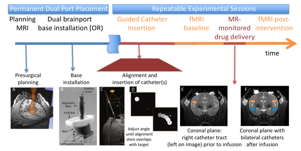
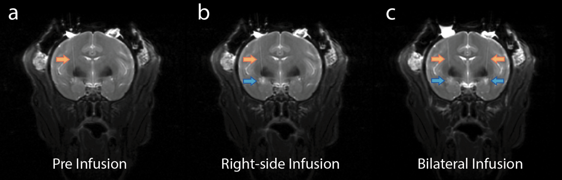
Two untrained monkeys slated for euthanasia were used for initial testing. As shown in Fig. 1A, pre-surgical MRI was used to determine skull locations for craniotomies for installation of NavigusTM brain ports. After installation (Fig. 1B), real-time control of the scanner was usurped with the RtHawk portal (HeartVista, CA)(5). The ports were aligned in real–time (Fig. 1D) to provide trajectories aimed at the central nucleus of the amygdala (CeA) using projection-based methods that determine the 3D orientation of the brain port central axis at 5 frames/sec(6, 7). Fused silica 0.7 mm catheters were then inserted into the CeA where 24 mg of muscimol (an inhibitory agent) was infused in 24 ml of buffered solution under pressure over 12 minutes, first on the right side of the brain in Fig. 1F and then later on the left side. Resting-state functional-connectivity MRI imaging (rs-fcMRI) was performed for 45 minutes prior to the unilateral infusion, 45 minutes after the unilateral infusion, and 45 minutes after the bilateral infusion.Results:
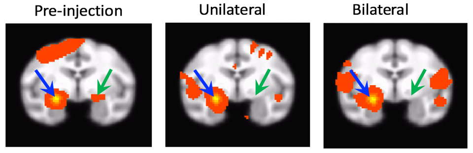
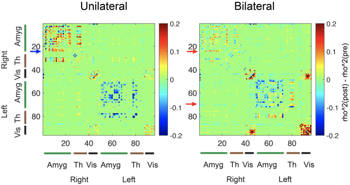
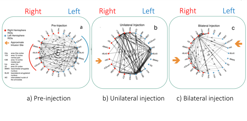
Bilateral catheters were successfully aligned and inserted into the CeA targets with sub-mm accuracy, as shown in Fig. 2. Accuracy was measured by comparing the location of tip of the catheter to the desired MR coordinates selected prior to insertion. A co-infused Gd tracer was not used to identify the infusion volume, as Gd could alter the fMRI measurements. Instead, T2-weighted imaging detected the enhanced T2 from the infusion’s saline buffer, as shown in Fig. 2. Pre-infusion rs-fcMRI provides results consistent with prior studies, which have shown that the CeA is most strongly connected to the contralateral CeA [2], as shown in Fig. 3 (left). This connectivity was significantly reduced following both unilateral (Fig. 3 center) and bilateral injections of muscimol (Fig. 3 right) into the CeA, demonstrating the effectiveness of the muscimol infusions.To depict changes over a broader region, R2 adjacency matrices in Fig. 4 show statistically significant alterations in connectivity from the pre-infusion to the post-infusion resting-state connectivity maps. After unilateral or bilateral infusions, connectivity between the left and right amygdala is decreased. After a bilateral infusion, connectivity within the ipsilateral and contralateral amygdala is further reduced. Significant controversies arise when deriving effective connectivity models (directionality) from BOLD fMRI time series using Conditional Grainger Causality (CGC). However, the ability to compare signals before and after inhibiting a node(s) allows us to provide estimates of directional influence, as shown in Fig. 5. Besides simply attenuating connectivity with the contralateral side, CGC analysis shows unexpected new connectivity after the unilateral infusion. Upon the bilateral infusion, global effective connectivity in the region is reduced.Discussion:
Though demonstrated in anesthetized NHP models for feasibility, we believe permanent brain ports can be affixed to skulls in a surgical setting which can provide long-term access for repeated catheter insertions for studies in awake models performing cognitive tasks. We have already trained other NHP models to remain still and perform cognitive tasks in the magnet.Conclusion:
The first feasibility studies for performing casual experiments to alter the functional organization and dynamics of brain networks have been completed. Expected and unexpected changes in resting state functional connectivity resulted from unilateral and bilateral infusions of inhibitory agents. Further development will allow us to test hypotheses about network topology and information flow, as well as to further the understanding of the mechanisms underlying the signals provided by fMRI.Acknowledgements
We graciously acknowledge support of research funding from the UW-Madison 2020 program and the UW-Madison Radiology Research and Development Fund.
References
1. Richardson RM, Kells AP, Rosenbluth KH, Salegio EA, Fiandaca MS, Larson PS, Starr PA, Martin AJ, Lonser RR, Federoff HJ, Forsayeth JR, Bankiewicz KS. Interventional MRI-guided putaminal delivery of AAV2-GDNF for a planned clinical trial in Parkinson's disease. Molecular therapy : the journal of the American Society of Gene Therapy. 2011;19(6):1048-57. doi: 10.1038/mt.2011.11. PubMed PMID: 21343917; PMCID: 3129792.
2. Kalin NH, Fox AS, Kovner R, Riedel MK, Fekete EM, Roseboom PH, Tromp do PM, Grabow BP, Olsen ME, Brodsky EK, McFarlin DR, Alexander AL, Emborg ME, Block WF, Fudge JL, Oler JA. Overexpressing Corticotropin-Releasing Factor in the Primate Amygdala Increases Anxious Temperament and Alters Its Neural Circuit. Biol Psychiatry. 2016;80(5):345-55. doi: 10.1016/j.biopsych.2016.01.010. PubMed PMID: 27016385; PMCID: 4967405.
3. Lonser RR, Warren KE, Butman JA, Quezado Z, Robison RA, Walbridge S, Schiffman R, Merrill M, Walker ML, Park DM, Croteau D, Brady RO, Oldfield EH. Real-time image-guided direct convective perfusion of intrinsic brainstem lesions. Technical note. Journal of neurosurgery. 2007;107(1):190-7. doi: 10.3171/JNS-07/07/0190. PubMed PMID: 17639894.
4. Sampson JH, Raghavan R, Provenzale JM, Croteau D, Reardon DA, Coleman RE, Rodriguez Ponce I, Pastan I, Puri RK, Pedain C. Induction of hyperintense signal on T2-weighted MR images correlates with infusion distribution from intracerebral convection-enhanced delivery of a tumor-targeted cytotoxin. AJR Am J Roentgenol. 2007;188(3):703-9. doi: 10.2214/AJR.06.0428. PubMed PMID: 17312057.
5. Santos JM, Wright GA, Pauly JM. Flexible real-time magnetic resonance imaging framework. Conf Proc IEEE Eng Med Biol Soc. 2004;2:1048-51. PubMed PMID: 17271862.
6. Olsen ME, Brodsky EK, Oler JA, Fekete EM, Riedel MK, N.H. K, Block WF, , editors. Rapid Localization for prospective stereotaxy: Using computation instead of imaging. Proc of ISMRM 24th Annual Meeting 2016; Singapore.
7. Brodsky EK, Olsen ME, Oler JA, Fox AS, Kovner RL, Riedel MK, Fekete EM, Roseboom PH, Tromp DPM, Grabow BP, Fudge JL, Alexander AL, Emborg ME, N.H. K, Block WF, editors. MRI-Guided Delivery of Viral Vectors for Targeted Alteration of Neurochemistry. National Center for Targeted Alteration of Neurochemistry. National Center for Image Guided Therapy Workshop; 2016; Bethesda, MD.
Figures