1050
Awake and behaving mouse fMRI in Go/No-go task.1Institute of Neuroscience, Chinese Academy of Sciences, Shanghai, China
Synopsis
The current study describes a novel awake and behaving mouse fMRI paradigm, which enables the functional imaging of mice brain during an olfaction based go/no-go (GNG) task. This novel paradigm incorporates a MR compatible behavioral apparatus (olfactometer, licking detection and water delivery) and awake head fixation mechanism, all specifically designed to be used with a cryogenic coil at high field. High-resolution functional images uncovered the large-scale networks that are differentially recruited in the hit vs. correct-rejection trials, with greatly limited motion artefacts. This method paved the way for future whole-brain, systematic mapping of cognitive processes in mice.
Introduction
The fMRI studies in rodents are largely performed under anesthesia1, which greatly limits the scope of application and the behavioral relevance. Recent studies have started to use awake mice under the resting state fMRI2 and other conditions3. However fMRI in the awake behaving rodents has not been achieved. In the current study, a novel fMRI paradigm for awake and behaving mice was developed, allowing functional imaging of the mouse brain in an olfaction based go/no-go (GNG) task. We present a novel awake mouse fMRI setup, includes a MRI compatible behavioral apparatus, which enables active behavior during fMRI. Functional imaging results demonstrated dynamic sequences of BOLD activities in a distributed network involving many cortical and subcortical regions in the GNG task.Methods
Animals preparations
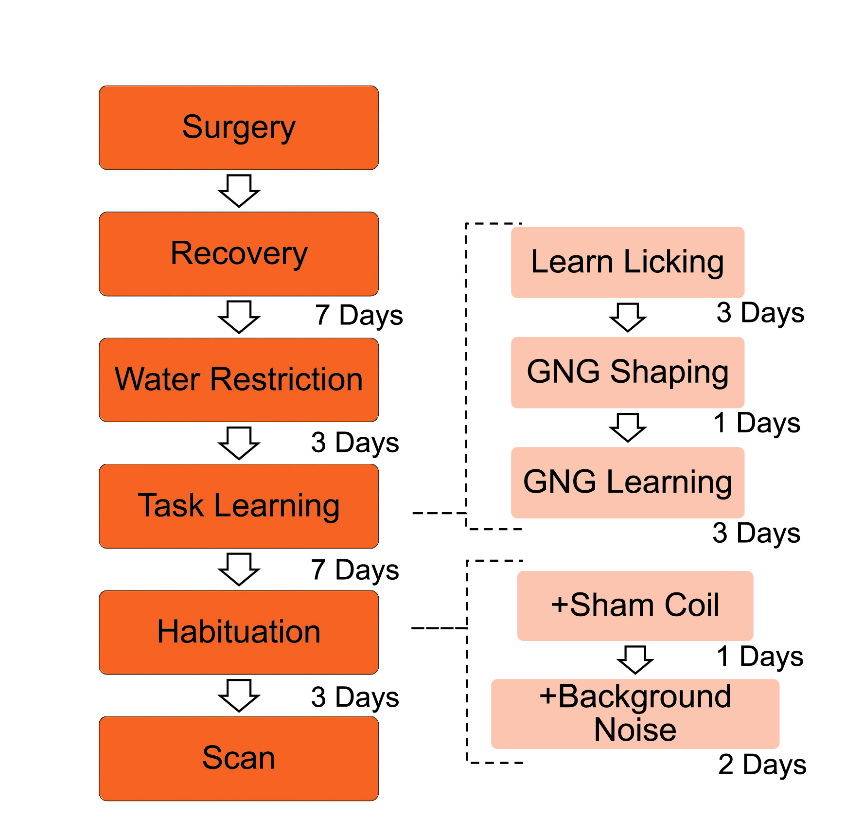
8 male C57BL/6 mice were used for experiment. After the surgery for head holder implantation and middle ear filling, mice were trained for the GNG task. Mice were trained to lick after the Go odor (3-methyl-2-buten-1-ol, O1) and not to lick after No-go odor (propyl acetate, O2). Two odors were delivered in a pseudo-random order4. “Hit” represents lick in a Go trial, “Correct rejection” (CR) represents no-lick in a No-go trial. The overall experimental procedure is shown in Figure 1.
MRI compatible behavior training setup
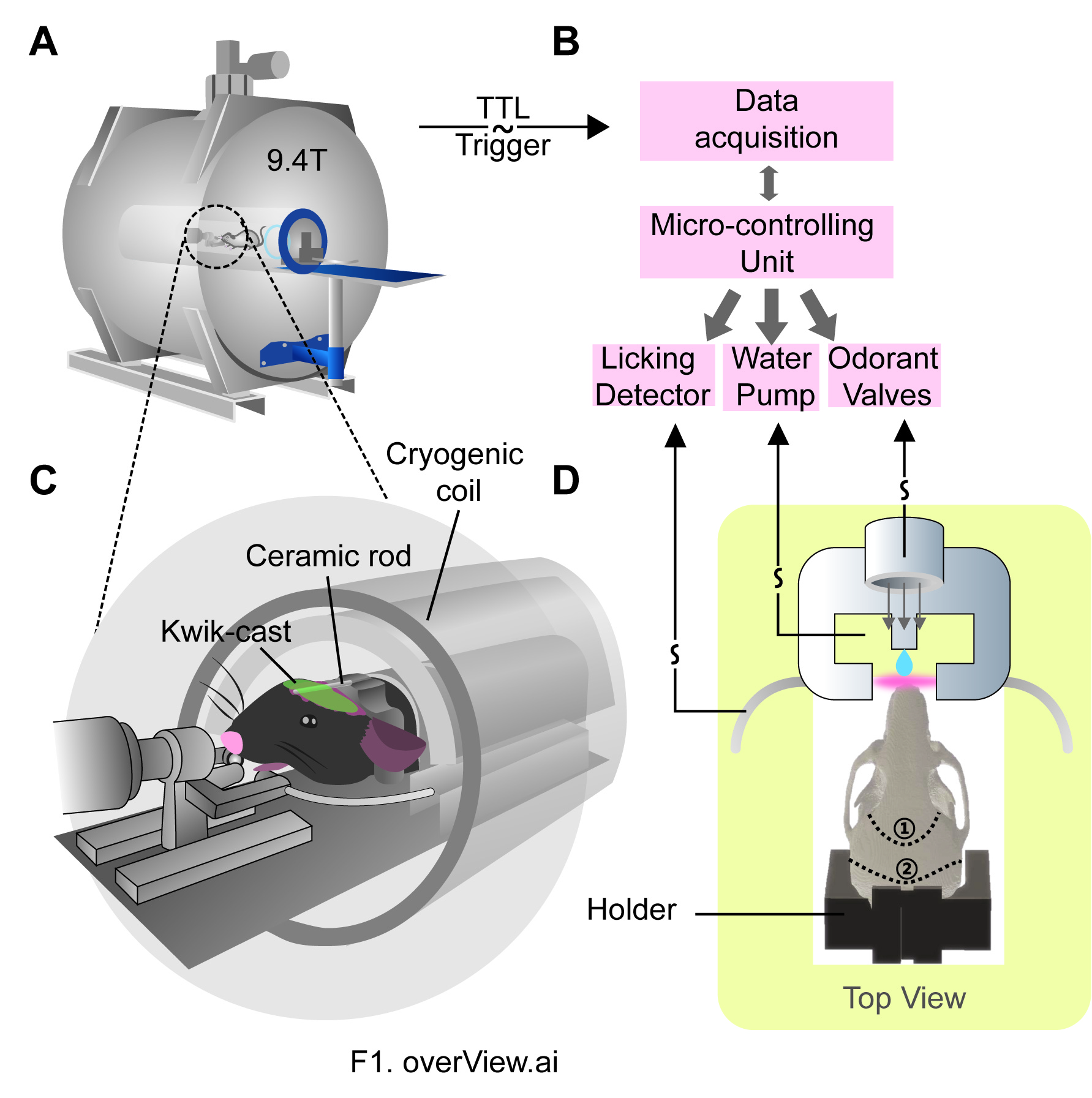
This setup includes five major components: the animal bed, the odor-delivery system, the peristaltic pump for water reward, the licking detector, and the Arduino controller (Figure 2).
fMRI experiment
Mice were secured in the olfaction behavior apparatus without any anesthesia, and placed inside the MRI scanner (9.4T Bruker BioSpec scanner with 4 channel phased array cryogenic mouse head coil). Single-shot gradient echo EPI images were acquired while mice were actively engaged in GNG task, with following parameters: TR/TE = 1500/15ms, FOV = 14.1×10 mm2, matrix = 94×67 (resolution = 0.15×0.15mm2), slice thickness = 0.4mm, 22 slices, for 6 min (240 volumes). In each EPI session, every 15s odor O1 or odor O2 was randomly delivered for 1 second around as behavioral cues, and any resulting licking was recorded. Typically 8-10 EPI sessions were acquired for each mouse. The olfaction behavior apparatus was synchronized with EPI data acquisition using TTL triggers from the scanner.
Image processing and data analysis
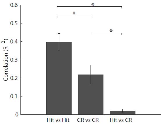
Image preprocessing were performed with SPM: motion correction, registration, normalization and spatial smoothing. EPI scans with maximum frame-wise displacement of more than 0.075mm were discarded. To further reduce the effect of motion, group independent component analysis(ICA) based de-noising strategy was used (GIFT toolbox). After regression of 12 motion parameters (6 motion parameters from SPM realignment and their derivatives) and time courses of motion related ICA components, time series of each voxel were detrended and normalized to their standard deviations. After normalization, epochs of Hit and CR trials were extracted and the whole-brain dynamic spatiotemporal patterns were thus generated by massive averaging approach5. A linear mixed effect model was utilized to examine the statistical significance of each time point of ROI time series, with subjects being the random effect. To evaluate the differential spatiotemporal patterns of brain activity between the two trials, epochs within same trial type or between Hit and CR trials were randomly divided into two groups, and correlation was calculated for averages of the two groups for each mouse.
Results and Discussion
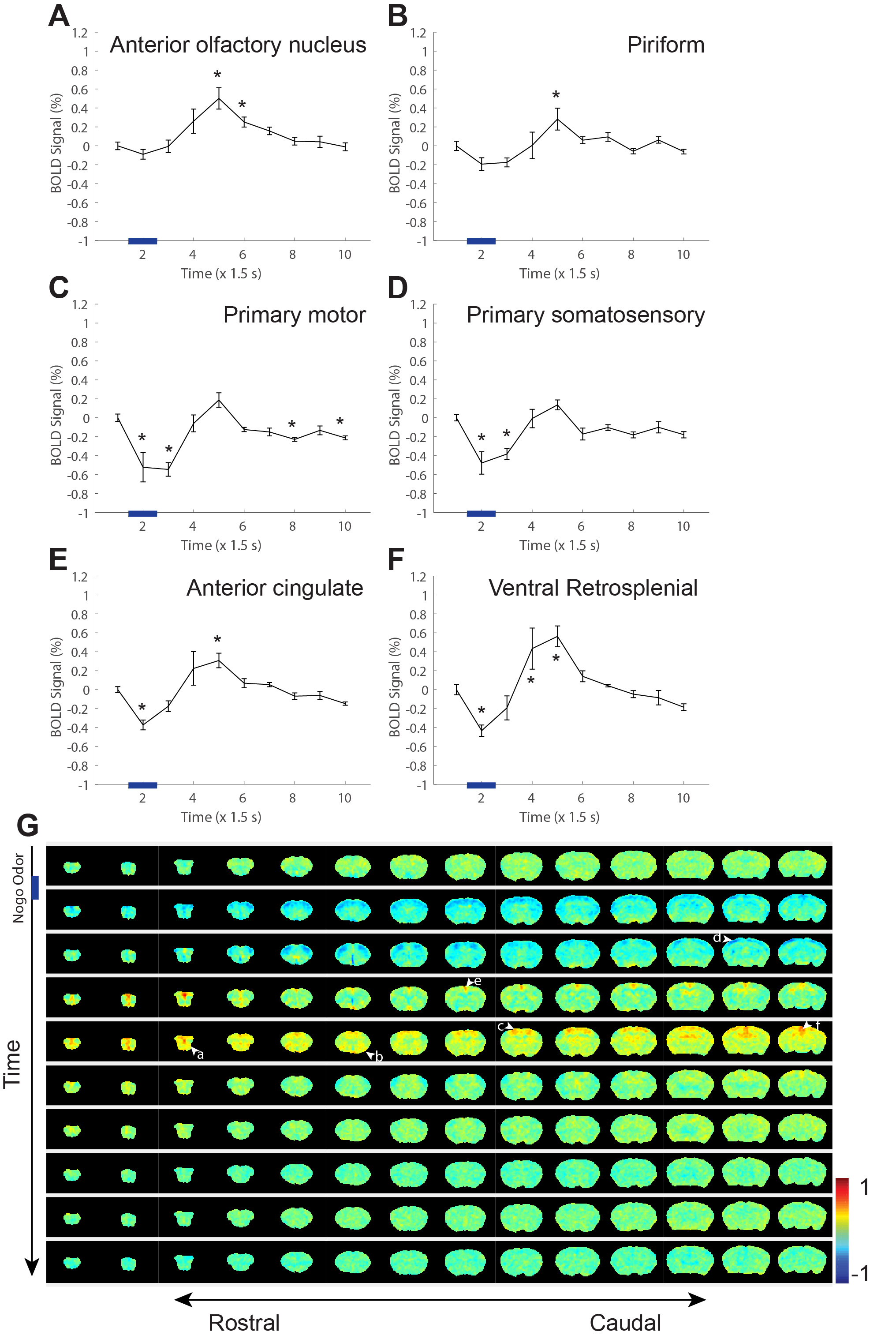
For awake imaging, the head holder based fixation mechanism was highly effective in reducing head motion. And the middle-ear Kwik-cast filling significantly reduced the susceptibility artifact in the brain regions close to the ear canal. In the hit trials, widespread cortical activation was observed (Figure 3), and some subcortical regions such as caudoputamen and medial dorsal thalamus were also clearly activated. Such wide-spread involvement was consistent with the fact that many cognitive processes are required in the hit trials, such as olfaction, decision making, motor output, and reward processing. In contrast, the spatiotemporal maps of CR trials showed a different pattern, with more limited activation spread. The whole-brain spatiotemporal patterns from the same trial type exhibited a high correlation coefficient (Hit vs. Hit, CR vs. CR), whereas those between the different trial types exhibited chance-level correlation coefficient (Hit vs. CR). Therefore the brain-wide activation pattern is drastically different between that of the hit and CR trials (Figure 5).Conclusion
Our novel fMRI paradigm in the awake and behaving mice is a great tool for mapping brain-wide activity modulation for behaviors. The current study expands the applicability of mouse fMRI, by demonstrating the feasibility of functional imaging of awake mice during a behavioral task.Acknowledgements
This work was supported by the General Program of National Natural Science Foundation of China (81771821 and 31471049), CAS Hundreds of Talents Program, the National Science Foundation for Distinguished Young Scholars of China (31525010, to C.T.L.), the Instrument Developing Project of the Chinese Academy of Sciences (Grant No. YZ201540), the Key Research Program of Frontier Sciences of the Chinese Academy Sciences (QYZDB-SSW-SMC009), the Key Project of the Shanghai Science and Technology Commission (No.15JC1400102, 16JC1400101), the China–Netherlands CAS-NWO Programme: The Future of Brain and Cognition (153D31KYSB20160106).References
1.Schroeter, A., Schlegel, F., Seuwen, A., et al. Specificity of stimulus-evoked fMRI responses in the mouse: the influence of systemic physiological changes associated with innocuous stimulation under four different anesthetics. NeuroImage. 2014; 94:1025-1029.
2. Madularu, D., Mathieu, A.P., Kumaragamage, C., et al. A non-invasive restraining system for awake mouse imaging. J Neurosci Methods. 2017; 287: 53-57.
3. Harris, A.P., Lennen, R.J., Marshall, I., et al. Imaging learned fear circuitry in awake mice using fMRI. Eur J Neurosci. 2015; 42: 2125-2134.
4.Han, Z., Zhang, X., Zhu, J., et al. High-Throughput Automatic Training System for Odor-Based Learned Behaviors in Head-Fixed Mice. Front Neural Circuits. 2018;12,15.
5. Gonzalez-Castillo, J., Saad, Z.S., Handwerker, D.A., et al. Whole-brain, time-locked activation with simple tasks revealed using massive averaging and model-free analysis.Proc Natl Acad Sci U S A. 2012; 109:5487-5492
Figures