1047
A Joint Recommendation for Optimized Preprocessing of Connectom Diffusion MRI Data1Max Planck Institute for Human Cognitive and Brain Sciences, Leipzig, Germany, 2Martinos Center for Biomedical Imaging, MGH, Boston, MA, United States, 3CUBRIC, Cardiff University Brain Research Imaging Centre, Cadiff, United Kingdom
Synopsis
The possibility of acquiring diffusion MRI data with extremely high resolution and strong diffusion weighting imposes new challenges on the preprocessing. In a joint effort, all research institutions with the availability of a human MRI system with 300
Introduction
The recent introduction of strong gradients (Gmax=300 mT/m) to human MRI systems enabled acquisitions of unprecedented diffusion MRI (dMRI) data with high contrast and resolution1,2. Advanced dMRI data are generally acquired with high resolution and strong diffusion-weighting and, hence, often suffer from low SNR and significant distortions. Therefore, an adapted preprocessing pipeline is required to retain data-quality3,4. In an attempt to optimize and standardize preprocessing, all MRI research sites with availability of a strong gradient human MRI system (CUBRIC, Cardiff, UK; Martinos Center for Biomedical Imaging, Boston, USA; MPI CBS, Leipzig, Germany) compared their data preprocessing and agreed upon one dMRI preprocessing pipeline which will be recommended for future studies. The pipeline applies a broad range of preprocessing-steps and combines multiple corrections into one single interpolation. It enables better comparability between research findings from different imaging sites and satisfies standards of high-quality dMRI data.Methods
In multiple discussions, teams of the involved research sites compared their respective dMRI preprocessing pipelines. The context of the discussion was provided by comparison of processing of a state-of-the-art dMRI dataset, acquired on a 3T MRI system with ultra-high gradient strength of Gmax=300 mT/m (Magnetom Connectom, Siemens Healthineers, Erlangen, Germany) and a 32-channel phased-array head coil. The acquisition parameters were: 1.2x1.2x1.2mm3 resolution, FoV=216x216mm2, 90 slices, TR=5400ms, TE=68ms, PF=6/8, EPI-BW=1544Hz/px, GRAPPA=2 and SMS=2. A total of 180 diffusion-weighted volumes were acquired at b=[1200,3000,5000]s/mm2 alongside four initial and 12 interspersed b=0 volumes. Three b=0 volumes with reversed phase-encoding were acquired to correct for off-resonance distortions5. Special emphasis was placed on evaluation of gradient-nonlinearity induced effects and preservation of data-quality and resolution. To evaluate the influence of gradient-nonlinearities on image-distortions and diffusion-contrasts, the nonlinearity displacement field was analyzed with respect to voxel-displacements and b-value deviations6,7. Through evaluation of each site’s respective preprocessing, a consensus pipeline was developed.Results
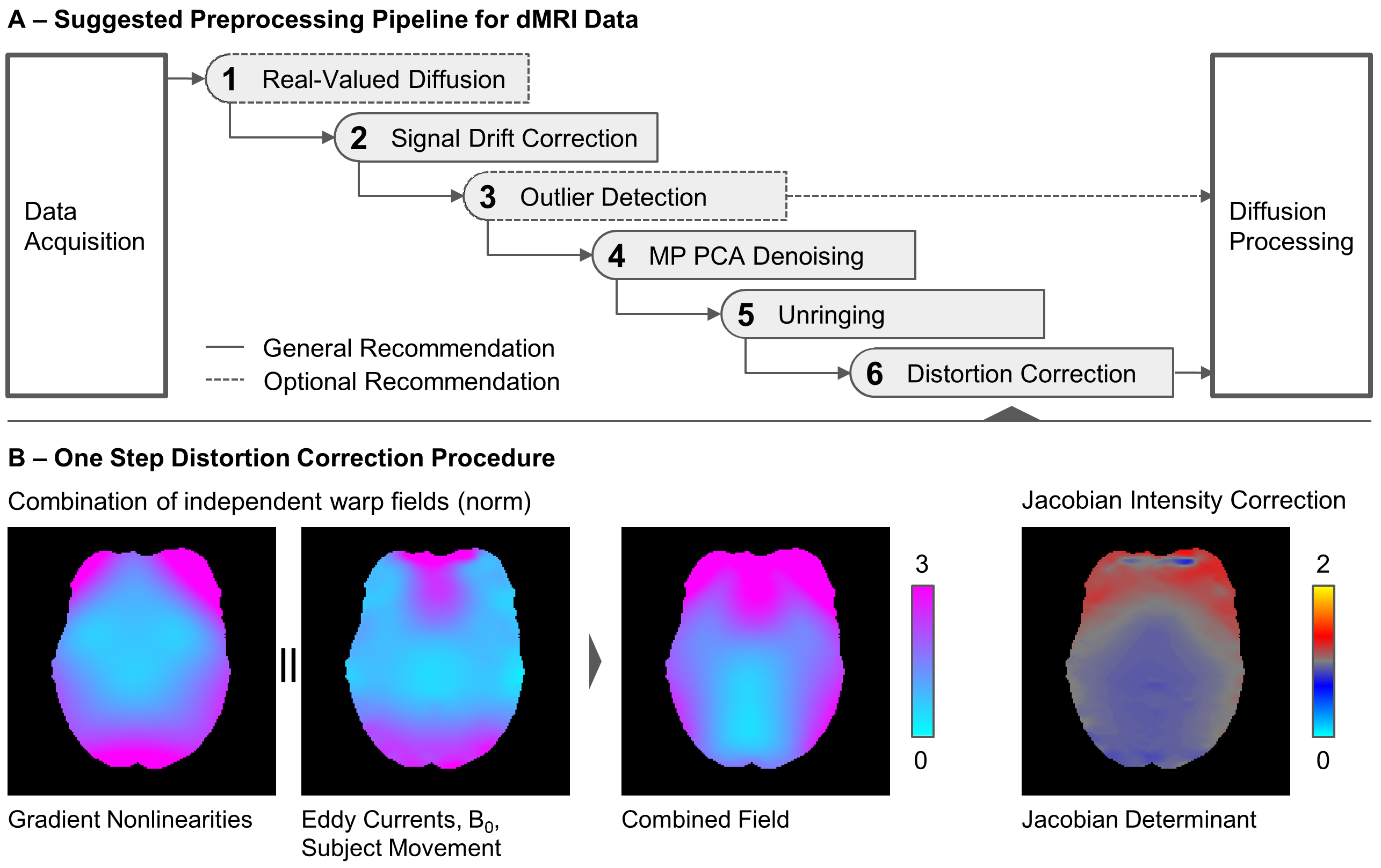
Discussions of preprocessing-steps converged toward an optimized pipeline which is summarized in Figure 1A. Transformation of complex data into real-valued dMRI data8 is recommended as the first optional processing step if phase-data are available. Real-valued dMRI data enable better image contrast and more accurate model-fits by avoiding bias introduced by the noise floor. Further research will aim to provide a more thorough understanding of the benefits of processing real-valued MRI data. The authors recommend a temporal drift-correction based on global intensity variations of b=0 volumes9. If diffusion-models support down-weighting of single data points, an outlier-detection can be employed to increase fitting stability10. The outlier-detection does not alter the data but informs diffusion analyses of potential outliers. MP-PCA denoising11 is recommended prior to further interpolation steps, in order to minimize potential noise coherences between neighboring voxels. To reduce influences of Gibbs-ringing artifacts, unringing using sub-voxel-shift is recommended as the fifth step12.
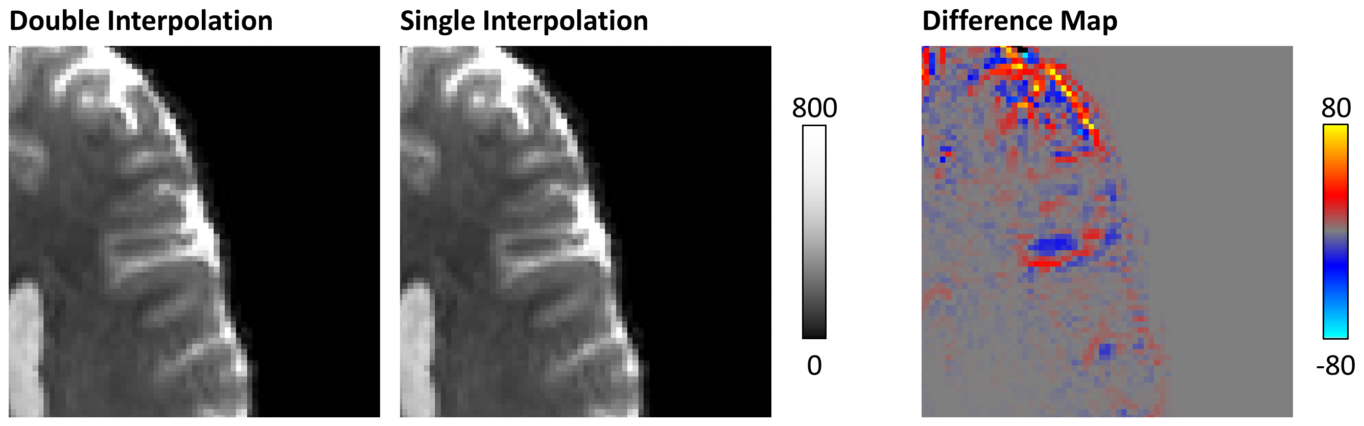
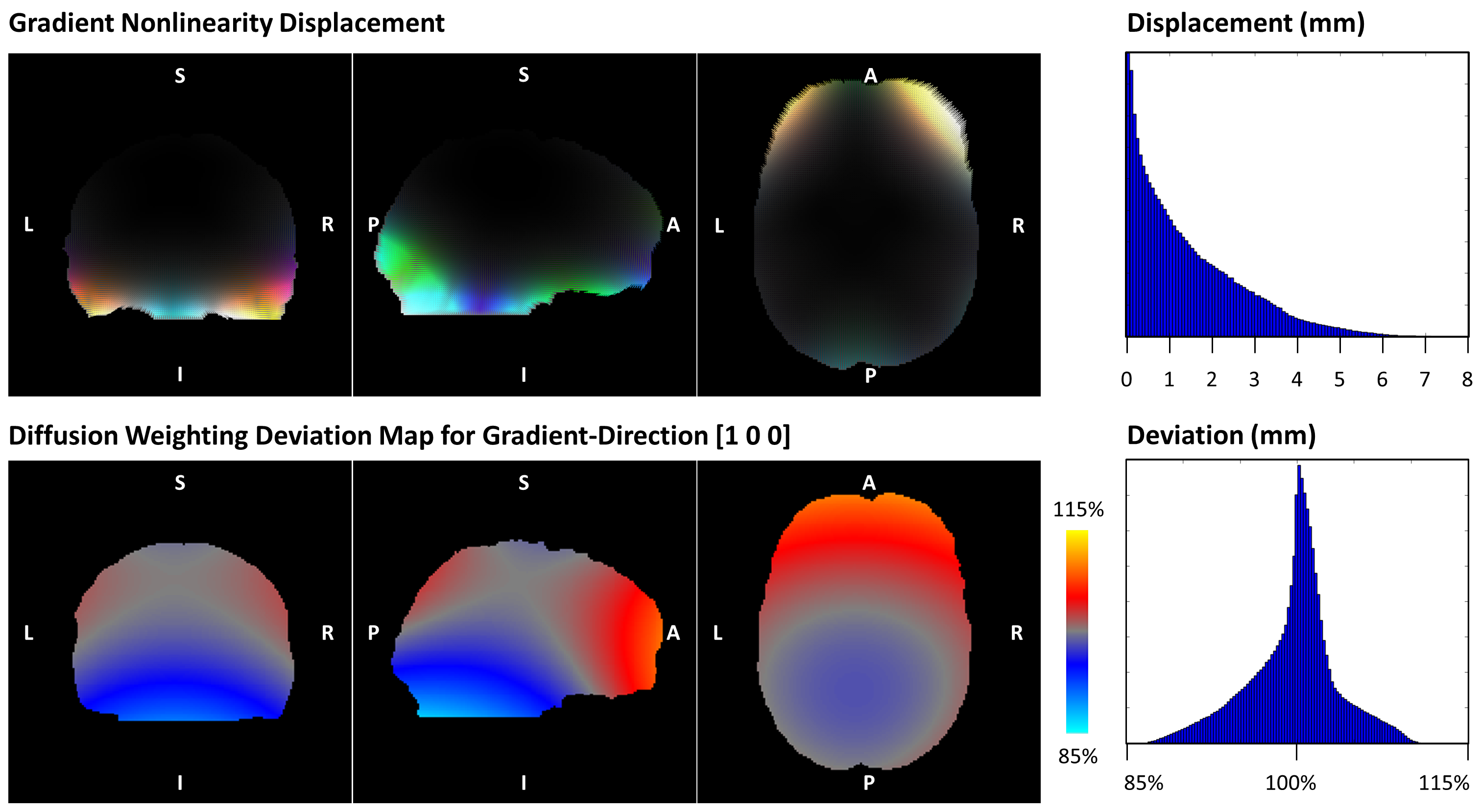
One distinguishing feature of the proposed pipeline is the combination of interpolations from multiple intermediate processing steps into a single step (Figure 1B). In this context, the distortions from B0-off-resonance, subject movement, and eddy currents were estimated for each volume13 and concatenated with a global gradient distortion field provided for the gradient system6. Jacobian determinants of the combined warp fields were evaluated to correct for signal pile-up due to image-warping. A comparison of unwarping with single and dual-interpolation is depicted in Figure 2. The analysis of gradient-nonlinearities revealed a voxel-displacement of up to 6mm within the analyzed brain volume. The diffusion-weighting variance within the brain volume revealed locally effective b-values deviations of ±15% (Figure 3). These results underline the importance of correcting for gradient-nonlinearity induced image-distortions and spatial and temporal b-value deviations6,7.
Discussion
High-amplitude gradient systems are essential for the acquisition of dMRI data with strong diffusion-weighting and high spatial resolution. However, such data generally require adapted preprocessing due to low SNR, significant gradient-nonlinearities and lengthy scan times. In a joint effort, all Connectom sites discussed and established a common preprocessing recommendation for future studies on this gradient MRI system. The resulting pipeline concatenates various preprocessing-steps and minimizes the number of required interpolation steps. Image-distortions and b-value deviations related to gradient-nonlinearities are corrected by means of warping and the output of b-value deviations. The benefit of using this preprocessing pipeline is not restricted to scanners with large gradient strengths but applies generally to all MRI systems which suffer from gradient-nonlinearities to some extent. The authors will share the code for the pipeline online to enable future studies using their recommended preprocessing.Acknowledgements
CE is supported by the SPP2041 program "Computational Connectomics" of the German Research Foundation (DFG)References
- Setsompop K et al. Pushing the limits of in vivo diffusion MRI for the Human Connectome Project. Neuroimage. 80:220-33, 2013
- Jones D et al. Microstructural imaging of the human brain with a 'super-scanner': 10 key advantages of ultra-strong gradients for diffusion MRI. Neuroimage. 182:8-38, 2018
- Glasser MF et al. The minimal preprocessing pipelines for the Human Connectome Project. Neuroimage. 80:105-24, 2013
- Jones D and Cercignani M, Twenty-five pitfalls in the analysis of diffusion MRI data. NMR in Biomedicine. 23:803-20, 2010
- Anderson JL et al. How to correct susceptibility distortions in spin-echo echo-planar images: application to diffusion tensor imaging. Neuroimage. 20:870-88, 2003
- Bammer R et al. Analysis and generalized correction of the effect of spatial gradient field distortions in diffusion‐weighted imaging. MRM. 50:560-9, 2003
- Rudrapatna U, Parker G, Roberts J, and Jones D. Can we correct for interactions between subject motion and gradient-nonlinearity in diffusion MRI? In Proceedings of the ISMRM, 2018, page 1206
- Eichner C et al. Real diffusion-weighted MRI enabling true signal averaging and increased diffusion contrast. Neuroimage. 22:373-84, 2015
- Vos SB et al. The importance of correcting for signal drift in diffusion MRI. MRM. 77:285-99, 2017
- Sairanen V et al. Fast and accurate Slicewise OutLIer Detection (SOLID) with informed model estimation for diffusion MRI data. Neuroimage. 181:331-46, 2018
- Veraart J et al. Denoising of diffusion MRI using random matrix theory. MRM. 76:1582-93, 2016
- Keller E et al. Gibbs‐ringing artifact removal based on local subvoxel‐shifts. MRM. 76:1574-81, 2016
- Andersson JL and Sotiropoulos SN. An integrated approach to correction for off-resonance effects and subject movement in diffusion MR imaging. Neuroimage. 125:1063-78, 2016
Figures