1016
High Gradient Amplitude and High Slew Rate Oscillating Gradient Diffusion-Encoding for Human Brain Imaging1GE Global Research, Niskayuna, NY, United States, 2GE Healthcare, Stockholm, Sweden, 3Department of Radiology, Mayo Clinic, Rochester, MN, United States, 4Walter Reed National Military Medical Center and Uniformed Services University of the Health Sciences, Bethesda, MD, United States
Synopsis
Two high amplitude and slew rate head-gradient MRI systems (C3T: 80mT/m, 700T/m/s, and MAGNUS: 200 mT/m, 500 T/m/s) with significantly better performance than clinical whole-body MRI have been developed. These allow for microstructure-sensitive, oscillating-gradient-spin-echo (OGSE) diffusion-encoding to be feasibly applied for human brain imaging. An analysis of waveforms at varying gradient performances reveals that peripheral nerve stimulation and gradient slew rates limit OGSE frequencies, while gradient strength limits b-values and echo times. In vivo human imaging was performed at 3T, demonstrating significantly increased measured diffusivity in OGSE diffusion tensor imaging (DTI) as compared to standard DTI.
Introduction
Oscillating gradient spin echo (OGSE) diffusion-encoding1,2 has been demonstrated to provide preferential selectivity of microstructure length scales. The demonstration of OGSE in vivo has been, however, mostly limited to preclinical MRI3, primarily due to the lack of high gradient amplitude and slew rates necessary to effect sufficiently high b-values and oscillating frequencies, while keeping echo times (TE) short for adequate SNR. A compact, asymmetric head-gradient MRI at 3T (C3T) with 80 mT/m gradient amplitude and 700 T/m/s slew rate4 has demonstrated rapid EPI with echo spacings up to half that of conventional clinical MRI scanners5. The key to achieving rapid EPI clinically is the significantly reduced electric field of the head-gradient, which allows for a much higher gradient slew rate dB/dt with equivalent peripheral nerve stimulation (PNS)6-7. Like EPI, OGSE waveforms are fast-switching but unlike EPI, both high gradient amplitude and slew rates are required. This work evaluates the feasibility of OGSE for human brain imaging with head-gradients.Methods
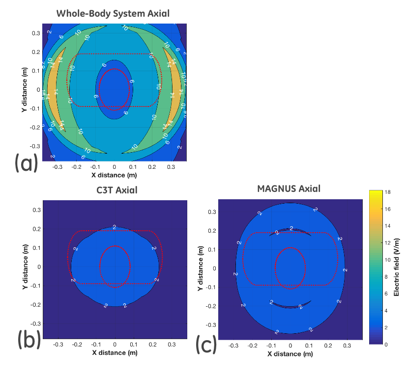
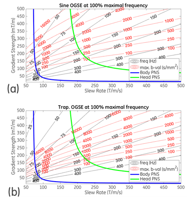
Sinusoidal OGSE waveforms were analyzed as a function of gradient amplitudes (30-500 mT/m) and slew rates (50-500 T/m/s) to obtain maximal b-values and OGSE frequencies. It can be shown that the maximum allowable frequency for a given maximum gradient amplitude and slew rate is simply:$$$f_{max}=SR_{max}/2\pi G_{max}.$$$ The effect of increasing b-value by replacing sinusoids with trapezoids8 was also evaluated. PNS thresholds from clinical whole-body MRI and a head-gradient4 were applied in the analysis. The electric-fields of these gradients (Fig. 1) were generated using custom-written Matlab code (Mathworks Inc., Natick MA, USA) for comparison to establish correspondence against the experimentally-derived PNS thresholds7.
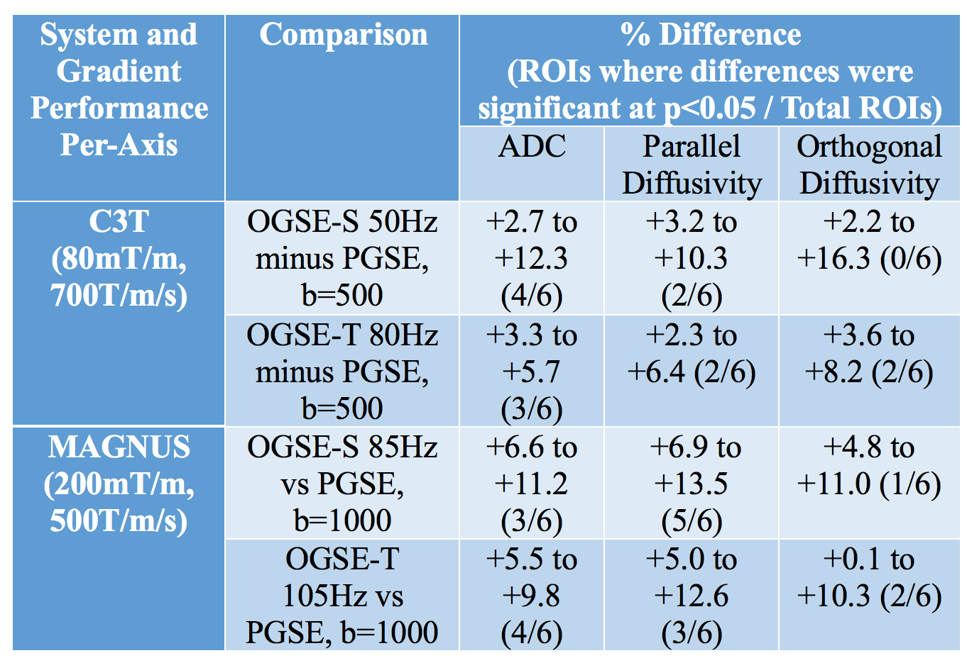
Two head-gradient systems at 3T with equivalent power per-axis (1MVA) – C3T4(80 mT/m, 700 T/m/s) and MAGNUS8(200mT/m, 500T/m/s) were used to image two subjects (M, Age=40-41 years) using an IRB-approved protocol. An isotropic diffusion phantom (25% PVP) with a nominal diffusivity of 1250um2/sec (at 19 deg C) was imaged to verify the quantitative accuracy of OGSE to standard pulse-field spin echo (PGSE). On C3T, sinusoidal OGSE (OGSE-S) with 50Hz, b=500sec/mm2, trapezoidal OGSE (OGSE-T) with 80Hz, b=500sec/mm2 and PGSE were acquired at similar TE=97ms. On MAGNUS, the higher gradient amplitude allowed for higher frequency and b-values (OGSE-S with 85Hz and OGSE-T with 105Hz at b=1000sec/mm2). DTI with 10 and 16 directions was acquired for C3T and MAGNUS, respectively, with 2mm-isotropic EPI and parallel imaging R=2. Gradient nonlinearity correction9 was applied to reduce spatial bias due to the nonlinear gradient fields. Six regions of interest (ROI) (176 to 408mm2) were selected in the white matter for comparison of mean (ADC), parallel and orthogonal diffusivity.
Results
In OGSE-S, higher gradient amplitudes increase b-value but decrease maximum frequencies; higher slew rates increase both b-value and maximum frequencies (Fig 2a). OGSE-T provides slightly higher b-values (Fig. 2b). In both OGSE-S/T, the body-PNS severely limits the ability to simultaneously achieve higher frequency and b-value; the head-PNS increases the obtainable frequency or b-value significantly. The higher PNS thresholds of the head-gradients correspond well to the electric fields of Fig. 1.
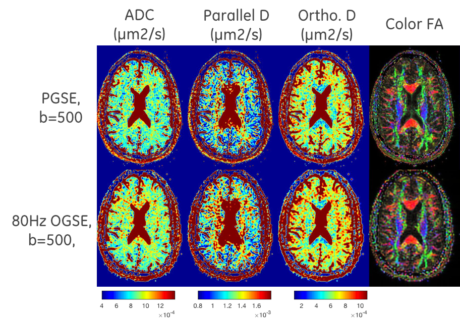
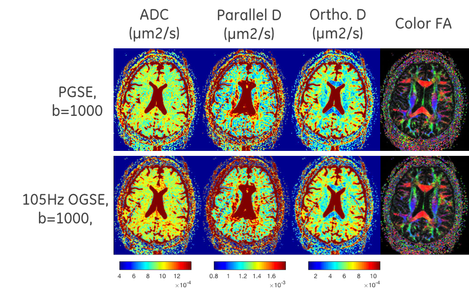
In phantom imaging, ADC was not significantly different between OGSE-S/T and standard PGSE sequences, (+0.6 to +2.2%). In vivo imaging on MAGNUS, however, resulted on average in higher diffusivity with OGSE than PGSE (ADC: +5.6 to +6.4%, parallel diffusivity: +5.5 to +6.0%, orthogonal diffusivity: +5.8 to +6.9%). Many individual ROIs showed statistically-significant differences (Table 1). This was also true on C3T, but by smaller margins (ADC: +3.8 to +4.6%, parallel diffusivity: +3.2 to +4.6%, and orthogonal diffusivity: +4.3 to +5.0%) with slightly fewer individual ROIs with statistically-significant differences. Visually, higher diffusivity could be seen in OGSE-diffusivity maps than in PGSE (Fig. 3-4).
Discussion and Conclusion
In this preliminary feasibility study of OGSE in high performance gradients for human imaging, the results of increased diffusivity corresponded well with pre-clinical results10, as well as lower frequency/b-value imaging performed on whole-body scanners11-12. While higher gradient performance was required for OGSE, our study found that a higher PNS threshold was equally crucial for OGSE. The same head-gradient PNS threshold was applied in both C3T and MAGNUS, with both subjects experiencing very-mild but not painful stimulation in the face. Future investigations into optimization of PNS would be carried out, along with waveform optimizations to maximize b-value and frequency. Future work would include an investigation into OGSE kurtosis, which could provide increased sensitivity to microstructural tissue changes from neurological diseases13.Acknowledgements
This work was supported in part by NIH U01-EB024450 and CDMRP W81XWH-16-2-005. The opinions or assertions contained herein are the private views of the authors and are not to be construed as official or reflecting the views of the NIH or the U.S. Department of Defense.References
1. Schachter M, et al. J. Magn Reson 2000. 147(2):232-237.
2. Does MD, Parsons EC, Gore JC. Magn Reson Med 2013. 49(2):206-215.
3. Xu J, et al. Magn Reson Med 2009. 61(4):828-833.
4. Foo TKF, et al. Magn Reson Med 2018. 80(5):2232-2245.
5. Tan ET, et al. JMRI 2016. 44(3):653-664.
6. Chronik BA, et al. Magn Reson Med 2001. 46(2):386-394.
7. Lee SK, et al. Magn Reson Med 2016. 76(6):1939-50.
8. Foo TKF, et al. Proc. 26th ISMRM 2018. 839.
9. Tan ET et al, JMRI 2012. 38(2):448-453.
10. Aggarwal M, et al. Magn Reson Med 2011. 67(1):98-109.
11. Baron CA, Beaulieu C. Magn Reson Med 2013. 72(3):726-736.
12. Andica C et al. Magn Reson Med Sci 2018. 17:269-272.
13. Wu D, et al. NMR in Biomed 2018. 31(6):3917-9.
Figures