1011
Multi-diffusion time DWI to detect altered microvessel structure in a rat model of hypertension1Division of Neuroscience and Experimental Psychology, University of Manchester, Manchester, United Kingdom, 2Henry Moseley X-ray Imaging Facility, School of Materials, University of Manchester, Manchester, United Kingdom, 3Bioxydyn Limited, Manchester, United Kingdom
Synopsis
The role of microvascular pathology in the development and progression of dementia is currently unclear. Non-invasive methods for imaging microvessel structure are needed to study cerebrovascular alterations in-vivo. The time for water molecules to change direction relative to the diffusion time alters the measured pseudo-diffusion coefficient of intra-voxel incoherent motion (IVIM). Using multi-diffusion time multi-b-value data and a velocity autocorrelation model, the capillary segment length (l) can estimated. In this study we validate hippocampal l against vascular corrosion casts, then apply the method to study vascular structure and function in a rat model of hypertension in comparison with age-matched controls.
Introduction
Increasing evidence shows that microvascular changes in the brain occur alongside, and may even precede, the development of dementia pathology1. These microvascular changes may contribute to disease pathogenesis and accelerate cognitive decline, thereby representing a potential treatment target. Chronic hypertension increases the risk of developing dementia and can cause increased capillary stiffness and tortuosity2. However, methods to measure microvessel structure in-vivo, are lacking. In this study, we develop a DWI method for estimating microvessel structure and validate using micro x-ray CT of a vascular corrosion cast. We test the method in a rat model of hypertension. Multi-diffusion time DW data is analysed with a velocity autocorrelation (VA) model3 and intra-voxel incoherent motion (IVIM)4, in order to estimate perfusion fraction (f), extravascular diffusion (D), pseudo-diffusion (D*), average blood velocity (v) and average vessel segment length (l).Methods
Corrosion Casting: A Wistar-Kyoto control rat was casted using Mercox agent. When the vasculature was fully perfused (through left ventricle), the resin was left for curing. Tissue was then dissolved and the cast freeze dried to remove moisture.
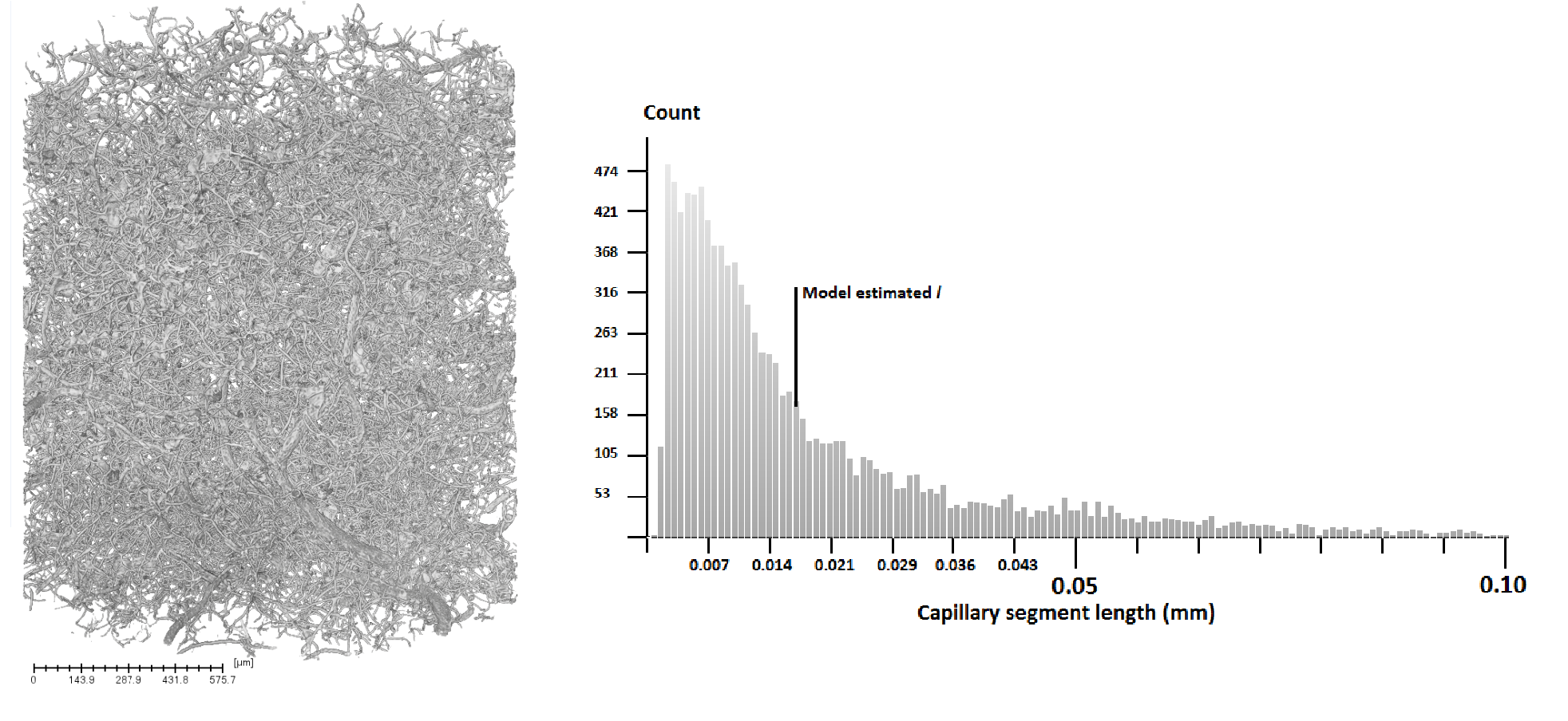
Micro-CT: Cast scanned using ZEISS Xradia 520 Versa 3D X-ray microscope. Source-to-sample: 25.01mm. Sample-to-detector: 87.65mm. Voxel size: 0.75µm. Binning: 2. Magnification: x4. Voltage: 80kV. Power: 7W. Exposure time: 18s. Reconstructed using filtered-back projection. Reconstructed image segmented and skeletonised using AVIZO software. Capillary segment lengths derived from skeletonised image are branch to branch length.
MR: Axial DW images were acquired for male rats aged 13 months (σ=0.29): 8 spontaneously hypertensive rats (SHRs) and 10 Wistar-Kyoto controls. b-values: 0, 10, 20, 50, 100, 200, 500, 1000mm2s-1. Δ (diffusion time) values: 11.6, 20, 30, 40 and 50ms were acquired for each b-value. δ=5.8ms. TR: 3000ms. TE: 66.9ms. FOV=30 x 30 x 30mm. Matrix size = 96 x 96 x 30. Rats were anaesthetised using 2-2.5% isoflurane in 100% O2 and scanned on a Bruker Avance III console interfaced with an Agilent 7T 16cm bore magnet, with maximum gradient strength Gmax= 300mTm-1. A Bruker transmit only resonator was used for transmission and a Bruker rat brain surface coil was used for reception. Least squares fitting of the IVIM model4 using a Levenberg-Marquardt algorithm provided estimates of f, D and D*. Fitting a VA model3 to multi-diffusion time signals provided estimates of v. Models were fitted in two-steps, first modelling the extravascular signal by estimating D and f using signals at b=500 and b=1000 mm2s-1. Vascular parameters were then estimated using the logarithm of the intravascular signal (total normalised signal minus extravascular signal) at remaining b-values and Δ-values. For IVIM, D* was equal to the gradient of the Δ=50ms data: this Δ allows blood flow to appear as pseudo-diffusion and be appropriately modelled as IVIM. Gradients were estimated for the remaining Δ values, and v estimated by fitting the VA model to these gradients as a function of Δ. The average vessel segment length (l) was calculated using the relationship: l = 6D*/v. A T2-weighted RARE anatomic image was acquired for region segmentation via registration with the Schwarz rat atlas5.
Results and discussion
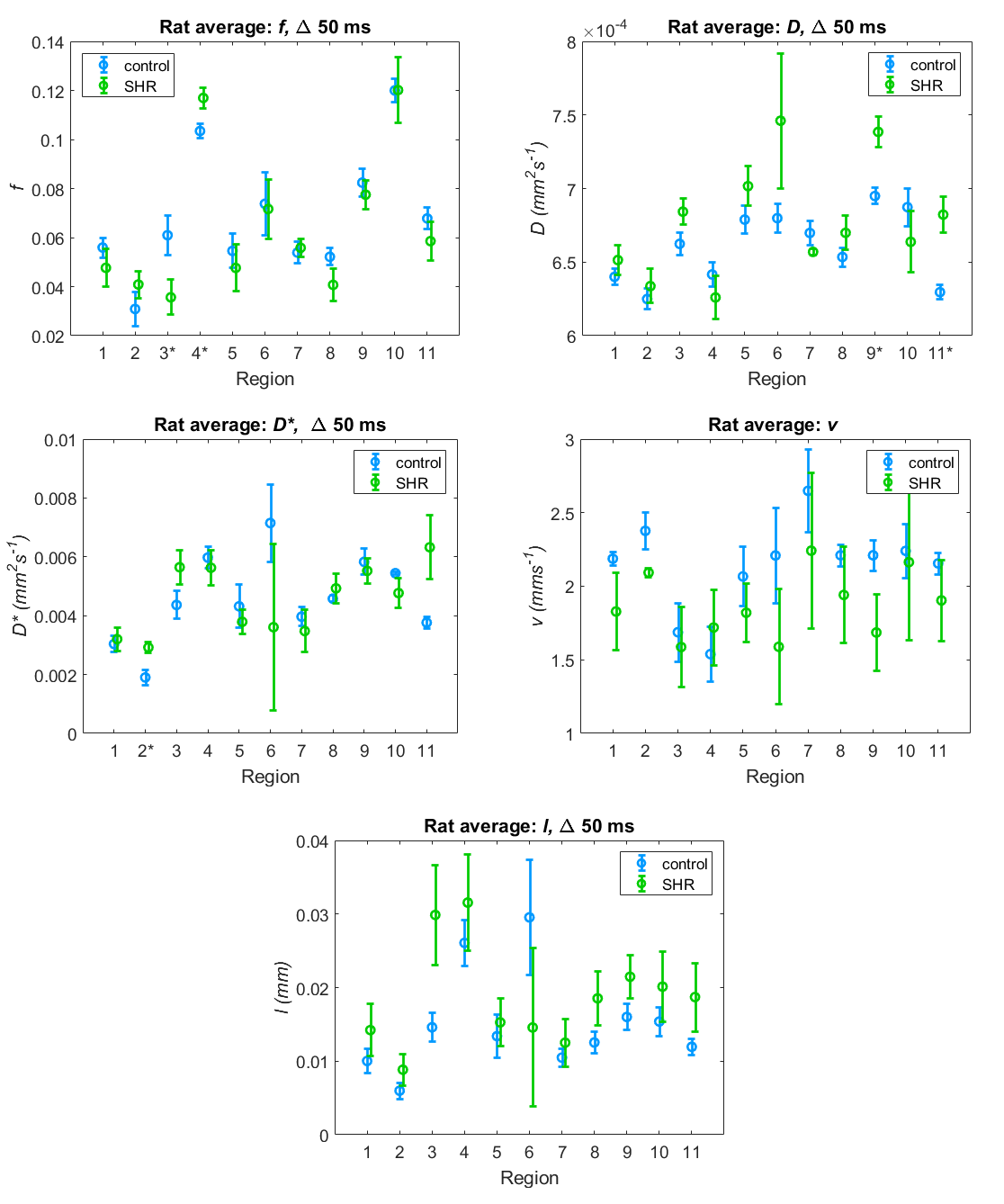
The MR estimate of average capillary segment length of the hippocampus in control rats (l=0.016mm) is in good agreement with the micro-CT of the corrosion cast (Figure 1). Estimates of f, D, D* and v also lie within the expected range6,7. Application of multivariate analysis of variance (MANOVA) showed significant effects of both genotype and region (p=0.008 and p<0.001 respectively). Unpaired t-tests assuming unequal variances (no correction for multiple comparisons) showed significant differences between SHR and control rats in IVIM estimated parameters (Figure 2 – significant results indicated by * on region number). Higher blood volume (f) is found in the cingulate cortex in SHRs (p=0.03) possibly indicating formation of micro-collaterals. Lower blood volume is found in the temporal cortex of the SHRs (p=0.03), possibly indicating damaged vessels. The diffusion coefficient (D) is significantly higher in the SHR group in the thalamus (p=0.004) and hippocampus (p=0.004) - this could represent neuronal loss reducing barriers to diffusion8. The pseudo-diffusion (D*) coefficient is significantly higher in the SHRs in the corpus callosum (p=0.01).Conclusion
Our DW approach produces reasonable estimates of f, D, D* and v 5,6: l was validated with micro-CT assessment for the hippocampus. Our results are also consistent with the expected alterations of vessel structure in a rat model of hypertension. In particular, alterations are seen in regions which are associated with dementia.Acknowledgements
UK Medical Research Council (MRC)References
1 Iadecola C, The overlap between neurodegenerative and vascular factors in the pathogenesis of dementia, Acta Neuropathol. 2010 Sep;120(3):287-96
2 Han HC, Twisted blood vessels: symptoms, etiology and biomechanical mechanisms, J Vasc Res. 2012;49(3):185-97
3 Kennan R, Gao J, Zhong J, Gore J, A general model of microcirculatory blood flow effects in gradient sensitized MRI, Med Phys 1994 Apr;21(4):539-45
4 Le Bihan D, Breton E, Lallemand D, Aubin ML, Vignaud J, Laval-Jeantet M, Separation of diffusion and perfusion in intravoxel incoherent motion MR imaging, Radiology. 1988 Aug;168(2):497-505
5 Schwarz AJ, Danckaert A, Reese T, Gozzi A, Paxinos G, Watson C, Merlo-Pich EV, Bifone A, A stereotaxic MRI template set for the rat brain with tissue class distribution maps and co-registered anatomical atlas: application to pharmacological MRI, Neuroimage. 2006 Aug 15;32(2):538-50
6 Unekawa M, Tomita M, Tomita Y, Toriumi H, Miyaki K, Suzuki N, RBC velocities in single capillaries of mouse and rat brains are the same, despite 10-fold difference in body size, Brain Res. 2010 Mar 12;1320:69-73
7 Federau C, O'Brien K, Meuli R, Hagmann P, Maeder P, Measuring brain perfusion with intravoxel incoherent motion (IVIM): initial clinical experience, J Magn Reson Imaging. 2014 Mar;39(3):624-32
8 Londoño A, Castillo M, Lee YZ, Smith JK, Apparent diffusion coefficient measurements in the hippocampi in patients with temporal lobe seizures, AJNR Am J Neuroradiol. 2003 Sep;24(8):1582-6
Figures