1008
Biophysical modeling of the white matter: from theory towards clinical practice1Champalimaud Research, Champalimaud Centre for the Unknown, Lisbon, Portugal, 2CUBRIC, School of Psychology, Cardiff University, Cardiff, United Kingdom, 3Champalimaud Centre for the Unknown, Lisbon, Portugal, 4iMinds - Vision Lab, University of Antwerp, Antwerp, Belgium
Synopsis
We address the degeneracy of the diffusion standard model and improve the precision and accuracy in parameter estimation, thus promoting clinical applicability. Acquisition of additional data is not required; instead, we introduce a more robust and accurate estimator by fitting the standard model directly to diffusion-weighted data rather than its rotational invariants. We are able to overcome the implicit assumption of data being shelled in terms of b-values. This enables the correction of gradient nonlinearities to avoid biases in model parameter estimation, whereas revising the optimal experimental design demonstrates that non-shelled encoding schemes are favorable in terms of achievable precision.
Introduction
The development of biophysical models aiming to relate diffusion MRI signal to brain tissue microstructure has seen an exponential growth in recent years. However, there is a rising awareness of the approach’s fundamental limitations1. A low precision, accuracy, and robustness of the parameters estimators of even the simplest biophysical models2 currently limit the clinical applicability of the developed techniques3,4. Factoring out the fiber orientation distribution function (fODF) using rotationally invariant metrics has reduced the complexity of the modeling problem, but in turn uncovered an intrinsic degeneracy in microstructural parameter estimation3. Moreover, such “powder averaging” approaches5,6,7 entail an implicit assumption that diffusion MRI data are perfectly shelled, i.e. different gradient directions for a finite set of $$$b$$$-values. However, this assumption is usually unmet due to gradient nonlinearities8 and poses an unnecessary constraint on experimental design9.Theory
We here adopt the widely used two-compartment model of water inside narrow impermeable “sticks” representing axons10, embedded in an extra-cellular matrix, coined the diffusion Standard Model (SM)2,11:
$$S_{SM}(b,\hat{g})=\int d\hat{n}\mathcal{P}\left(\hat{n}\right)\mathcal{K}\left(b,\hat{g}\cdot\hat{n}\right)\;\;\;\;\left(1\right)$$
with
$$\mathcal{K}\left(b,\xi\right)=fe^{-bD_a\xi^2}+(1-f)e^{-bD_e^\perp-b\left(D_e^\parallel -D_e^\perp\right)\xi^2}\;\;\;\;\left(2\right)$$
being the signal response kernel of an individual fiber fascicle parameterized by the intra-axonal signal fraction $$$f$$$ and parallel diffusivity $$$D_a$$$, and the extra-axonal radial and axial diffusivities $$$D_e^\parallel$$$ and $$$D_e^\perp$$$, respectively. The fODF $$$\mathcal{P}\left(\hat{n}\right)$$$ is parameterized by its spherical harmonic coefficients $$$p_{lm}$$$, up to order $$$L$$$.
The $$$4+(L+1)(L+2)/2$$$ parameters can be estimated by minimizing the following non-linear object function: $$\|S(b,\hat{g})-S_{SM}(b,\hat{g})\|^2\;\;\;\;\left(3\right)$$
If data is acquired with several $$$b$$$-shells, this system can also be solved by first representing each $$$b$$$-shell by its spherical harmonic (SH) coefficients $$$S_{lm}(b)$$$:
$$\|S_{lm}(b)-p_{lm}K_l (b)\|^2\;\;\;\;\left(4\right)$$
with $$$K_l(b)$$$ the projection of $$$\mathcal{K}\left(b,\xi\right)$$$ onto Legendre polynomials3. The dimensionality of this optimization problem can be reduced by adopting the $$$l^{\mathrm{th}}$$$ rotational invariants of the signal and fODF, $$$S_l^2=\sum_{m=-l}^l|S_{lm}|^2/4\pi(2l+1)$$$ and $$$p_l^2=\sum_{m=-l}^l |p_{lm}|^2/4\pi (2l+1)$$$, respectively, thereby factoring out the full fODF3: $$\|S_{l}(b)-p_{l}K_l(b)\|^2\;\;\;\left(5\right).$$
Although all three fitting approaches, i.e. Eqs. (2), (3), and (4) are derived from the same standard model, we will demonstrate their robustness, accuracy, and precision is significantly different.
We will refer to the parameter estimators based on Eqs. (2), (3), and (4) as “SM”, “SM-SH”, and “SM-RotInv”, respectively. The maximal SH order $$$L$$$ is 6 unless stated differently.
Data
Simulations: Synthetic signals were produced by solving Eq. (1) with ground truth parameters $$$f\,=\,0.75,\,D_a\,=\,2.1\,\mathrm{\mu\,m^2/ms},\,D_e^\parallel\,=\,1.5\,\mathrm{\mu\,m^2/ms}$$$, and $$$D_e^\perp\,=\,0.5,\mathrm{\mu\,m^2/ms}$$$ and a crossing fiber geometry for $$$\mathcal{P}\left(\hat{n}\right)$$$ using the diffusion-weighted encoding scheme of the MR data. Gaussian noise was added (SNR$$$_{b=0}=50$$$) to evaluate the robustness, accuracy and precision of the different estimators.
MRI experiments: Four volunteers were scanned on a Connectom 3T MR scanner. Diffusion-weighting was applied along 30 gradient directions for $$$b = 1$$$ and $$$2\,\mathrm{ms/\mu\,m^2}$$$, and 60 gradient directions for $$$b\,=\,3,\,5,\,7,\,9,\,11,\,12.1,\,13.5$$$ and $$$15\,\mathrm{ms/\mu\,m^2}$$$. Following parameters were kept constant: $$$\Delta/\delta\,=\,30/13\,\mathrm{ms}$$$, $$$\mathrm{TR/TE}=3500/62\,\mathrm{ms}$$$ and resolution $$$3\,\times\,3\times\,3\,\,\mathrm{mm}^3$$$. Data was denoised12 and Gibbs-13, eddy current-14, and Rician bias-15 corrected prior to analyses. Moreover, the $$$b$$$$-values were corrected for spatially varying gradient nonlinearities16.
Results
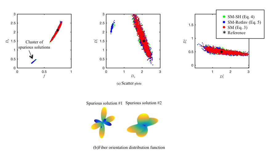
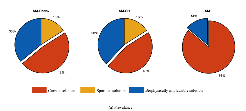
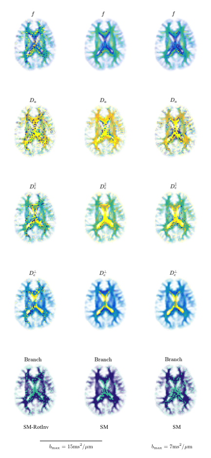
Degeneracy: Simulations demonstrate that, unlike “SM-SH" and “SM-RotInv", “SM” does not show the notorious cluster of biophysically plausible, yet spurious solutions, which occur even at high $$$b$$$-values (Fig.$$$\,1$$$). The results are independent from the starting point (Fig. 2). Although the degeneracy is technically not resolved, the basin of attraction is strongly reduced, leaving an apparent lack of degeneracy. The findings are confirmed in the MR data (Fig. 3). Although fits were performed from random starting points, “SM” shows to produce smooth parametric maps, even for lower $$$b$$$-values, whereas ``SM-RotInv"-derived maps show speckled noise, indicating the degeneracy.
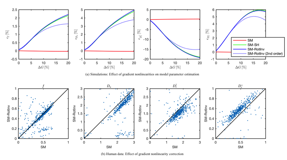
Accuracy: Uncorrected gradient-nonlinearities bias the shell-based “SM-RotInv” and “SM-SH”- estimators (Fig.$$$\,4a$$$). Some parameters, e.g. $$$D_e^\perp$$$, show errors up to $$$15\%$$$. These biases were also observed in the MR data by comparing “SM-RotInv” and “SM” estimates (Fig.$$$\,4b$$$).
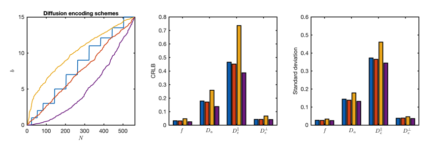
Precision: Cramer-Rao lower bound (CRLB) analysis shows that non-shelled encoding schemes improve the precision of the parameter estimators, especially for more densely sampled low $$$b$$$-values (Fig. 5).
Discussion
We here show that the full “SM” fitting provides parameter estimates with higher:
- Robustness: no observed degeneracy in our simulated and clinical data, even if only relatively low $$$b$$$-values were selected;
- Accuracy: Correcting for gradient nonlinearities is required to avoid bias in the estimated model parameters, especially for the extra-axonal space, but prevents the use of estimated that rely on shelled data;
- Precision: Non-shelled gradient encodings might have a higher predicted precision, especially with a denser sampling of low b-values as shown in a CRLB analysis, thereby promoting the clinical applicability of the standard model. The simultaneous estimation of the fODF and the microstructural kernel does not complicate the estimation problem, nor does it lower the precision of the parameter estimator. Instead, it enables fiber tractography without relying on arbitrarily chosen –often global– signal kernels.
Acknowledgements
JV is a Postdoctoral Fellow of the Research Foundation - Flanders (FWO; grant number 12S1615N). The data was acquired at the UK National Facility for In Vivo MR Imaging of Human Tissue Microstructure funded by the EPSRC (grant EP/M029778/1), and The Wolfson Foundation.References
- Novikov DS, Kiselev VG, Jespersen SN. On modeling. Magn. Reson. Med. 2018; 79(6):3172-3193.
- Jespersen, S. N., Kroenke, C. D., Ostergaard, L., Ackerman, J. J., Yablonskiy, D. A.. Modeling dendrite density from magnetic resonance diffusion measurements. Neuroimage 2007; 34 (4), 1473–1486.
- Novikov DS, Veraart J, Jelescu IO, Fieremans E. Rotationally-invariant mapping of scalar and orientational metrics of neuronal microstructure with diffusion MRI. Neuroimge 2018; 174: 518-538.
- Jelescu IO, Veraart J, Fieremans E, Novikov DS. Degeneracy in model parameter estimation for multi‐compartmental diffusion in neuronal tissue. NMR Biomed 2016; 29(1): 33-47.
- Jespersen, S. N., Lundell, H., Sønderby, C. K., Dyrby, T. B. Orientationally invariant metrics of apparent compartment eccentricity from double pulsed field gradient diffusion experiments. NMR in Biomedicine 2013; 26 (12), 1647–1662.
- Kaden, E., Kruggel, F., Alexander, D. C.. Quantitative mapping of the per-axon diffusion coefficients in brain white matter. Magnetic Resonance in Medicine 2016; 75 (4), 1752–1763.
- Lasic, S., Szczepankiewicz, F., Eriksson, S., Nilsson, M., Topgaard, D. Microanisotropy imaging: quantification of microscopic diffusion anisotropy and orientational order parameter by diffusion MRI with magic angle spinning of the q-vector. Frontiers in Physics 2014; 2, 11.
- Romeo F, Hoult DI. Magnet field profiling: analysis and correcting coil design. Magnetic Resonance in Medicine 1984;1(1):44–65
- Jones, D., Horsfield, M., Simmons, A.: Optimal strategies for measuring diffusion in anisotropic systems by MRI. Magnetic Resonance in Medicine 1999; 42(39) 515 – 525
- Kroenke, C. D., Ackerman, J. J., Yablonskiy, D. A.. On the nature of the NAA diffusion attenuated MR signal in the central nervous system. Magnetic Resonance in Medicine 2004; 52 (5), 1052–1059.
- Novikov D.S., Fieremans E., Jespersen S.N., Kisilev V.G. , Quantifying brain microstructure with diffusion MRI: Theory and parameter estimation. NMR in Biomedicine 2018; https://doi.org/10.1002/nbm.3998
- Veraart J, Novikov DS, Christiaens D, Ades-Aron B, Sijbers J, Fieremans E. Denoising of diffusion MRI using random matrix theory. Neuroimage 2016; 146: 394-406.
- Kellner E, Dhital B, Kiselev VG, Reisert M. Gibbs-ringing artefact removal based on local subvoxel-shifts. Magnetic Resonance in Medicine 2016; 76(5): 1574-1581.
- Andersson JLR, Sotiropoulos SN. An integrated approach to correction for off-resonance effects and subject movement in diffusion MR imaging. NeuroImage 2016; 125:1063-1078.
- Koay CG, Basser P J. Analytically exact correction scheme for signal extraction from noisy magnitude MR signals. Journal of Magnetic Resonance 2006; 179(2):317–322.
- U.S. Rudrapatna, G. Parker, J. Roberts, D.K. Jones. Can we correct for interactions between subject motion and gradient-nonlinearity in diffusion MRI? ISMRM 2018,p 1026
Figures