0999
Joint evidences for a posterior parieto-motor cortex pathway in humans using diffusion MRI tractography and direct cortical stimulation1CNRS/ISC, Bron, France, 2CNRS/INCIA, Bordeaux, France, 3Department of Pediatric Neurosurgery, Hôpital Mère Enfant, Bron, France
Synopsis
This study aims to identify projections from the posterior parietal "reach area" to the primary motor cortex, in healthy humans, using diffusion MRI tractography and direct cortical stimulation information. We analyzed multiple-shell data from 20 subjects of the Human Connectome Project and found significant ipsilateral projections connecting the identified region to the primary motor cortex, especially the hand-knob area which shows the highest streamlines density on both hemispheres. Strikingly, we also identified a density peak in the left (language-related) hemisphere, within the dorsolateral part of the precentral gyrus related to mouth control.
Introduction
In monkeys, converging evidence links a specific region in the posterior parietal cortex to hand/reach activity1. Recently, a potential counterpart of this parietal reach region (PRR) has been identified in humans2. When stimulated, this region disrupts ongoing hand movements with short latency and specificity. However, to date, it remains unclear in humans whether this influence relies on direct projections from PRR to primary motor cortex (M1) or whether it is routed through indirect pathways involving, for instance, the premotor cortex. Here, we used state-of-the-art diffusion MRI (dMRI) tractography methods on the multiple-shell Human Connectome Project (HCP) dataset (b=1000, 2000 and 3000 s/mm2, 90 directions per b-value, 1.25mm isotropic) to address this issue.Material and methods
Based on a previous study2, we identified the 95% confidence area of the cortical region that disrupts hand motor activity when stimulated (shown in Figure 1a for the left hemisphere mapped onto the template pial surface). Using the individual white and pial surfaces computed with FreeSurfer3, we generated the corresponding PRR for each subject on both hemispheres (shown for one subject in Figure 1b), and we inferred a volumic region of interest (ROI) located within the parietal lobe representing the individual confidence ellipse.
In order to better quantify the putative anatomical
connectivity between this region and the primary motor area located in the
precentral gyrus, we used the MRtrix3 software4 on the dMRI data
available in the HCP to perform state-of-the-art tractography methods. The white
matter fiber Orientation Distribution Functions (fODF) were obtained using the Constrained
Spherical Deconvolution (CSD) technique5, and we performed a whole
brain tractography using probabilistic and deterministic algorithms, with a grey
matter white matter interface seeding allowed by the Anatomically Constrained
Tractography (ACT) approach6. This modular addition takes advantage
of the tissue segmentation done with Freesurfer in order to influence the
streamlines reconstruction, and has been shown to give more accurate results
when dealing with connectomics. By using deterministic and probabilistic algorithms,
and by varying some tractography parameters (the step size, the cutoff and the
angle), we obtained multiple tractograms before filtering them to get the
individual white matter pathways. We made the step size vary among [0.3, 0.6,
1.25] based on the spatial resolution of the dMRI data, we made the angle vary among
[30°, 45°, 60°] and the cutoff among [0.05, 0.1, 0.15], according to the advice
given by the MRtrix3 community for the HCP post-processing pipeline. Finally,
we set the maximum length to 80 mm based on the Euclidean distance between the two
ROIs centers. Thus, we filtered the 54 whole brain tractograms (1 million
streamlines per tractogram) to detect only streamlines of which endpoints are
located within the parietal ROI and within the precentral gyrus (given by the
Destrieux’s parcellation7).
Results
The main virtually reconstructed white matter pathways between the two ROIs seem visually very reproducible among subjects according to their ending points and the streamlines count. Thus, to obtain a more accurate quantification of the connectivity between those two areas, we computed the density maps using the Track Density Imaging (TDI) method8 for each subject and for both hemispheres ipsilaterally. We kept only streamlines ending in the precentral gyrus to identify their distribution along this region. Figure 2 displays the mean density map projected onto the average pial surface where we can see the streamlines density peak located within the hand-knob area for both hemispheres. In the left hemisphere, a second significant peak was also found in the dorsal region of the precentral gyrus where facial movements are typically represented9.Discussion
These results show that PRR projects directly to M1 in humans. In particular, we found the major ipsilateral streamlines density to be located in the so-called hand-knob region. Moreover, for the left hemisphere, we identified additional ipsilateral streamlines in the dorsal area where facial movements are represented9. These observations confirm clinical per-operative data showing that stimulating the counterpart of PRR in humans can disrupt hand movements ipsilaterally, irrespective of the hemisphere. They also shed light on speech perturbations that can occur in response of left hemisphere parietal stimulations11.Conclusion
This study identifies, for the first time, in humans, direct pathways from PRR to M1 with such precision. One, ipsilateral, found in both hemispheres, links PRR to the hand-knob area of M1 and could mediate on-line hand motor control10. Another ipsilateral, found only in the left hemisphere, links PRR to the facial control region of M1 and could mediate speech control. These projections might represent specific anatomo-functional segments of the Superior Lateral Fasciculus (SLF)12,13,14.Acknowledgements
Data were provided by the Human Connectome Project, MGH-USC Consortium (Principal Investigators: Bruce R. Rosen, Arthur W. Toga and Van Wedeen; U01MH093765) funded by the NIH Blueprint Initiative for Neuroscience Research grant; the National Institutes of Health grant P41EB015896; and the Instrumentation Grants S10RR023043, 1S10RR023401, 1S10RR019307.References
1: Andersen, R.A., Andersen, K.N., Hwang, E.J., and Hauschild, M. (2014). Optic ataxia: from Balint's syndrome to the parietal reach region. Neuron 81, 967-983.
2: Desmurget et al., 2018, Current Biology 28, 1–7.
3: Fischl B. FreeSurfer. Neuroimage 2012;62(2):774-781.
4: J.-D. Tournier, F. Calamante, A. Connelly. MRtrix: Diffusion tractography in crossing fiber regions. INT J IMAG SYST TECH, 22 (2012), pp. 53-66.
5: J.-D. Tournier, F. Calamante, and A. Connelly. Robust determination of the fibre orientation distribution in diffusion MRI: non-negativity constrained super-resolved spherical deconvolution. Neuroimage, 35 (2007), pp. 1459–72.
6: R.E. Smith, J.-D. Tournier, F. Calamante, A. Connelly. Anatomically-constrained tractography: improved diffusion MRI streamlines tractography through effective use of anatomical information. NeuroImage 62 (2012), pp. 1924–1938.
7: Destrieux C, Fischl B, Dale A, Halgren E. Automatic parcellation of human cortical gyri and sulci using standard anatomical nomenclature. Neuroimage. 2010;53(1):1-15.
8: Calamante, F., Tournier, J. D., Jackson, G. D., & Connelly, A. (2010). Track-density imaging (TDI): super-resolution white matter imaging using whole-brain track-density mapping. Neuroimage, 53(4), 1233-1243.
9: Penfield, W., and Boldrey, E. (1937). Somatic motor and sensory representation in the cerebral cortex of man as studied by electrical stimulation. Brain 60, 389-443.
10: Desmurget, M., and Grafton, S.T. (2000). Forward modeling allows feedback control for fast reaching movements. Trends Cogn. Sci. 4, 423-431.
11: Sanai, N., Mirzadeh, Z., and Berger, M.S. (2008). Functional outcome after language mapping for glioma resection. N. Engl. J. Med. 358, 18-27.
12: Catani, Marco, et al. "Virtual in vivo interactive dissection of white matter fasciculi in the human brain." Neuroimage 17.1 (2002): 77-94.
13: Kamali, Arash, et al. "Tracing superior longitudinal fasciculus connectivity in the human brain using high resolution diffusion tensor tractography." Brain Structure and Function 219.1 (2014): 269-281.
14: Hecht, Erin E., et al. "Virtual dissection and comparative connectivity of the superior longitudinal fasciculus in chimpanzees and humans." Neuroimage 108 (2015): 124-137.
Figures
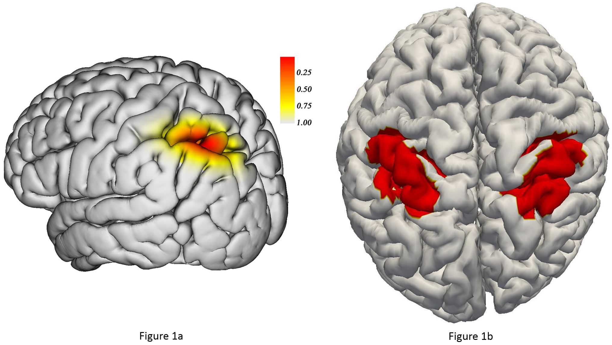
Figure 1: a) Anatomical distribution of the motor inhibition sites shown as a confidence ellipsoid mapped onto the template pial surface. b) The corresponding
individual confidence
areas for both hemispheres projected onto the pial surface for one subject.
