0990
High Resolution T2W imaging using Multi-shot Reduced Field-of-View PROPELLER1Global MR Applications & Workflow, GE Healthcare, Houston, TX, United States, 2Global MR Applications & Workflow, GE Healthcare, New York, NY, United States, 3Global MR Applications & Workflow, GE Healthcare, Menlo Park, CA, United States, 4Global MR Applications & Workflow, GE Healthcare, Madison, WI, United States, 5Global MR Applications & Workflow, GE Healthcare, Boston, MA, United States, 6M. D. Anderson Cancer Center, Houston, TX, United States
Synopsis
Multi-shot fast-spin-echo based PROPELLER (FSE-PROPELLER) combines the robustness to off-resonance and bulk motion and has been increasingly used in body imaging. However, FSE-PROPELLER methods require longer acquisition times compared with conventional FSE methods, especially in high resolution imaging with small FOV, since the oversampling is not confined to a single axis. Outer volume suppression alone can provide a modest reduction in scan time. In this work, we optimized the rotating outer volume suppression method and combined it with variable refocusing flip angles to further reduce scan time for high resolution T2W imaging.
Introduction
High-resolution T2W imaging can depict fine anatomical details and small lesions. However, high-resolution T2W imaging methods often suffer from long scan times and are sensitive to motion, especially in body imaging. While multi-shot FSE-PROPELLER is useful for correcting bulk motion, it requires longer acquisition times compared with conventional FSE methods, since the phase-encoding direction in PROPELLER is changed with each blade, and the oversampling is not confined to a single axis. Although inner-volume imaging (IVI)1 and outer-volume suppression (OVS)2,3 have been incorporated into PROPELLER to reduce the FOV, they cannot dramatically reduce the scan time, because interleaved slice acquisition is not compatible with IVI, and OVS increases the TR, which partially offsets the reduced oversampling factor. Thus, the purpose of this work was to optimize the rotating outer volume suppression method3 and to use variable refocusing flip angles4 to further reduce the scan time for high resolution T2W imaging by reducing SAR and increasing echo train length (ETL).Materials and Methods
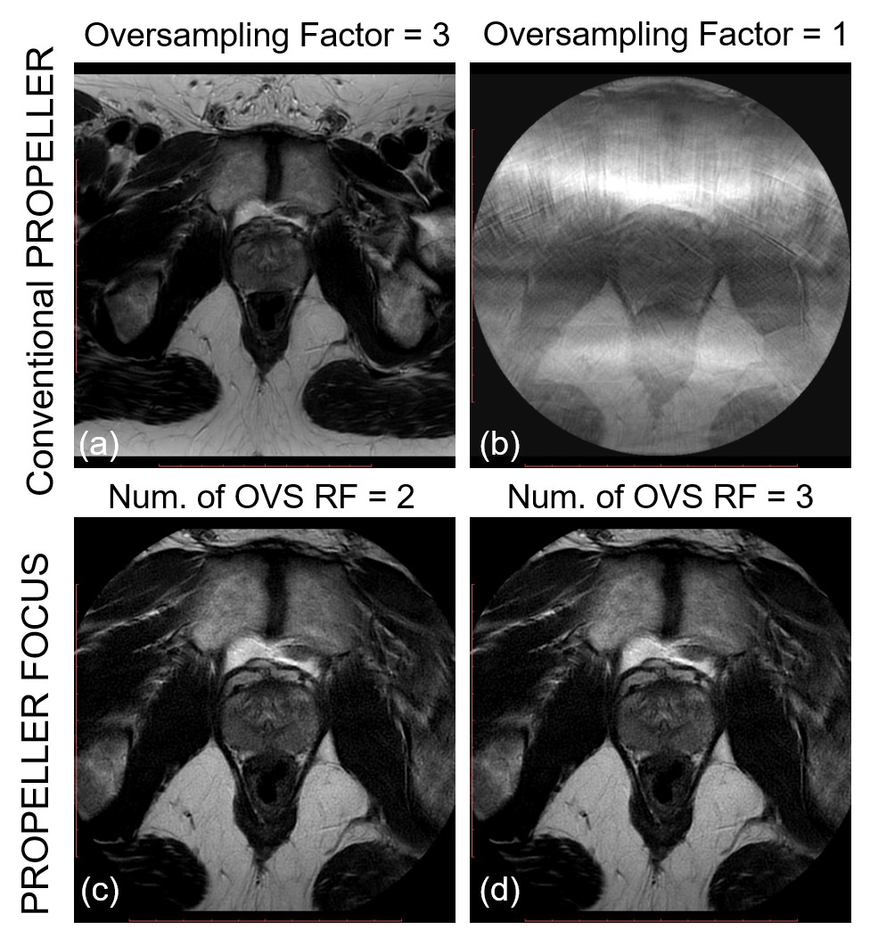
PROPELLER with rotating outer volume suppression (PROPELLER FOCUS)3 and variable refocusing flip (VRF) angles acquisition4 was implemented on a 3.0T MRI scanner (Discovery MR750, GE Healthcare, Waukesha, WI) and is shown in Figure 1a, 1b. The proposed method was optimized and evaluated in phantoms and prostate of 2 healthy volunteers with IRB approval and written informed consent. The number of OVS saturation pulses was varied from 1 to 3 to evaluate saturation efficiency, SAR and scan time. The typical in-vivo imaging parameters included: axial orientation; FOV = 280 × 280 mm2 (Large FOV) / 140 × 140 mm2 (Small FOV); Resolution = 1 × 1 mm2 (Low Resolution) / 0.5 × 0.5 mm2 (High Resolution); slice thickness = 4 mm; number of slices = 22; Oversampling Factor (OSF) = 1.5 (Large FOV) /3 (Small FOV); FA = 120 degrees; ETL = 28; TE = 103 ms and auto TR. Same parameters were used in PROPELLER with VRF, except ETL is 46 for VRF = 130 (first), 60 (minimum), 120 (middle),160 (last) degrees to match the TE. For conventional PROPELLER, the scan times for T2W low-resolution imaging and high-resolution image were 2:31 min and 5:40 min respectively. For PROPELLER FOCUS, the scan times were 3:53 min (3 OVS pulses), 3:30 min (2 OVS pulses) and 2:36 min (2 OVS pulses with VRF).Results and Discussion
The spatial profile of saturation band using the rotating outer volume suppression is shown in Figure 1d. Using conventional PROPELLER, the bright spots in the ACR phantom were well separated in the high-resolution images with reduced FOV (Fig. 2b vs 2a), but the scan time was doubled due to the increased oversampling factor. With VRF angles, the ETL was increased to 46 to match the TE, and images with similar quality were generated with reduced scan times (Fig. 2c). The aliasing artifacts due to reduced FOV (Fig. 2d) were suppressed using the rotating OVS. However, minor aliasing artifacts were observed in images acquired using 1 OVS pulse (Fig. 2e) and not observed using 2 or 3 OVS pulses (Fig. 2f, 2g). Figure 3 also shows efficient outer-volume suppression using 2 or 3 OVS pulses in prostate imaging. The conventional PROPELLER images with reduced FOV (Fig. 4b) demonstrate a shaper profile of the prostate compared to the low-resolution large-FOV images (Fig. 4a), but significantly increased the scan time (2:31 min vs 5:40 min). PROPELLER FOCUS with 2 OVS pulses (Fig. 4c) also achieved high resolution, but with reduced scan time (3:30 min). The scan time of high-resolution imaging was further reduced by using VRF with PROPELLER FOCUS (Fig. 4d, 2:36 min). While images acquired using PROPELLER FOCUS showed reduced SNR, the SNR could be probably improved by combining VRF, low receiver bandwidth and post-processing methods in the future. Figure 5 shows the scan time of the in-vivo protocol with different acquisition parameters. Smaller FOV often requires a larger oversampling factor, where the PROPELLER FOCUS can save more scan time (Fig. 5b). Compared to standard PROPELLER with oversampling factor of 3, PROPELLER FOCUS with VRF can save at least 60% scan time (Fig. 5a, 5c, 5d). However, PROPELLER FOCUS can save more scan time if <10 slices are acquired, because the scan time is more limited by the repetition time than the SAR.Conclusion
PROPELLER with rotating outer volume suppression and variable refocusing flip angles can be used to generate high-resolution T2W images in shorter scan times by effectively suppressing outer volume signals, and mitigating SAR.Acknowledgements
No acknowledgement found.References
[1] Deng J and Larson A. Multishot targeted PROPELLER magnetic resonance imaging: description of the technique and initial applications. Invest Radiol. 2009; 44(8): 454-462.
[2] Kojima S, Morita S, Ueno E, et al. Aliasing artifacts with the BLADE technique: Causes and effective suppression. J Magn Reson Imaging. 2011; 33(2):432-440.
[3] Litwiller DV, Taviani V, Banerjee S, et al. Rotating Outer Volume Suppression for Reduced Field of View PROPELLER Imaging. Joint Annual Meeting ISMRM-ESMRMB. 2685 (2018).
[4] Busse RF, Hari H, Anthony V, et al. Fast spin echo sequences with very long echo trains: design of variable refocusing flip angle schedules and generation of clinical T2 contrast. Magn Reson Med. 2006; 55(5):1030-7
Figures