0986
Validation of prostate tissue composition measurement using Hybrid Multidimensional MRI: Correlation with quantitative histology1Department Of Radiology, University of Chicago, Chicago, IL, United States, 2Faculty of Health Sciences, University of Sydney, Sydney, Australia, 3Human Tissue Resource Center, University of Chicago, Chicago, IL, United States, 4Department Of Pathology, University of Chicago, Chicago, IL, United States
Synopsis
This study validates prostate tissue composition measured non-invasively using Hybrid Multidimensional MRI (HM-MRI) with ground truth reference standard quantitative histology results from whole mount prostatectomy. There was no significant difference in prostate tissue composition measured using HM-MRI and quantitative histology with excellent correlation (0.89) and agreement on Bland-Altman analysis (bias <5%). There is significant difference in epithelium and lumen volume between cancer and normal tissue with high area under the ROC curve (0.87-0.95). This study demonstrates that the tissue composition measured using HM-MRI matches very closely with ground truth quantitative histology measures and can be used for non-invasive prostate cancer diagnosis.
Introduction
Prostate tissue composition of gland components: stroma, epithelium, and lumen change with the presence (1) and Gleason grade of prostate cancer (2). Therefore, the distinct MR properties of these tissue components (3) can be exploited to measure tissue composition changes non-invasively using MRI and be used as biomarker for non-invasive prostate cancer (PCa) detection. A recent feasibility study showed that prostate tissue composition can be measured non-invasively using Hybrid Multidimensional MRI (HM-MRI) and that this approach has the potential to improve PCa diagnosis and determine its aggressiveness (4). However, this previous study lacked validation with ground truth pathology results. Therefore, the present study aimed to validate the tissue composition measured using pre-operative HM-MRI with ground truth reference standard quantitative histology results from whole mount prostatectomy.Methods
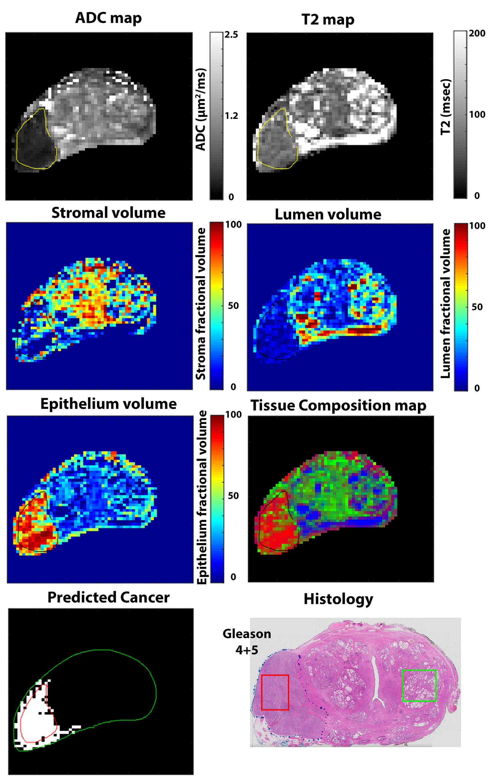
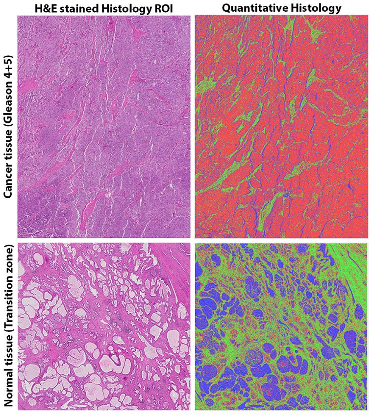
The IRB approved prospective study recruited consenting patients with biopsy confirmed PCa. Patients (n=17, age = 59 years, PSA = 10.9 ng/ml) underwent preoperative 3T prostate MRI between December 2016 and February 2018 prior to undergoing radical prostatectomy. Axial images using HM-MRI were acquired with all combinations of TE = 57, 70, 150, 200 ms and b-values of 0, 150, 750, 1500 s/mm2. Fractional volumes of tissue components- stroma, epithelium and lumen were calculated by fitting the HM-MRI data to a three compartment signal model, with distinct, paired ADC and T2 values associated with each compartment, similar to the previous study (4). The individuals subsequently underwent radical prostatectomy and histology slides of H&E stained whole mount prostate sections were scanned at 20× magnification using Olympus VS120 whole mount digital microscope. Histology and MRI images were co-registered. ROIs for cancer and benign tissue were marked on histology and ADC maps on sites of prostatectomy verified malignancy and normal tissue. Quantitative histology was performed to calculate volumes of tissue components in ROIs from regions corresponding to the MR ROIs (n=55, 23 PCa, 32 normal tissue) using Image Pro Premier on the basis of color, intensity, morphology and background with the “Smart Segment” tool similar to a previous study (2). The tissue composition measures from HM-MRI and quantitative histology were correlated (Pearson correlation) and compared using Bland Altman analysis along with linear regression analysis. The difference in tissue composition between PCa and normal tissue was assessed by t-test and receiver operating characteristic (ROC) analysis was performed to evaluate the performance of fractional volume of tissue components in differentiating cancer from normal prostatic tissue.Results
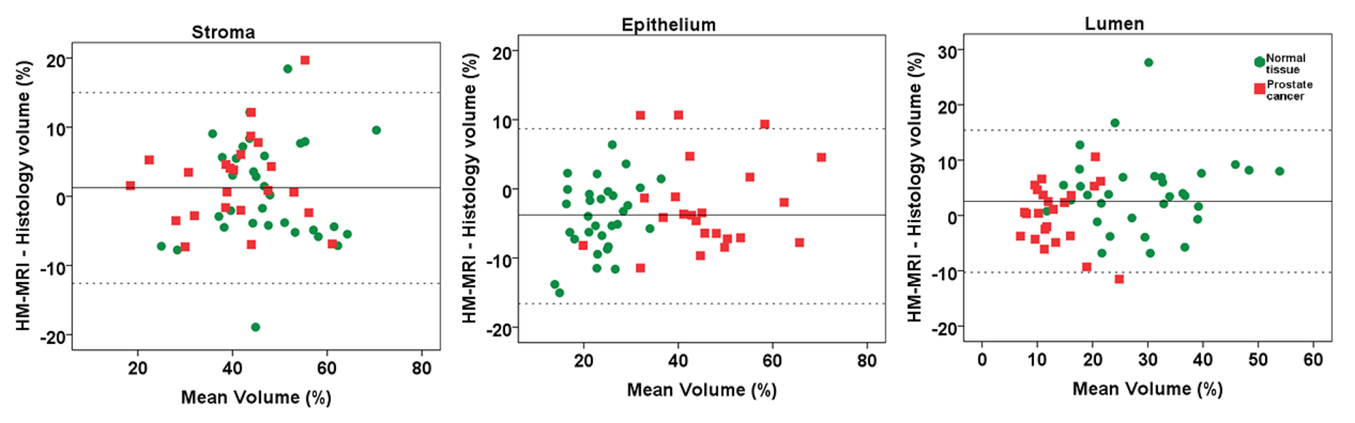
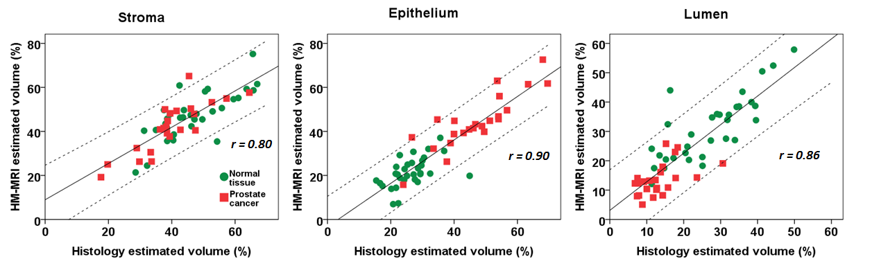
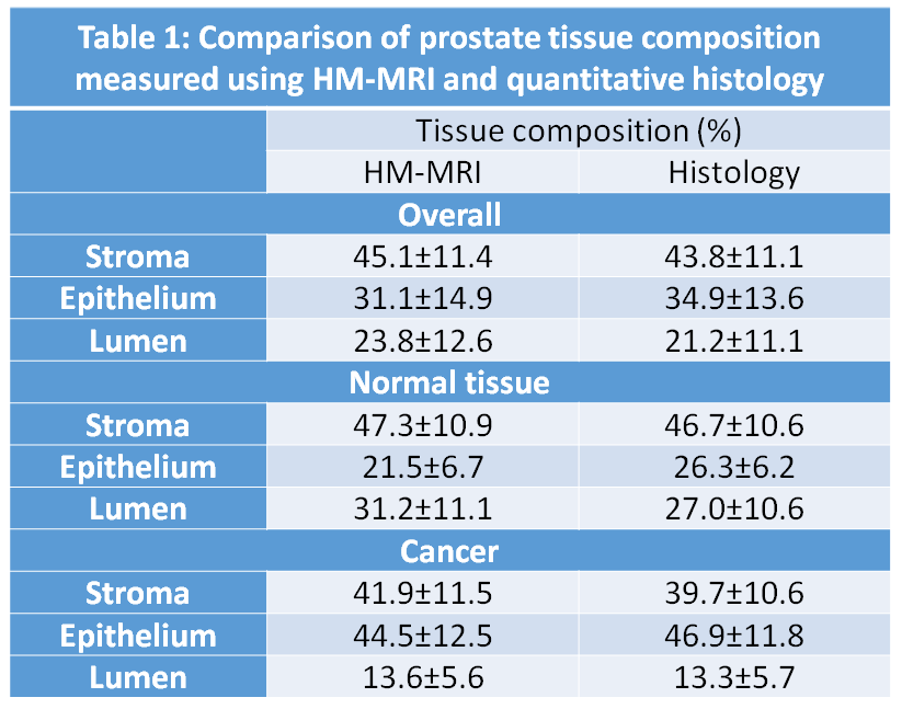
An example of tissue composition measured using HM-MRI is shown in Figure 1, along with corresponding quantitative histology for PCa and normal tissue ROI in Figure 2. There was no significant difference in prostate tissue composition: stroma (45.1±11.4 vs 43.8±11.1, p=0.29), epithelium (31.1±14.9 vs 34.9±13.6, p=0.18) and lumen (23.8±12.6 vs 21.3±11.1, p=0.54) measured using HM-MRI and quantitative histology. Minimal bias which is within the acceptable limit with no proportional bias on linear regression analysis (stroma: 1.2±7.0%, p=0.75; epithelium: -3.8±6.4%, p=0.14; lumen: 2.5±6.6%, p=0.07) was found between the tissue composition estimated using HM-MRI and histology (see Figure 3). There was excellent Pearson correlation (overall=0.89, stroma=0.80, epithelium=0.90, lumen=0.86, p<0.05) between prostate tissue composition measured using these methods (Figure 4).
There is significant difference (p<0.05) in epithelium and lumen volume between PCa and normal tissue but not for stroma volume (see Table 1). ROC analysis showed high area under the curve for differentiating between cancer and benign tissue using HM-MRI (epithelium=0.95, lumen=0.93) and histology (epithelium=0.94, lumen=0.87) results.
Discussion
This study demonstrates that the tissue composition measured using HM-MRI matches very closely with ground truth quantitative histology measures and can be used for non-invasive PCa diagnosis. There was no significant difference in prostate tissue composition: stroma, epithelium and lumen measured using HM-MRI and quantitative histology. There was excellent Pearson correlation between prostate tissue composition measured using these two methods. In addition, the measured tissue composition matched values from previous studies that performed quantitative histology (1,2). ROC analysis showed high area under the curve using HM-MRI and histology results, suggesting PCa can be differentiated from normal tissue due to increased epithelium and reduced luminal volume in PCa compared to normal tissue. Therefore, tissue composition measured using compartmental analysis of HM-MRI data can be used for diagnosing PCa and to guide targeted biopsies and decisions regarding treatment, and has the potential to reduce the number of unnecessary procedures and increase treatment efficiency and efficacy. This will result in significant benefits for patients while dramatically reducing costs.Conclusion
This study demonstrates that the tissue composition measured using HM-MRI matches very closely with ground truth quantitative histology measures and can be used for non-invasive PCa diagnosis.Acknowledgements
No acknowledgement found.References
1. Langer DL, van der Kwast TH, Evans AJ, Plotkin A, Trachtenberg J, Wilson BC, Haider MA. Prostate tissue composition and MR measurements: investigating the relationships between ADC, T2, K(trans), v(e), and corresponding histologic features. Radiology 2010;255(2):485-494.
2. Chatterjee A, Watson G, Myint E, Sved P, McEntee M, Bourne R. Changes in Epithelium, Stroma, and Lumen Space Correlate More Strongly with Gleason Pattern and Are Stronger Predictors of Prostate ADC Changes than Cellularity Metrics. Radiology 2015;277(3):751-762.
3. Bourne RM, Kurniawan N, Cowin G, Stait-Gardner T, Sved P, Watson G, Price WS. Microscopic diffusivity compartmentation in formalin-fixed prostate tissue. Magn Reson Med 2012;68(2):614-620.
4. Chatterjee A, Bourne R, Wang S, Devaraj A, Gallan AJ, Antic T, Karczmar GS, Oto A. Diagnosis of Prostate Cancer with Noninvasive Estimation of Prostate Tissue Composition by Using Hybrid Multidimensional MR Imaging: A Feasibility Study. Radiology 2018;287(3):864-872.
Figures