0964
Structural and functional organization of the brain connectome in patients with different motor neuron diseases: a multicenter studyCamilla Cividini1, Federica Agosta1, Silvia Basaia1, Francesca Trojsi2, Nilo Riva3, Cinzia Femiano2, Cristina Moglia4, Edoardo G. Spinelli1, Maria Rosaria Monsurrò2, Yuri Falzone3, Andrea Falini5, Giancarlo Comi3, Adriano Chiò4, Gioacchino Tedeschi2, and Massimo Filippi1,3
1Neuroimaging Research Unit, Institute of Experimental Neurology, Division of Neuroscience, San Raffaele Scientific Institute, Vita-Salute San Raffaele University, Milan, Italy, 2Department of Medical, Surgical, Neurological, Metabolic and Aging Sciences, University of Campania "Luigi Vanvitelli", Naples, Italy, 3Department of Neurology, Institute of Experimental Neurology, Division of Neuroscience, San Raffaele Scientific Institute, Vita-Salute San Raffaele University, Milan, Italy, 4ALS Center, ‘Rita Levi Montalcini’ Department of Neuroscience, University of Torino, Torino, Italy, 5Department of Neuroradiology and CERMAC, Division of Neuroscience, San Raffaele Scientific Institute, Vita-Salute San Raffaele University, Milan, Italy
Synopsis
We investigated structural and functional brain network topology in amyotrophic (ALS), primary lateral sclerosis (PLS) and progressive muscular atrophy (PMA) patients and in healthy controls (HC), using graph analysis and connectomics. ALS and PLS patients showed widespread microstructural alterations in sensorimotor network, basal ganglia area and prefrontal cortex and posterior brain regions compared to HC, while PMA subjects did not show significant brain damages. All groups had a relatively preserved global and local functional connectome properties compared to each other. Graph analysis and connectomics might represent a powerful approach to understand the pathophysiological process associated with motor neuron diseases.
Introduction
Graph analysis and connectomics might represent a powerful approach to assess brain network degeneration in motor neuron disease. In this study, structural and functional neural organization was investigated in amyotrophic lateral sclerosis (ALS), primary lateral sclerosis (PLS) and progressive muscular atrophy (PMA) patients.Methods
173 ALS, 38 PLS, and 28 PMA patients and 79 healthy controls (HC) were recruited from three different centers. Subjects underwent Diffusion Tensor (DT) and resting-state functional (RS-fMRI) MRI. The brain was parcellated into 220 similarly-sized cortical and subcortical regions. DT data were skull-stripped using Brain Extraction Tool implemented in FSL and were corrected for distortions caused by eddy currents and movements. This correction algorithm combined a rigid-body 3D motion correction with a constrained non-linear warping. Deterministic DT tractography was performed using Diffusion Toolkit/Trackvis, reconstructing the whole-brain tractogram. Fiber tracking followed in each white matter (WM) voxel the principal diffusion direction, stopping if the reconstructed fiber entered a voxel with FA<0.15, if the streamline made a turn with an angle of 45° or when the trajectory exceeded the brain mask. Structural matrices for each subject were reconstructed: for each tract passing between the 220 segmented regions of interest (ROIs), number of streamlines (NOS) was calculated and inserted in an adjacency matrix (M-NOS). A zero was inserted into the M-NOS matrix in case no streamline connected a couple of nodes or when connections had less than 3 fibers. Finally, to avoid spurious structural connections, a zero was set in those connections that were present in less than 40% of the sample. For connections surviving such a threshold, graph analysis metrics and microstructural integrity and organization were measured considering average FA. RS-fMRI was preprocessed by removing the first 4 volumes, minor head movement and non-brain tissue. Functional connectome was reconstructed extracting mean time series from each ROI by averaging the signal from all voxels within each region. RS-fMRI data were masked with subject’s structural map in order to obtain functional connections only where an anatomical connection occurred in each subject. Functional connectivity (FC) matrices were obtained based on correlation analysis. Pearson’s correlation coefficients of each node pair, as measures of functional connectivity, entered into the FC matrix. The coefficients were then converted to z-scores using Fisher’s transformation and negative values were set to ‘NaN’ to mark no-connections. Graph analysis metrics and functional connectivity at global and lobar level were calculated. Network-based Statistics (NBS) analyses were performed both for structural and functional evaluations.Results
Compared to controls, ALS and PLS patients showed lower structural nodal strength, local efficiency and clustering coefficient, longer mean path length, while PMA patients did not show altered global structural alterations. All groups of patients had a relatively preserved global and local functional connectome properties compared to controls and between each other. ALS and PLS patients showed local changes in microstructural properties in the sensorimotor, basal ganglia, frontal and parietal areas relative to controls. PLS patients demonstrated lower local efficiency and clustering coefficient and longer mean path length at a local structural level in the sensorimotor network relative to PMA group. Widespread structural changes were observed in ALS and PLS patients relative to controls (Figure): decreased fractional anisotropy within the sensorimotor network, the basal ganglia area, and in connections to the prefrontal cortex. ALS and PLS patients also showed decreased fractional anisotropy relative to PMA cases within the sensorimotor network and frontal lobe. ALS patients showed increased FC in the sensorimotor network and middle/superior frontal gyri. PLS patients had increased FC in sensorimotor, basal ganglia and temporal networks relative to HC.Discussion
This study showed widespread structural motor/extra-motor network degeneration in ALS and PLS compared to controls, while global and local functional connectome properties were relatively preserved. PMA did not show significant microstructural and functional alterations.Conclusions
Graph analysis and network-based advanced MRI analyses hold the promise to provide an objective in vivo assessment of motor neuron disease-related pathological changes, delivering potential diagnostic and prognostic markers.Acknowledgements
This study was partially supported by a grant from the Italian Ministry of Health (#RF-2011-02351193).References
No reference found.Figures
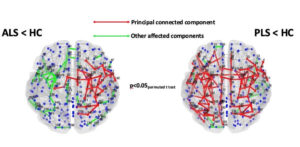
Figure 1. Affected structural
connections in patients with ALS relative to HC and in patients with PLS
compared to HC (network based statistics).