0963
Improving strain diagnosis of prion disease by diffusion MRI and biophysical modelling1Centre for Medical Image Computing, Department of Computer Science, University College London, London, United Kingdom, 2Neuroradiology, Fondazione IRCCS Istituto Neurologico “Carlo Besta”, Milano, Italy, 3Neurology, Fondazione IRCCS Istituto Neurologico “Carlo Besta”, Milano, Italy, 4Philips United Kingdom, Guildford, United Kingdom, 5Queen Square MS Centre, UCL Institute of Neurology, Faculty of Brain Sciences, University College London, London, United Kingdom, 6Department of Brain and Behavioural Sciences, University of Pavia, Pavia, Italy, 7Brain MRI 3T Research Center, IRCCS Mondino Foundation, Pavia, Italy
Synopsis
Sporadic Creutzfeldt–Jakob disease (sCJD) is the most common form of prion disease, characterized by five different strains, presenting intracellular vacuoles with different diameter/distribution. Unfortunately, no reliable non-invasive method for strain identification currently exists. Here we provide the first quantitative maps of MR-measured vacuolar diameter/density in five sCJD patients, using multishell diffusion MRI and biophysical modelling. Results show distribution of small and larger vacuoles in the brain lesions of each patient, presumably corresponding to different sCJD strains, and absence of vacuoles in five age-matched healthy controls. If validated, this method would be extremely valuable for non-invasive diagnosis of sCJD strain
Introduction
The purpose of this study is to improve the strain diagnosis in prion disease by estimating the distribution of vacuoles size from DW-MRI in the brain of Sporadic Creutzfeldt–Jakob disease (sCJD) patients. sCJD is the most common human form of prion disease, a rare, transmissible, rapidly progressive and fatal neurological disease1. sCJD is a heterogeneous disease with five different strains, having different lesion distribution, prognosis and requiring different treatment. Fine spongiosis is characteristic of the most common sCJD forms (MM1 and MV1), having a survival time of few months, while others (MM2 and MV2C) have large confluent vacuoles2,3 and longer survival time. One fifth of the patients may host more than one strain at the time. Unfortunately, no reliable non-invasive method for strain identification is currently available. Diffusion MRI (DW-MRI) identifies sCJD lesions in the cortex, striatum and thalami with a diagnostic accuracy greater than 90%4. The neuropathological basis of DW-MRI hyperintensities is unknown, but a few studies suggested that the main determinant may be spongiform degeneration: the formation of intracellular vacuoles in brain tissues5-8. In vivo, non-invasive strain diagnosis will become very important once a treatment will be found.Methods
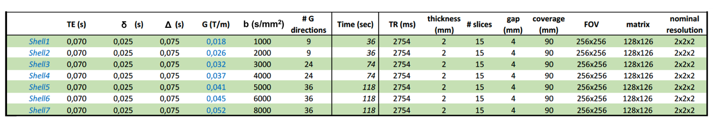
DW-MRI images from five patients with probable sCJD and five age-matched healthy controls were acquired on a 3T MRI scanner (Philips Achieva) using a Pulsed-Gradients-Stimulated-Echo (STEAM) sequence with parameters summarized in Tab.1. All the data were denoised using MP-PCA9, Gibbs ringing10, eddy-current and motion artifacts corrected using FSL11. The signal in each b shell was normalized by the corresponding b=0 and averaged across directions to compute the direction-averaged normalized signal S as a function of b. A tissue-inspired three compartment model, developed starting from previous work8:
S(b)/S(b=0) = fsticks Ssticks(b,Da) + fsphere Ssphere(b,D0=3 μm2/ms,dsphere) + (1- fsticks- fsphere)S(b,Diso)
was fitted to measured S(b) to estimate fsticks (measure of neurites MR signal fraction), axial diffusivity Da, fsphere (measure of vacuoles MR signal fraction), dsphere (MR measure of vacuoles diameter) and extra-cellular isotropic diffusivity Diso. Note that the data were acquired at long diffusion time (D-δ/3=67 ms) in order to minimise the possible confounding signal from cell bodies12,13, and that the MR signal fractions are T1-T2 weighted estimates. Bilateral regions of interest (ROIs) were manually drawn in eleven regions: precuneus, parietal, frontal, temporal, occipital and anterior cingulate cortex, insula, hippocampus, caudate, putamen, dorso-medial thalamus. Thresholds on fractional anisotropy (FA < 0.3) and mean diffusivity (MD < 2x10⁻³ mm2/s) were used to exclude partial volume from white matter and CSF. The distribution of vacuole diameters, weighted according to the fraction of the spherical compartment, was evaluated in the hyperintense part of each ROI.
Results
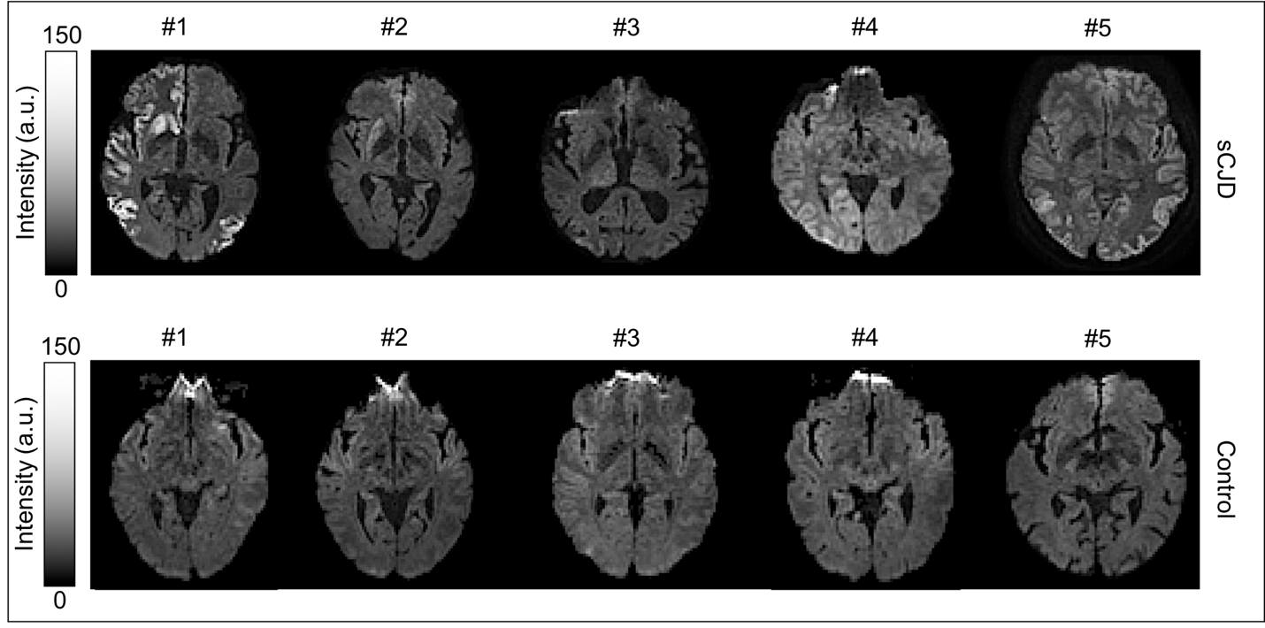
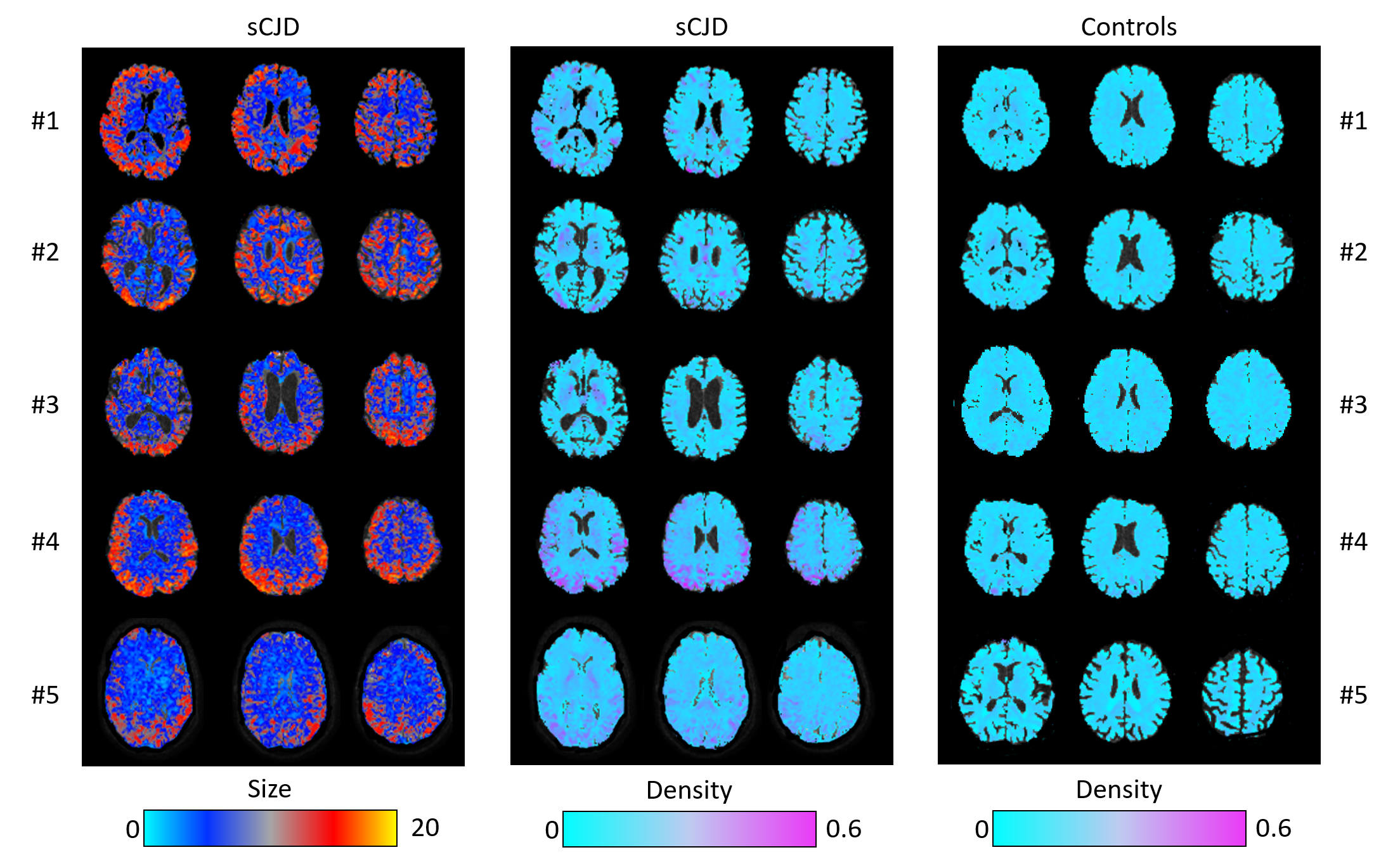
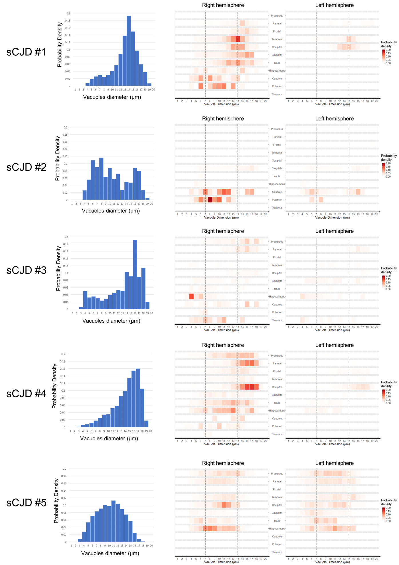
DW-MRI showed the typical hyperintensities in all five sCJD patients, with variable involvement of the cortical areas and striatum (Fig.1). The maps of the volumetric MR signal fraction of the spherical restricted compartment show values up to 0.35 in sCJD, with focal areas of high density largely corresponding to hyperintensities on DW-MRI, while they are uniform and generally below 0.15 in controls (Fig.2, right). The MR-measured diameter size of the spheres (vacuoles) in the restricted compartment was in the range between 4-18 µm, in agreement with histological values reported in literature14 (Fig.2, left). Vacuolar size distribution varied among patients: larger diameters were found in patient #1 and #4, with a peak around 13 and 15 µm respectively; small diameters were found in the striatum of patient #2; a bimodal distribution was estimated in patient #3, with prevalence of larger vacuoles; a broader distribution of diameters was found in #5 (Fig.3, left). The contribution of each anatomic region to these distributions is shown in the right columns of Fig.3. So far, autopsy brains were collected in patients #2 and #3 and eventually more brains will be collected as patients will pass away. Point to point correlation with histopathology results is in progress and will be presented at the ISMRM meeting.Discussion and Conclusion
We showed the feasibility of generating quantitative maps of MR-measured vacuolar size and density in the brain of sCJD patients. The range of vacuolar sizes measured is compatible with histopathology results14. We found different distributions in patients with presumably different sCJD strains. In this small group of patients, larger diameters were estimated in the cortex than in the striatum. If these results will be validated by neuropathology and confirmed in a larger group of patients, this method will become extremely valuable for the non-invasive diagnosis of sCJD strain, with an impact on more accurate prognosis and personalised therapy.Acknowledgements
This work was supported by the European Union’s Horizon 2020 research and innovation programme under grant agreement nr. 666992 and by EPSRC (EP/G007748, EP/I027084/01, EP/L022680/1, EP/M020533/1, N018702, EP/M507970/1)References
1 Gambetti P, Kong Q, Zou W, et al. Sporadic and familial CJD: classification and characterisation. Br. Med. Bull. 2003, 66, 213-239
2 Parchi P, De Boni L, Saverioni D, et al. Consensus classification of human prion disease histotypes allows reliable identification of molecular subtypes: an inter-rater study among surveillance centres in Europe and USA. Acta Neuropathol. 2012, 124 (4), 517-529
3 Iwasaki Y. Creutzfeldt‐Jakob disease. Neuropathology 2017, 37, 174–188.
4 Puoti G, Bizzi A, Forloni G, et al. Sporadic human prion diseases: molecular insights and diagnosis. The Lancet Neurology 2012, 11(7), 618-628.
5 Geschwind M D, Potter C A, Sattavat M, et al. Correlating DWI MRI with pathological and other features of Jakob-Creutzfeldt disease. Alzheimer Dis. Assoc. Disord. 2009, 23 (1), 82-87.
6 Manners D N, Parchi P, Tonon C, et al. Pathologic correlates of diffusion MRI changes in Creutzfeldt-Jakob disease. Neurology 2009, 72 (16), 1425-1431
7 Lodi R, Parchi P, Tonon C, et al. Magnetic resonance diagnostic markers in clinically sporadic prion disease: a combined brain magnetic resonance imaging and spectroscopy study. Brain 2009, 132 (10), 2669-2679
8 Figini M, Alexander D C, Redaelli V, et al. Mathematical models for the diffusion magnetic resonance signal abnormality in patients with prion diseases. Neuroimage: Clinical 2015, 7, 142-154.
9 Veraart, J., et al. Denoising of diffusion MRI using random matrix theory. Neuroimage 2016, 142, 394-406.
10 Kellner E, Dhital B, Kiselev V G, et al. Gibbs‐ringing artifact removal based on local subvoxel‐shifts. Magn. Reson. Med. 2016, 76(5), 1574-1581.
11 https://fsl.fmrib.ox.ac.uk/fsl
12 Palombo, M, Shemesh N, Ianus A, et al., A compartment based model for non-invasive cell body imaging by diffusion MRI. Proc. Int. Soc. Magn. Reson. Med. 2018, #0892.
13 Palombo M, Shemesh N, Ianus A, et al., Abundance of cell bodies can explain the stick model’s failure to describe high b-value diffusion signal in grey matter. Proc. Int. Soc. Magn. Reson. Med. 2018, #1096.
14 Kovacs GG, Budka H. Prion diseases: from protein to cell pathology. Am J Pathol. 2008 Mar;172(3):555-65
Figures