0960
Advanced brain aging in patients with right mesial temporal lobe epilepsy: A machine learning approach based on white matter tract integrity1Institute of Medical Device and Imaging, College of Medicine, National Taiwan University, Taipei, Taiwan, 2Institute of Biomedical Engineering, National Taiwan University, Taipei, Taiwan, 3Department of Neurology, National Taiwan University Hospital and College of Medicine, Taipei, Taiwan, 4Graduate Institute of Brain and Mind Sciences, College of Medicine, National Taiwan University, Taipei, Taiwan, 5AcroViz Technology Inc., Taipei, Taiwan, 6Molecular Imaging Center, National Taiwan University, Taipei, Taiwan
Synopsis
It is unclear whether left and/or right side lesions of mesial temporal lobe epilepsy (MTLE) exhibit different degrees of brain aging. Therefore, we developed machine-learning-based brain age models to quantify the brain aging of patients with unilateral MTLE and of healthy controls. The significantly overestimated brain age was found in the right but not left MTLE patients. Also, the degree of overestimated brain age was correlated with the clinical factors. Moreover, the right uncinate fasciculus was the most contributing feature to the overestimated brain age. This study uncovered the underpinning of advanced brain aging in right MTLE patients.
Introduction
Mesial temporal lobe epilepsy (MTLE) causes brain structural alterations and potentially induces aberrant brain aging[1,2]. The right and left types of MTLE manifest distinct patterns of structural impairments[3], but it is unclear whether they are associated with different degrees of brain aging. To address this issue, we developed brain age predictive models based on white matter integrity using machine learning approach to investigate brain aging in patients with unilateral MTLE. The models provided the predicted age difference (PAD) that reflected the degree of brain aging. The higher the PAD, the more degree of the overestimated brain age is. Also, we investigated the association between PAD scores and the clinical factors, i.e. duration of illness and age of onset, to validate the clinical relevance. Furthermore, we quantified the tract-specific alteration into the standardized score and used it to explore which altered tracts would highly contribute to the overestimated brain age.Methods
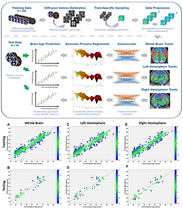
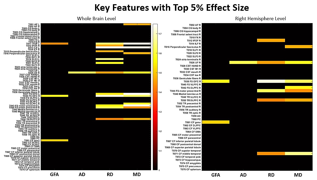
To develop predictive models of brain age, cerebral T1-weighted images and diffusion spectrum imaging (DSI) datasets of 300 and 40 healthy individuals whose age ranged across lifespan were used as the training and testing sets, respectively (Fig.1, upper). Also, we enrolled the left-side MTLE (L-MTLE, n=18) and right-side MTLE (R-MTLE, n=17) patients, and the matched healthy controls (n=37). All images were acquired on a 3T MRI system (TIM Trio, Siemen). To obtain white matter features, the regularized version of diffusion MAP-MRI framework was used to reconstruct DSI datasets into 7 diffusion indices such as generalized fractional anisotropy, mean diffusivity, etc[4]. Whole brain tract-specific analysis was conducted to sample the features according to the predefined 76 tracts from each diffusion index[5]. These tract-specific features from the training data were used to create whole-brain-based (WB), left-hemisphere-based (LH), and right-hemisphere-based (RH) brain age models by Gaussian process regression (Fig.1, upper). Pearson’s correlation and mean absolute error (MAE) were used to evaluate the performance in training and testing sets. PAD scores from WB, LH and RH brain age model were calculated by subtracting chronological age from predicted age and used to test group differences among the L-MTLE, R-MTLE and the control groups using multivariate analysis of covariance (MANCOVA), controlling age, sex and handedness. Within the MTLE groups, PAD scores were also assessed for association with age of disease onset and duration of illness by correlation analysis. To explore which altered tracts highly contributed to the overestimated brain age, we quantified the tract alterations by using normative model to transform diffusion indices into the z-scores which represented the tract deviation against normal population. Then, the tracts with the top 5% effect size were compressed using principal component analysis (PCA). The first component was used to regress the overestimated PAD score. The tract occupied the highest weight in the first component represented the most contributed feature.Results
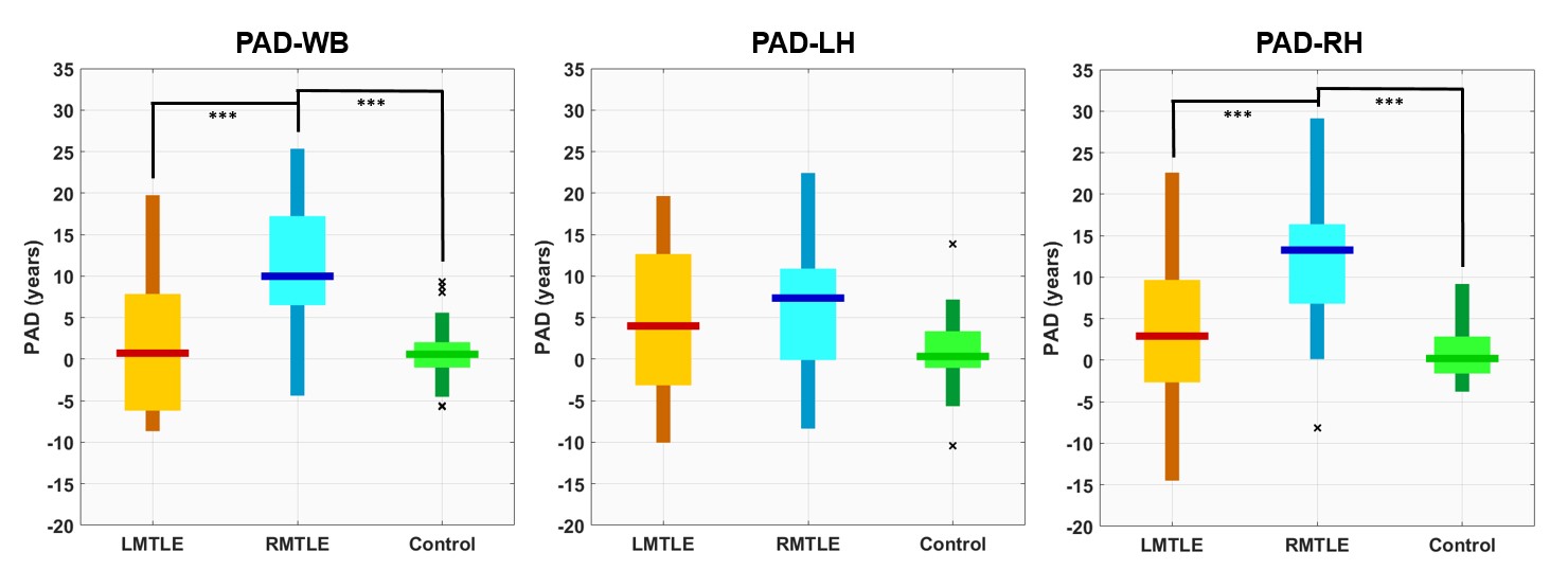
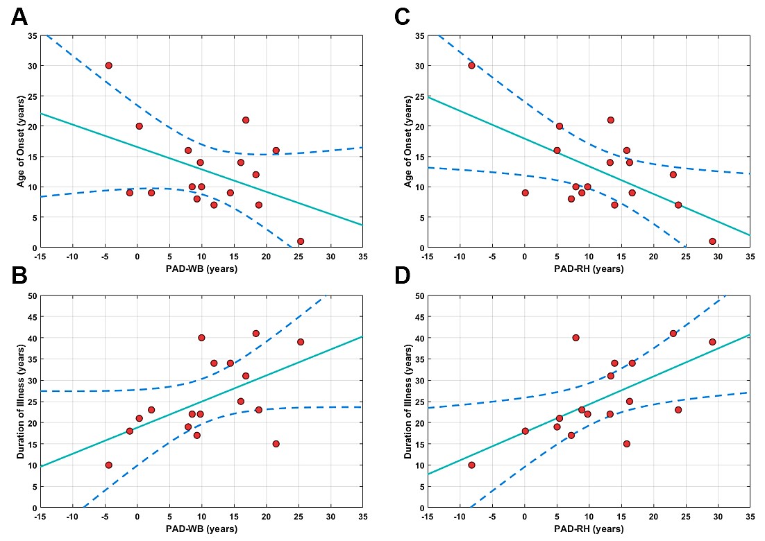
The models predicted each individual’s age for both the training and testing sets with satisfactory performance (training: avg. correlation=0.951, avg. MAE=4.92years; testing: avg. correlation=0.954, avg. MAE=5.12years) (Fig.1, lower). The results of MANCOVA showed that there was a significant difference (F(6,130)=6.653, p<0.001) in PAD scores among R-MTLE, L-MTLE and control groups. The post hoc analysis showed the R-MTLE had significantly increased PAD scores at the WB and RH levels as compared with L-MTLE and control groups (Fig.2). In the R-MTLE group, there was a negative correlation between age of onset and PAD scores for both PAD-WB (rho:−0.560, corrected-p (p*)<0.05) and PAD-RH (rho:−0.722, p*<0.01). By contrast, duration of illness and PAD scores were positively correlated for both PAD-WB (rho:0.535, p*<0.05) and PAD-RH (rho:0.684, p*<0.05)(Fig.3). Additionally, we selected 27 and 16 features at the WB and RH levels to represent the most altered tracts from the R-MTLE (Fig.4). After PCA, the first components from the WB and RH levels significantly explained the variance in PAD-WB (F(1,15)=13.7, p<0.01; adjusted R-squared=0.442) and in PAD-RH (F(1,15)=14.5, p<0.01; adjusted R-squared=0.447), respectively. The tract with the most contribution to these first components was the right uncinate fasciculus at both WB (22.7%) and RH (33.2%) levels in the R-MTLE.Discussion and Conclusion
Patients with R-MTLE exhibited significantly older brain age in the WB and RH than the L-MTLE patients and the controls, suggesting a more aggravated white matter alteration in R-MTLE. The high contribution of disease-affected white matter tracts and strong correlation between PAD scores and clinical factors (i.e., age of onset and disease duration) revealed the structural and clinical relevance of advanced brain aging in R-MTLE. This study uncovered the underpinning of advanced brain aging in R-MTLE patients and potentially provides a neuroimaging reference for clinical prognosis.Acknowledgements
This research was partially supported by Ministry of Science and Technology (MOST) Taiwan (grant: 107-2314-B-002-006).References
[1] Focke, N.K., Yogarajah, M., Bonelli, S.B., Bartlett, P.A., Symms, M.R., Duncan, J.S. (2008) Voxel-based diffusion tensor imaging in patients with mesial temporal lobe epilepsy and hippocampal sclerosis. NeuroImage, 40:728-737.
[2] Dabbs, K., Becker, T., Jones, J., Rutecki, P., Seidenberg, M., Hermann, B. (2012) Brain structure and aging in chronic temporal lobe epilepsy. Epilepsia, 53:1033-43.
[3] Besson, P., Dinkelacker, V., Valabregue, R., Thivard, L., Leclerc, X., Baulac, M., Sammler, D., Colliot, O., Lehericy, S., Samson, S., Dupont, S. (2014) Structural connectivity differences in left and right temporal lobe epilepsy. Neuroimage, 100:135-44.
[4] Hsu, Y.C., Tseng, W.Y. (2018) An efficient regularization method for diffusion MAP-MRI estimation. 2018 ISMRM-ESMRMB Joint Annual Meeting.
[5] Chen, Y.J., Lo, Y.C., Hsu, Y.C., Fan, C.C., Hwang, T.J., Liu, C.M., Chien, Y.L., Hsieh, M.H., Liu, C.C., Hwu, H.G., Tseng, W.Y. (2015) Automatic whole brain tract-based analysis using predefined tracts in a diffusion spectrum imaging template and an accurate registration strategy. Hum Brain Mapp, 36:3441-58.
Figures