0958
The alteration of white matter tract of patients with Parkinson's disease combined with the changes in tract-connected gray matter : a longitudinal report1Department of Medical Imaging and Radiological Sciences, Chang Gung University, Taoyuan City, Taiwan, 2Neurology, Chang Gung Memorial Hospital, Taoyuan City, Taiwan, 3Radiology, Keelung Chang Gung Memorial Hospital, Keelung City, Taiwan
Synopsis
Parkinson's disease (PD) is a progressive nervous system disorder as a result from the loss of cell in basal ganglia. Fixel-Based Analysis can qualify the fibre density and fibre-bundle cross-section in the nerve fibre. Further, gray matter degeneration was determined by using ROI-based Analysis to investigate Mean Diffusivity (MD) changes. Therefore, the current study proposed to investigate the structural connectivity of atrophy cortical regions in the PD patients with axonal injury over a period of 2 years.
INTRODUCTION
Parkinson's disease (PD) is a long-term degenerative disorder of the nervous system. Since the nature of slow disease progression,the microstructural changes in the brain of patients during the disease course is of great interest. Fixel-Based Analysis (FBA) allows to quantify the white matter characters such as the combination of fiber density and fiber-bundle cross-section.The current study aims to investigate the comprehensive brain changes in PD progression. A ROI-based analysis was performed to reveal the changes in tract-connected gray matter.METHODS
Image acquisition Multi-shell diffusion weighted images with the whole brain coverage were acquired from 87 PD patients (Male/Female=49/38, aged 60.7±7.2 years old) using 3T scanner (Trio, Magnetom, Siemens, Erlangen Germany) in the baseline and two years later (the third year). T1-weighted images were acquired from following image parameters: repetition time (TR) /echo time (TE) /inversion time/flip angle = 2000 ms/2.63 ms/900 ms/9°, voxel size=1×1×1 mm3, number of slices = 160. Diffusion weighted images (DWI) were acquired using a spin-echo echo planar imaging (EPI) sequence with the following parameters: the diffusion weighted gradient applied along 30 non-collinear directions.
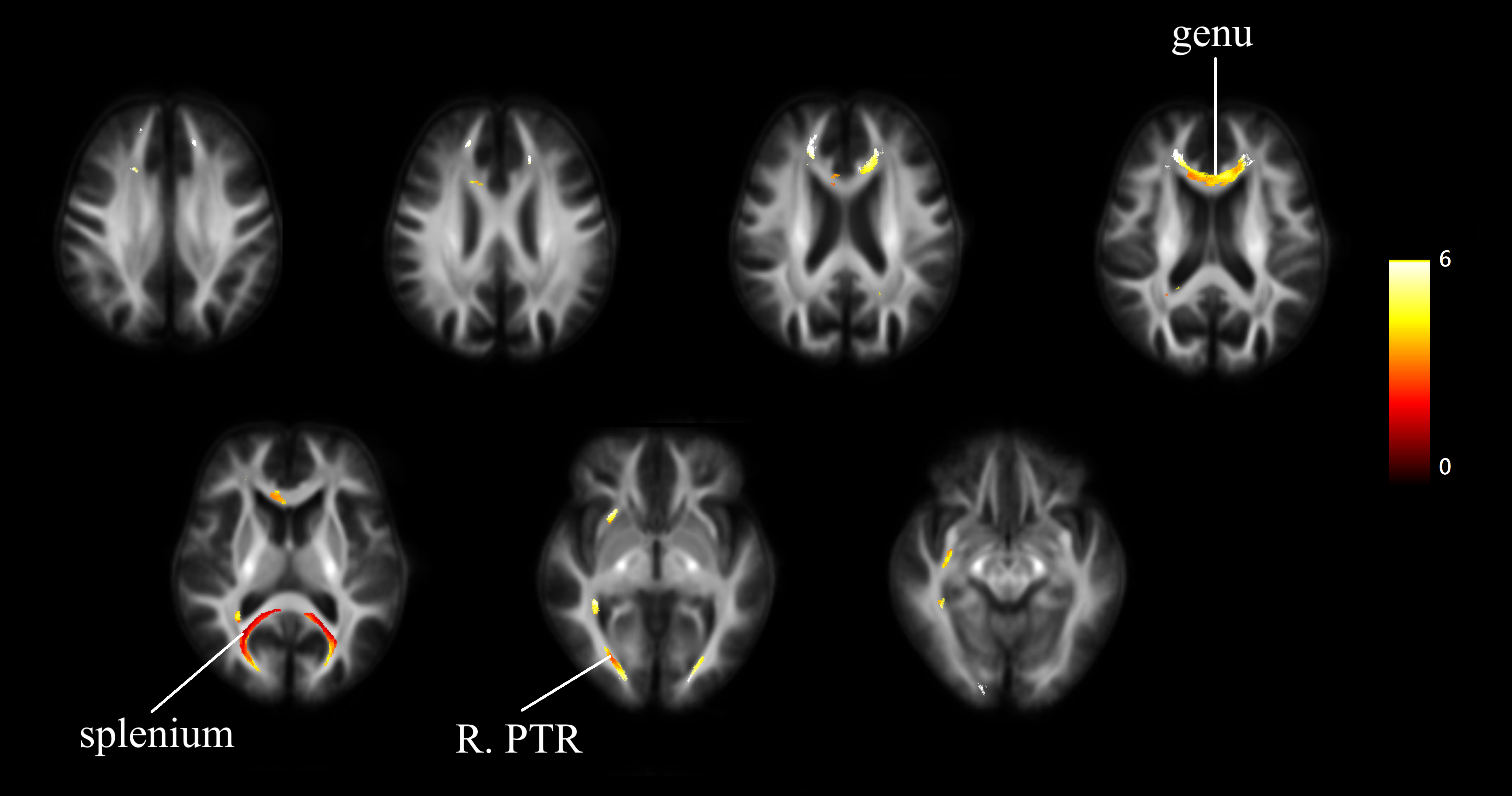
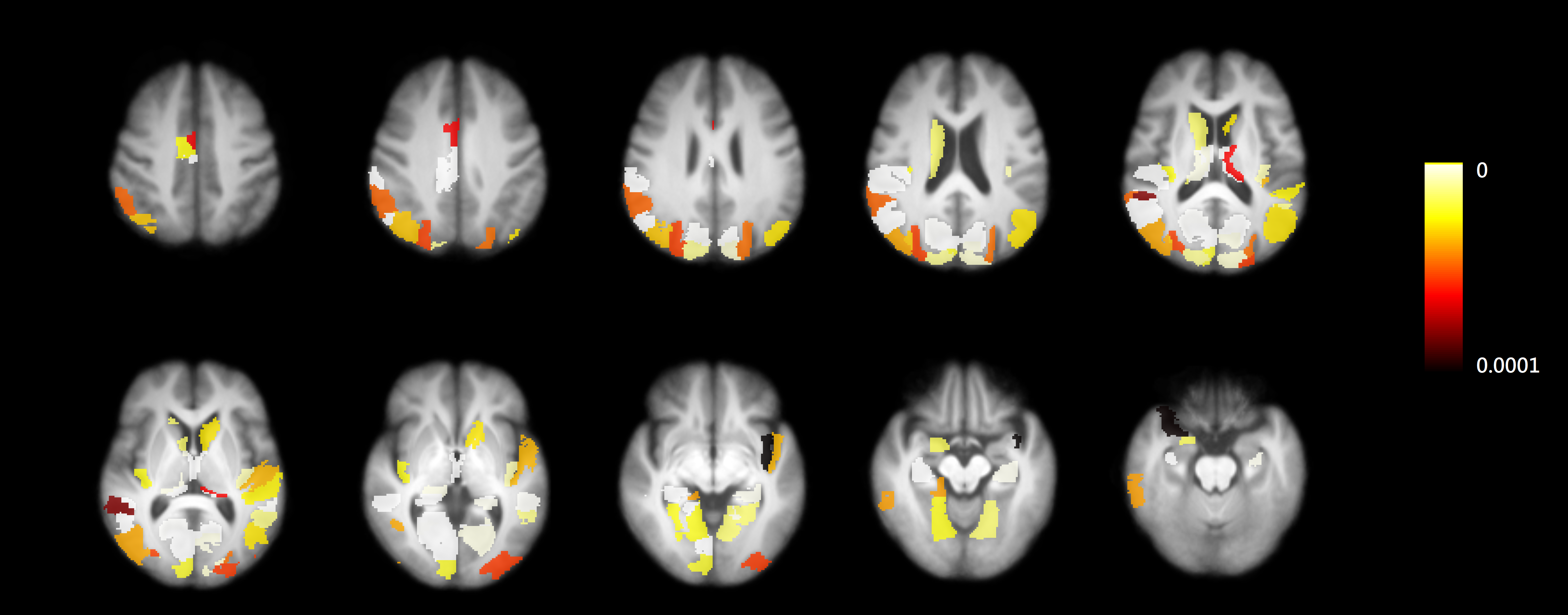
Diffusion Tensor Imaging (DTI) model was applied to reconstruct DWI for all subjects, DTI derived index (Mean Diffusivity, MD) was parcellated into 246 region of interest according to the Human Brainnetome Atlas. The mean, median, 10th and 90th percentiles from each ROI were extracted follow the procedure by refering the paper in european radiology by Lu et al 1. The significant level of p-values < 0.000203 (after correction for multiple comparisons) was chosen. FBA was performed using MRtrix3 following the recommended pipeline 2, including MP-PCA denoising, Gibbs ringing removal, motion and distortion correction, and bias field correction. The distribution of fiber orientations within each imaging voxel was estimated using multi-tissue constrained spherical deconvolution. A study-specific template was then created by spatial normalization over all subjects using FOD-based registration. Within each voxel, fixel-specific measures of fibre density & cross-section (FDC) was calculated. Significant white matter changes over the follow-up period was determined using fixel-wise paired t-test, with significance (FWE p<0.05). The significant regions were selected as seed of further ROI-based analysis, which including the genu, splenium of the corpus callosum. Regions with MD changes in gray matter and also contact to the fiber tracts with significant FDC reduction were identified.
RESULTS
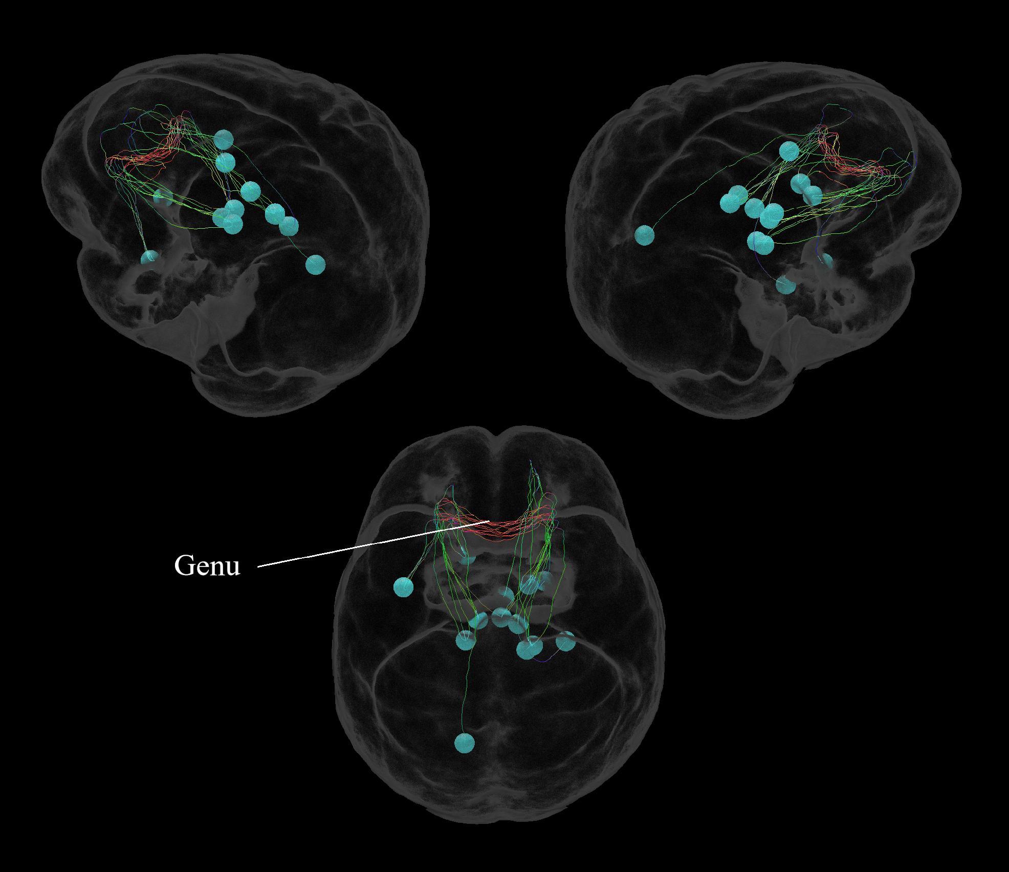
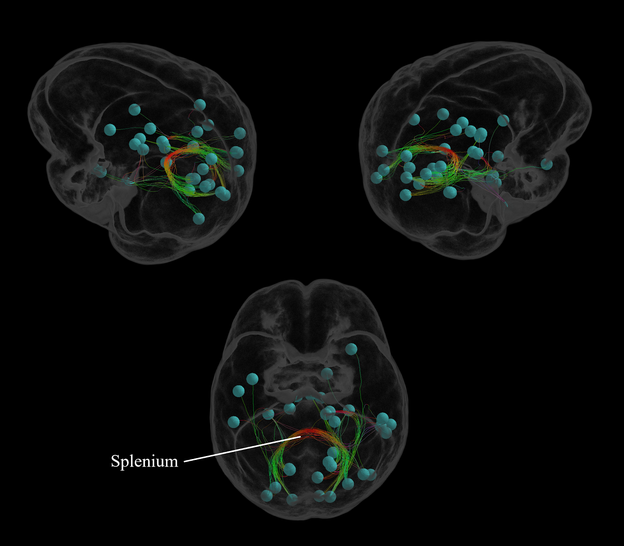
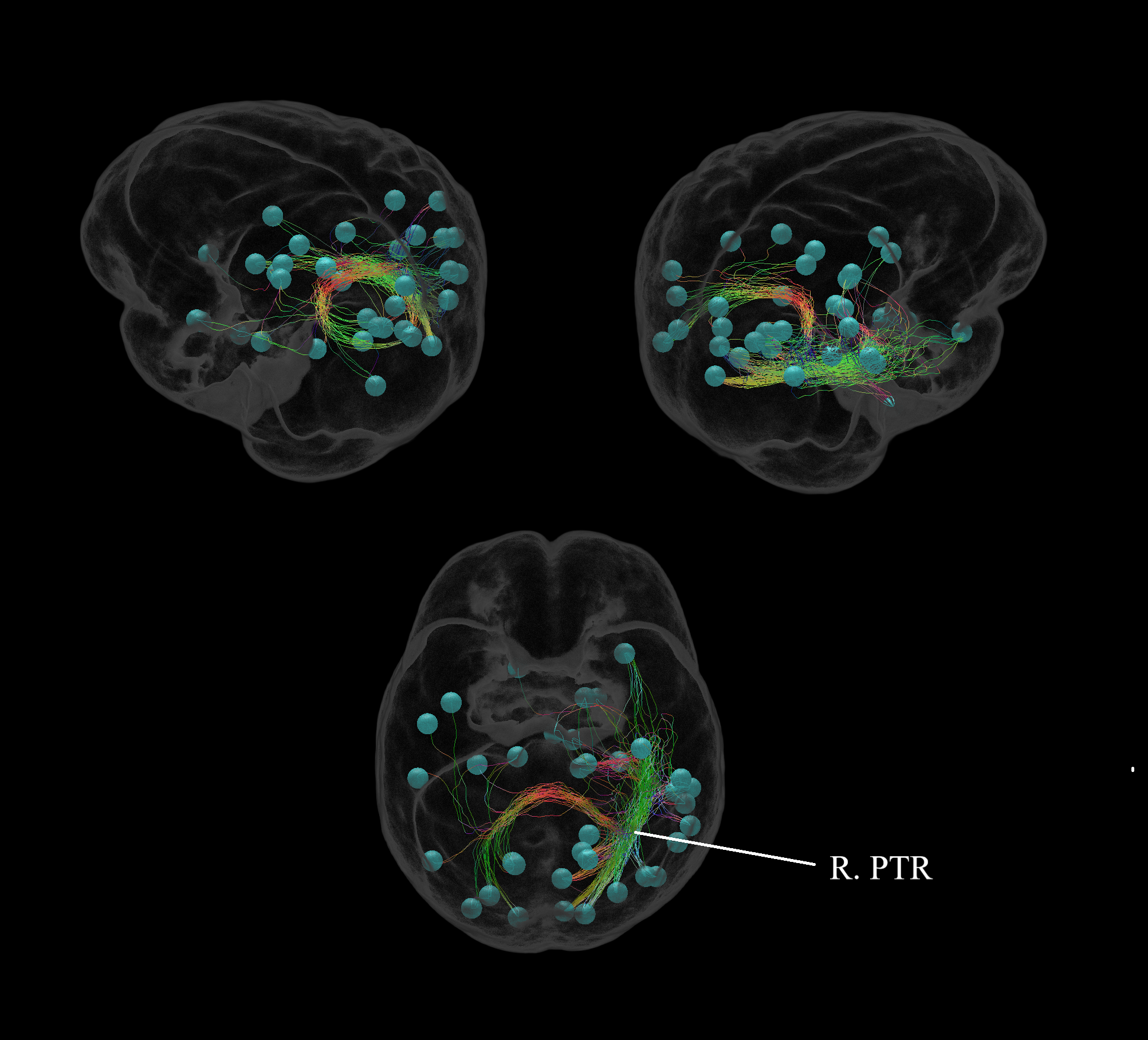
Changes in FDC during the follow-up period was overlaid on the study-specific template, noticeably in genu and splenium of corpus callosum, and right posterior thalamic radiation (Figure 1). 45 of 246 regions were observed with significantly increased MD mainly in Temporal lobe, Parietal lobe, Occipital lobe and Subcortical nuclei (Figure 2). The damaged white matter tracts passed the genu of corpus callosum mainly affected the connectivity in subcortical nuclei regions (Figure 3). The network of occipital lobe and subcortical nuclei regions was affected by the damaged white matter tracts of splenium of corpus callosum (Figure 4). The damaged white matter tracts passed posterior thalamic radiation on the right affected the connectivity of temporal lobe and parietal lobe (Figure 5).DISCUSSION & CONCLUSIONS
Previous study noticed that the regions of gray matter atrophy and the more extensive areas of increased MD was overlapped 3. As a result, it was found that during over the course of PD, damage of the fiber bundle and atrophy of the cortical regions can be observed simultaneously. The axonal damage of white matter may cause the degeneration of adjacent gray matter 4. Damaged tract-connected cortical regions can be observed to have neurons degeneration from patients with PD over a two year period. These changes observed using a longitudinal analysis match observations from other studies that used cross-sectional analyses.Acknowledgements
Processing was performed using the MRtrix3 package (www.mrtrix.org).References
1. Lu CS, Ng SH, Weng YH, et al. “Alterations of diffusion tensor MRI parameters in the brains of patients with Parkinson's disease compared with normal brains: possible diagnostic use” European radiology. 2016;26:3978-88
2. Raffelt, David A et al. “Investigating white matter fibre density and morphology using fixel-based analysis” NeuroImage vol. 144,Pt A (2017): 58-73.
3. Lin, Sung-Han et al. “Increased Water Diffusion in the Parcellated Cortical Regions from the Patients with Amnestic Mild Cognitive Impairment and Alzheimer's Disease” Frontiers in aging neuroscience vol. 8 325. 11 Jan. 2017, doi:10.3389/fnagi.2016.00325
4. Rektor, Ivan et al. “White matter alterations in Parkinson's disease with normal cognition precede grey matter atrophy” PloS one vol. 13,1 e0187939. 5 Jan. 2018, doi:10.1371/journal.pone.0187939
Figures