0955
Density-Weighted Concentric Ring Trajectory using simultaneous multi-slice (SMS) acceleration: 3D Metabolite-cycled Magnetic Resonance Spectroscopy Imaging at 3 T1School of Health Sciences, Purdue University, West Lafayette, IN, United States, 2Weldon School of Biomedical Engineering, Purdue University, West Lafayette, IN, United States, 3Wellcome Centre for Integrative Neuroimaging, University of Oxford, Oxford, United Kingdom, 4Department of Radiological Sciences, University of California, Los Angeles, Los Angeles, CA, United States, 5Department of Radiology and Imaging Sciences, Indiana University School of Medicine, Indianapolis, IN, United States
Synopsis
In this study, we proposed a novel simultaneous multi-slice (SMS) density weighted (DW) concentric ring trajectory (CRT) metabolite-cycling Magnetic Resonance Spectroscopy Imaging (MRSI) sequence to alleviate some conventional MRSI drawbacks, e.g. long acquisition time, eddy current artifacts, and side lobe artifacts. The sequence was tested on 5 healthy subjects, showing the feasibility of acquiring three slices of high-quality water-only and metabolite spectra simultaneously with a resolution of 5mm X 5mm X 10mm within 20 minutes.
Introduction
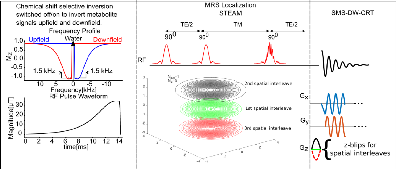
Magnetic Resonance Spectroscopy Imaging (MRSI) allows neurochemical profiles to be acquired from multiple voxels simultaneously over substantial regions of the brain. However, this technique has been hampered by several factors, including relatively long acquisition time, side lobe artefacts, eddy-current-induced artefacts, B0 drifts due to subject motion or thermal fluctuation, and lipid contamination. In this work, we demonstrated a novel method that alleviates these issues by acquiring simultaneous multi-slice (SMS) MRSI using metabolite-cycling density-weighted (DW) concentric ring trajectory(CRT)1.Methods
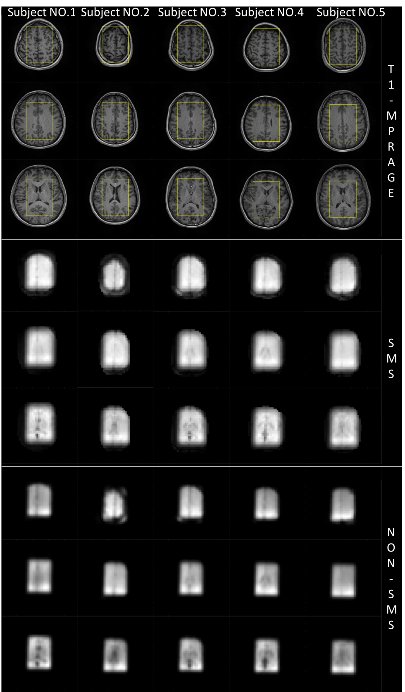
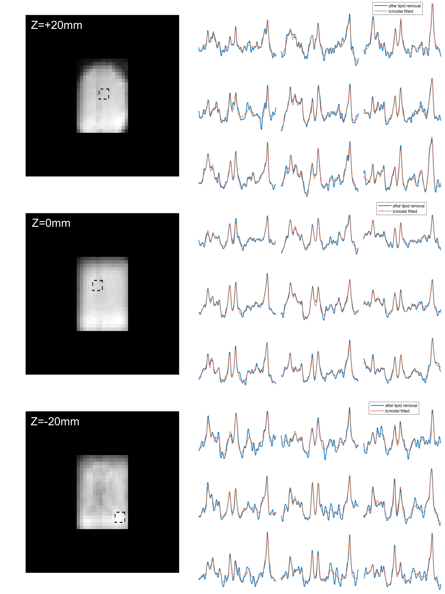
Phantom and in-vivo scans were acquired using a Siemens Prisma 3-Tesla (Siemens, Germany) scanner and a 20-channel head array receive coil. Five healthy volunteers (2 males/3 females, aged 22.6 1.67 year) participated. The metabolite-cycling MRSI acquisition was achieved by the inversion of the upfield and downfield spectral ranges before the STEAM localization with a gap of 9.6 ms so that water-only and metabolite spectra can be acquired simultaneously2,3. Time-shifted multi-slice excitation RF pulse4 was simply used in place of last single-slice (z-direction) pulse of STEAM localization (TR = 1 s, TE = 16 ms) to excite three 110 mm x 80 mm x 10 mm slices (FOV= 240 mm x 240 mm) with an inter-slice distance of 20 mm. For DW-CRT, three spatial interleaves (96 unique rings) resulted in an acquisition duration of 192 s with a resolution of 5 x 5 x 10 mm3. 160 k-space sampling points were evenly distributed on each ring so that azimuthal Nyquist sampling criteria is met. Hanning‐window density weighted acquisition was used to reduce the side lobe artefact, which determines the 96 different radii of three sets of x-y rings. A blipped z-gradient scheme for each spatial interleave (0, 1, -1) was created so that an adequate FOV and resolution in the z-direction were obtained5. The number of averages was 6, corresponding to a total acquisition duration of 19.2 minutes. Corresponding single-slice data were acquired with the same parameters for the inversion of the upfield (96 s for each slice) only. SENSE method was used for reconstructions of SMS-DW-CRT data6. HLSVD is used for lipid and residual water removal7.Results and Discussion
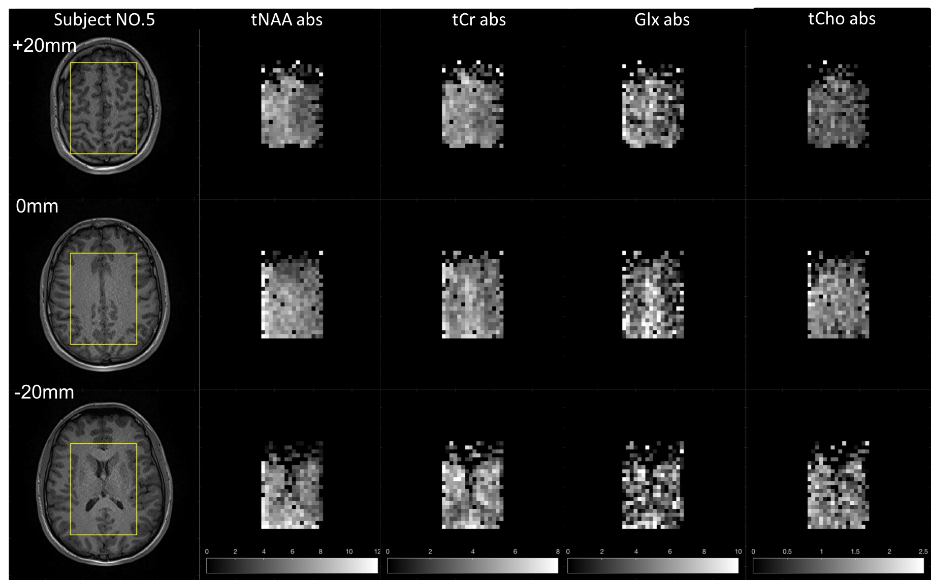
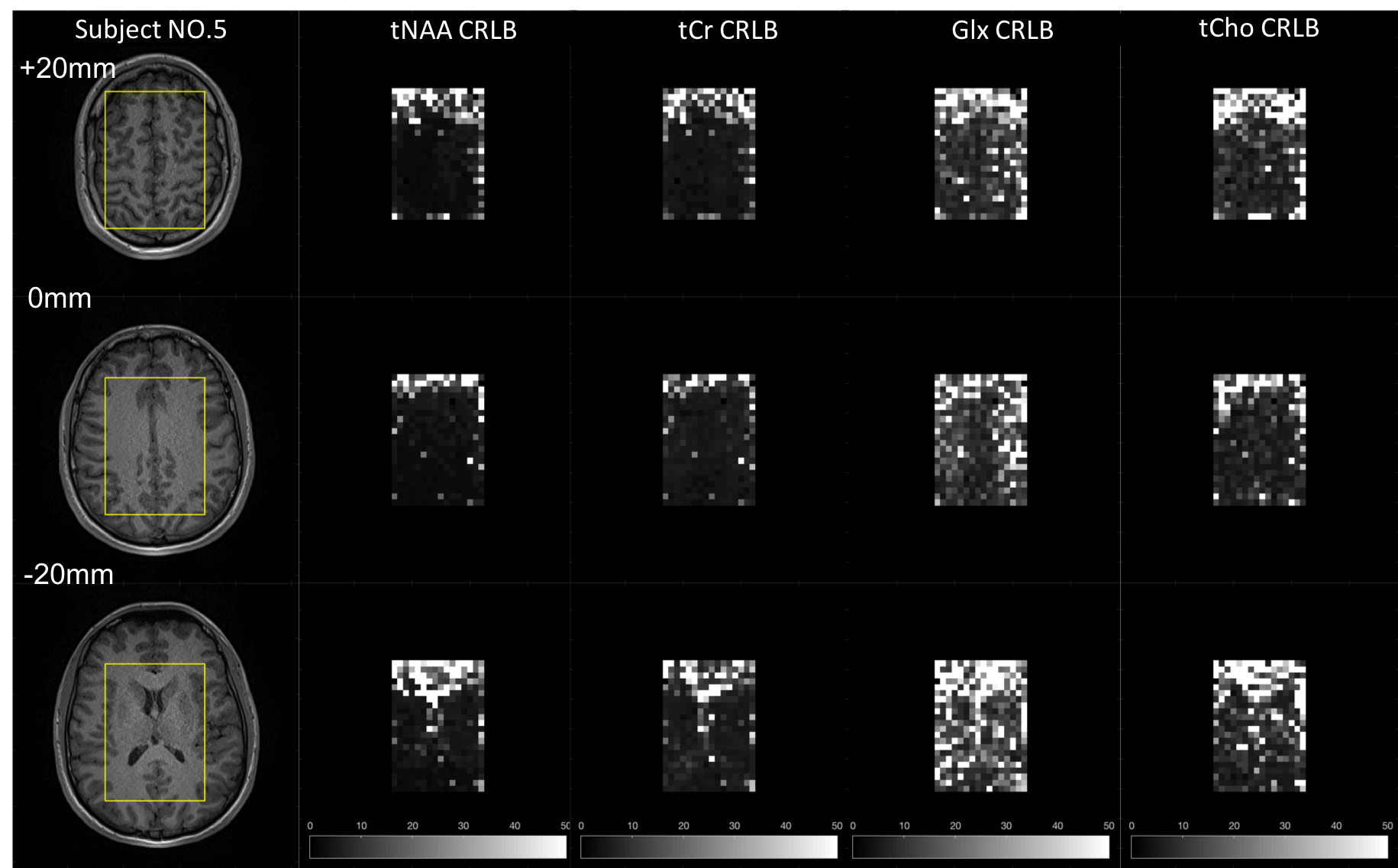
Figure 2 shows the image derived from the SENSE reconstructed SMS and non-SMS MRSI data overlaid on the conventional MPRAGE images. The non-water-suppressed metabolite-cycling MRSI generated water images with structural information similar to that of MPRAGE for all 5 subjects. Ventricles, basal ganglia, and longitudinal fissure can be clearly distinguished. The metabolite spectra extracted from the SMS DW-CRT MRSI data are shown in Figure 3. The input spectra (blue) for LCModel have gone through eddy current correction, phase correction, zero filling, apodization, water and lipid removal, and baseline correction. The LCModel fitting results (red) indicate overall good quality of spectra for all three slices. The resulting metabolite concentration maps are displayed in Figure 4. All the voxels with tNAA or tCr CRLB higher than 50 have been discarded. Low levels of tNAA, tCr, and Glx signals (slice z=-20mm) near ventricles and elevation of Glx in the longitudinal fissure grey matter region are observed as expected (slice z=0mm). CRLB maps are displayed in Figure 5. tNAA and tCr have low CRLB values except for the highly inhomogeneous regions nearby the skull, nasal cavity, and ventricles. Glx and tCho have higher CRLB, especially in slice z=-20mm. Nevertheless, the CRLBs are relatively low in the central region of slice z=0mm and z=+20mm.
In conclusion, it is shown that the SMS DW-CRT MRSI acquisition in combination with metabolite-cycled STEAM pulse localization allows fast, robust, and high-resolution 3D MRSI at 3T.
Acknowledgements
Supported by the Indiana CTSI, funded in part by grant #UL1TR001108 from the NIH, NCATS, CTS Award.References
1. Chiew M, Jiang W, Burns B, Larson P, Steel A, Jezzard P, Albert Thomas M, Emir UE. Density‐weighted concentric rings k‐space trajectory for 1H magnetic resonance spectroscopic imaging at 7 T. NMR in biomedicine. 2018 Jan;31(1):e3838.
2. Emir UE, Burns B, Chiew M, Jezzard P, Thomas MA. Non‐water‐suppressed short‐echo‐time magnetic resonance spectroscopic imaging using a concentric ring k‐space trajectory. NMR in biomedicine. 2017 Jul;30(7):e3714.
3. Steel A, Chiew M, Jezzard P, Voets NL, Plaha P, Thomas MA, Stagg CJ, Emir UE. Metabolite-cycled density-weighted concentric rings k-space trajectory (DW-CRT) enables high-resolution 1 H magnetic resonance spectroscopic imaging at 3-Tesla. Scientific reports. 2018 May 17;8(1):7792.
4. Auerbach EJ, Xu J, Yacoub E, Moeller S, Uğurbil K. Multiband accelerated spin‐echo echo planar imaging with reduced peak RF power using time‐shifted RF pulses. Magnetic resonance in medicine. 2013 May;69(5):1261-7.
5. Setsompop K, Gagoski BA, Polimeni JR, Witzel T, Wedeen VJ, Wald LL. Blipped‐controlled aliasing in parallel imaging for simultaneous multislice echo planar imaging with reduced g‐factor penalty. Magnetic resonance in medicine. 2012 May 1;67(5):1210-24.
6. Pruessmann KP, Weiger M, Scheidegger MB, Boesiger P. SENSE: sensitivity encoding for fast MRI. Magnetic resonance in medicine. 1999 Nov 1;42(5):952-62.
7. Cabanes E, Confort-Gouny S, Le Fur Y, Simond G, Cozzone PJ. Optimization of residual water signal removal by HLSVD on simulated short echo time proton MR spectra of the human brain. Journal of Magnetic Resonance. 2001 Jun 1;150(2):116-25.
Figures