0950
Next generation Crusher Coil for suppressing extra cranial lipid signals at 7 Tesla1Imaging Division, University Medical Center Utrecht, Utrecht, Netherlands, 2WaveTronica, WaveTronica B.V., Utrecht, Netherlands
Synopsis
We previously presented a crusher coil for suppression of extra-cranial lipids in brain Magnetic Resonance Spectroscopic Imaging (MRSI). To improve crushing performance and the reproducibility of the production of crusher coils, a new crusher coil was designed, created and tested. By doubling the amount of wires and improving their spatial distribution around the coil (consistent meandering), we could achieve efficient lipid suppression with as little as 30% of the power, used with the proof of principle coil. The presented design is easy to reproduce, repair and modify due to a modular build.
Introduction
Lipid signals in the skull distort brain MR Spectroscopic Images. With a crusher coil, lipid signals can be suppressed quickly, without the need of high power RF saturation pulses (and concomitant increases in TR) (1). This proof of principle was built with many mechanical inconsistencies and it required high current to achieve complete crushing of lipid signals.
In this work, we attempt to build an improved crusher coil through a new mechanical and electrical design to allow deeper crushing for more complete lipid suppression. The greater dynamic range of the crushing field makes this prototype suitable for use in individuals with smaller head sizes. The prototype consists out of reproducible components that will result in the possibility to build more crusher coils with the same specifications.
Methods
A helmet was designed in solid modeling CAD program SolidWorks. The same program was used to design the foundation of flexible circuit boards with powerlines. Final draft of the circuit boards was done on Target 3001 PCB design software. The helmet was designed to fit into an existing 32 channel receive coil (NOVA Medical).
Flexible circuit boards can only bend in one direction and therefore will not fit on a spherical helmet. To address this, the helmet was designed with seven slightly curved surfaces. Seven circuit boards were placed on these surfaces and the powerlines were soldered together to create one powerline This process facilitated a modular construction (see fig. 1). Parts can easily be modified by 3D printing of new mechanical parts or replacing the flexible circuit boards. The helmet was 3D printed by MR Coils (Zaltbommel, The Netherlands), the flexible circuit boards were produced by Multi Leiterplatten GmbH (Hofolding, Germany).
The flexible circuit boards contain two double-sided wires (four in total). The addition of two lines of wires enables creating a higher magnetic field or the same field with less power. The lines meander over the 3D printed helmet, which create a solid consistent crushing field.
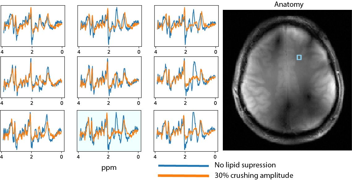
Data acquisition for testing: MRI and MRSI data was collected from a healthy 27 year old male on a Philips 7T system, in combination with a 2 channel transmit - 32 channel receive coil (NOVA Medical) and the new crusher coil. The crusher performance of the coil was tested at various current amplitude settings; from 0 to its maximum of 10A. Anatomical scans were used to assess signal suppression at various power levels (fig. 2).
Results
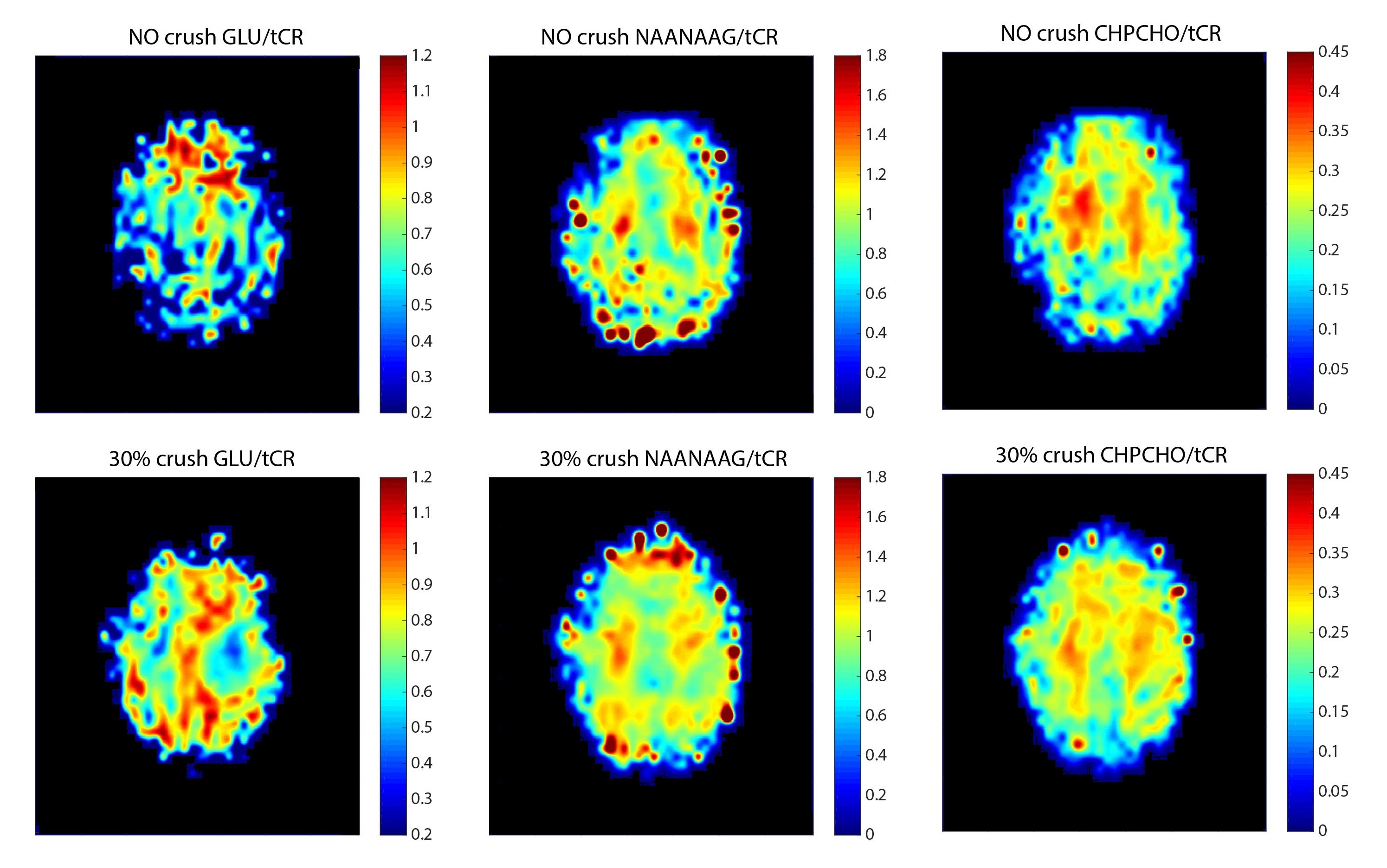
The extent of lipid suppression while using various power levels is shown in figure 2. Crushing at 60 to 100% results in an “overshoot” where parts of the brain tissue are also crushed. The amount of crushing of both skull and brain is visible in figure 3. Figures 2 and 3 both show that optimal crushing is reached at 30 to 50% of the maximum current. Figure 4 shows reconstructed spectra from two acquisitions, one with and one without crushing. Extra-cranial lipid signals are visible and are significantly reduced upon suppression. Figure 5 shows metabolite maps of glutamate, NAA+NAAG and GPC+Cho, generated by fitting of the MRSI data (2) without crushing and with crushing at 30%.
Discussion & conclusion
With the new crusher coil design there is larger spatial coverage of lipid crushing compared to the prototype (1). This enables more effective crushing of lipids in smaller diameter heads, e.g. tailor the level of crushing to the individual subject which will improve data quality of MRSI. In future designs, more wires, different meander patterns or more channels could improve the performance further. A multichannel design can facilitate image based 'lipid shimming' approaches which can be optimized for whole brain MRSI applications.
Acknowledgements
No acknowledgement found.References
1. Boer VO, van de Lindt T, Luijten PR, Klomp DW. Lipid suppression for brain MRI and MRSI by means of a dedicated crusher coil. Magnetic resonance in medicine : official journal of the Society of Magnetic Resonance in Medicine / Society of Magnetic Resonance in Medicine 2015;73(6):2062-2068. https://www.ncbi.nlm.nih.gov/pubmed/24947343
2. Estimation of metabolite concentrations from localized in vivo proton NMR spectra. Provencher SW. Magn Reson Med. 1993 Dec;30(6):672-9.
Figures
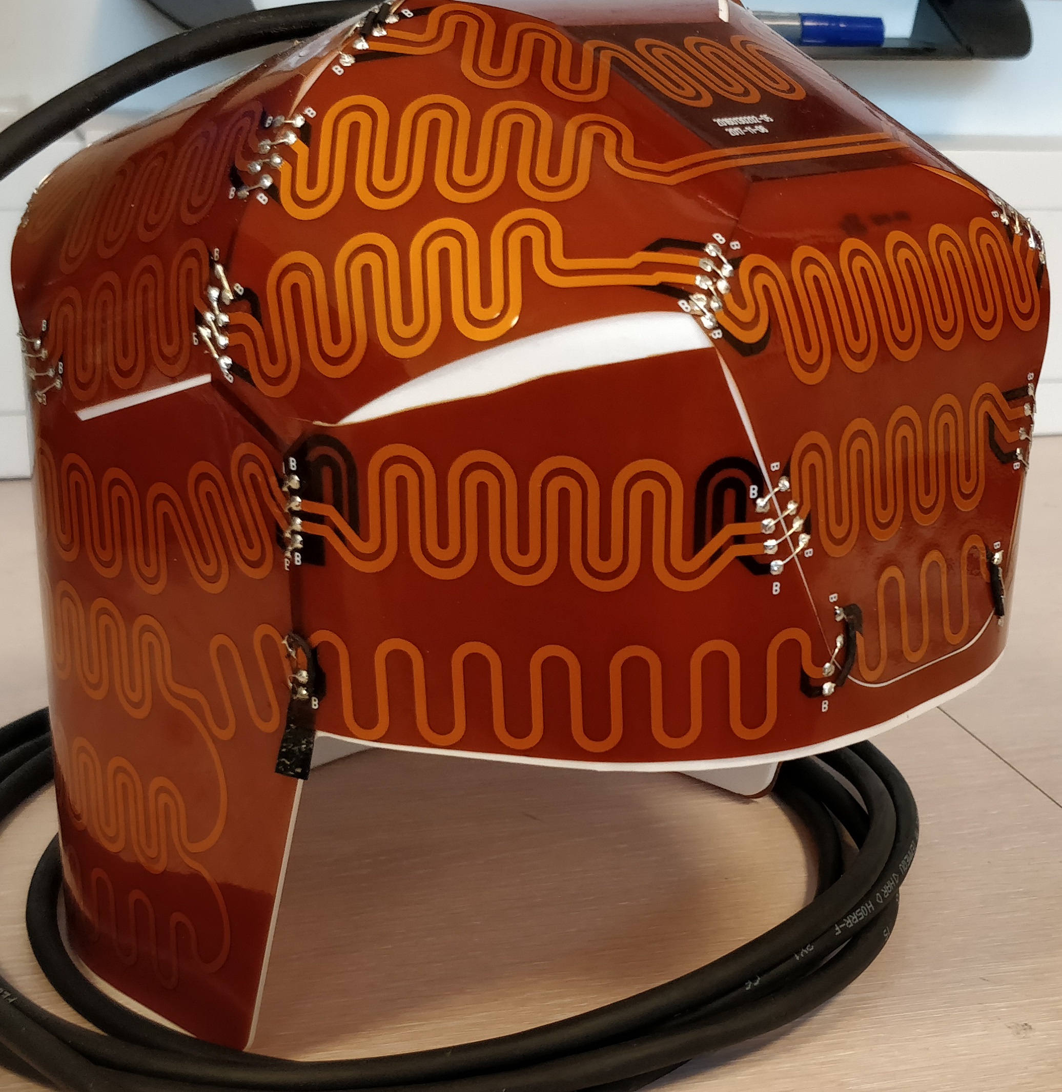
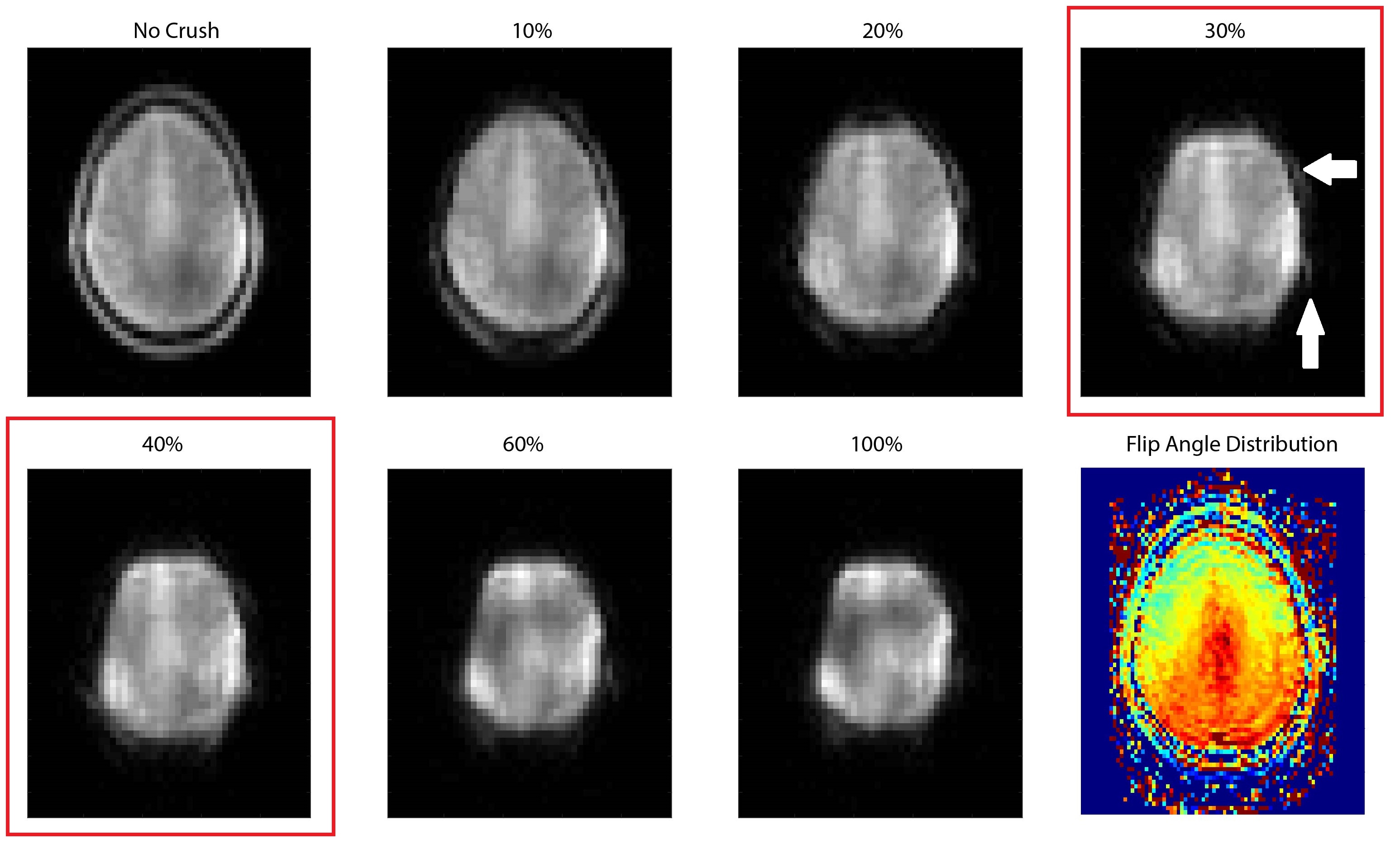
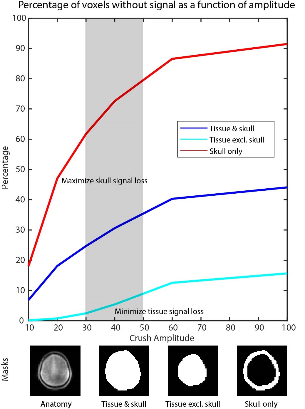
Fig 3: To evaluate crushing performance we created 3 masks based on anatomical scans: skull only, brain tissue only and skull + brain. The plots show the percentage of voxels without signal as a function of crushing amplitude. For optimal MRSI quality, the aim is to minimize signal loss in tissue voxels while maximizing signal loss in voxels in the skull region. Based on this reasoning, optimal crushing occurs in the range of 30-50% crushing amplitude. Note that the final choice of crushing amplitude will be subject dependent.