0948
Using an AC/DC Coil to Improve the Line Width and Lipid Suppression for measuring 2HG in Glioma Patients at 3T1Department of Radiology, A. A. Martinos Center for Biomedical Imaging, Boston, MA, United States, 2Department of Electrical Engineering and Computer Science, Massachusetts Institute of Technology, Cambridge, MA, United States, 3Harvard Medical School, Boston, MA, United States, 4Fetal Neonatal Neuroimaging and Developmental Science Center, Boston Children's Hospital, Boston, MA, United States, 5Department of Radiology, Harvard Medical School, Boston, MA, United States, 6Department of Radiology, Medical University of Vienna, Vienna, Austria, 7Department of Neurosurgery, Massachusetts General Hospital, Boston, MA, United States, 8Department Neurology, Division of Neuro-Oncology, Massachusetts General Hospital, Boston, MA, United States, 9Institute for Medical Engineering and Science, Massachusetts Institute of Technology, Cambridge, MA, United States
Synopsis
An AC/DC 32-channel coil, which can detect signal while simultaneously alter the B0-field, was used to improve lipid inversion and the spectral linewidth for measuring 2HG. Two patients and two volunteers were measured with a spiral-based 3D sequence once with the standard scanner shim only, and once with the additional shimming of the integrated B0/Rx coil. Data quality was strongly improved, leading to improved 2HG-detection in one patient, and a decreased variability of 2HG in tumor-void areas. This can reduce the likelihood of false-positive 2HG detection. Quantification of other metabolites was additionally improved by the AC/DC shim.
Introduction
MR Spectroscopy has recently been shown to enable the non-invasive detection of 2-hydroxyglutarate (2HG)1 , a biomarker for the genetic mutation of the IDH genes. These are mutated in many brain tumors, and their mutation status has been shown to correlate with the survival rate.2 Thus, 2HG-measurements have great value. However, due to its low concentration, low resolutions are usually used to increase the SNR3, but at the cost of increased spectral linewidths, and increased lipid contamination. These two problems can in turn prevent reliable 2HG-detection. AC/DC coils are novel array coils which can detect signal, and simultaneously alter the B0-field4. With an appropriate B0-alteration (=shim) during lipid suppression, the subcutaneous lipids are shifted further upfield than the metabolites inside the brain. This increases their spectral separation and results in an improved performance of frequency selective lipid inversion. By applying another shim during read-out, the spectral linewidths can be decreased. Both shim states can be dynamically switched.4Therefore, we propose using an AC/DC coil to combine the high SNR of low resolution MRSI while still maintaining low lipid contaminations and narrow linewidths. To evaluate these benefits, we compare using an AC/DC coil for shimming against the standard scanner shim for 2HG detection.Methods
Two volunteers and two patients were measured on a Siemens Skyra 3T MR scanner with a 32-channel AC/DC coil using a 3D spiral MRSI sequence. Patient 1 had a confirmed IDH1-R132H-mutated glioblastoma, which was partially resected two weeks prior the measurement. Patient 1 posed challenges for MRSI due to the surgery: Large accumulation of blood products in the resection cavity; a large titanium metal plate for skull repair. Patient 2 had a small diffuse astrocytoma with confirmed IDH1-R132H mutation, but the 2HG measurement using biopsy failed twice, suggesting very little 2HG.The MRSI sequence consisted of an EPI-based navigator5, a frequency selective lipid inversion pulse, WET water suppression, an adiabatic excitation pulse, and two adiabatic refocusing pulses. The sequence parameters were: TI/TR/TE1/TE2 210/1800/32/65 ms, 4 averages, FoV 240×240×100 mm³, VoI 240×240×50 mm³, matrix size 24×24×10, 1100 Hz spectral bandwidth, acquisition time 5:24 min. The MRSI sequence was measured twice: With the standard scanner shim, and with the standard scanner shim plus the AC/DC shim. FLAIR, MPRAGE, and B0-fieldmapping data were acquired additionally.All MRSI data were processed with LCModel. The 2HG, myo-Inositol, Glx, and tNAA concentration maps and spectra were compared qualitatively between the two shim conditions. The lipid suppression improvement, linewidth improvement, and the standard deviation of 2HG outside of tumors were compared between both conditions.Results
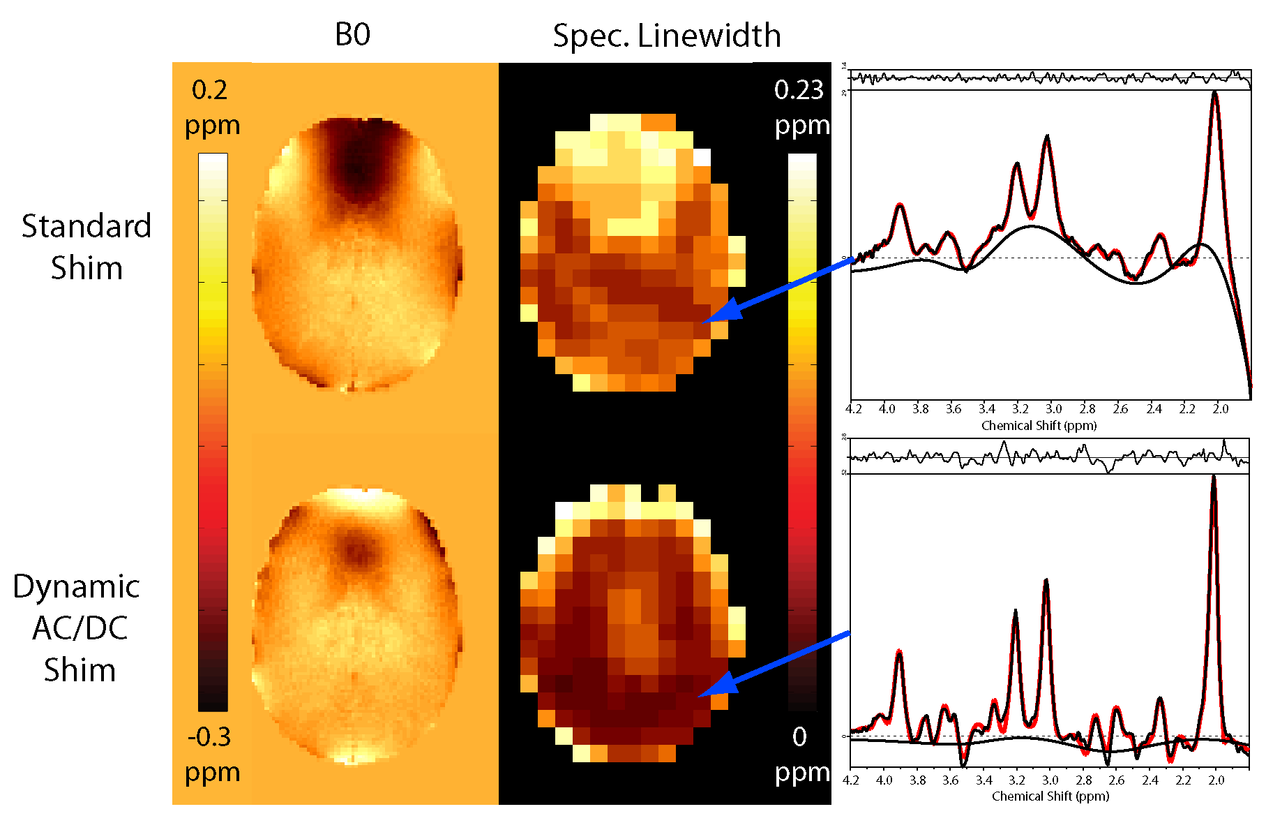
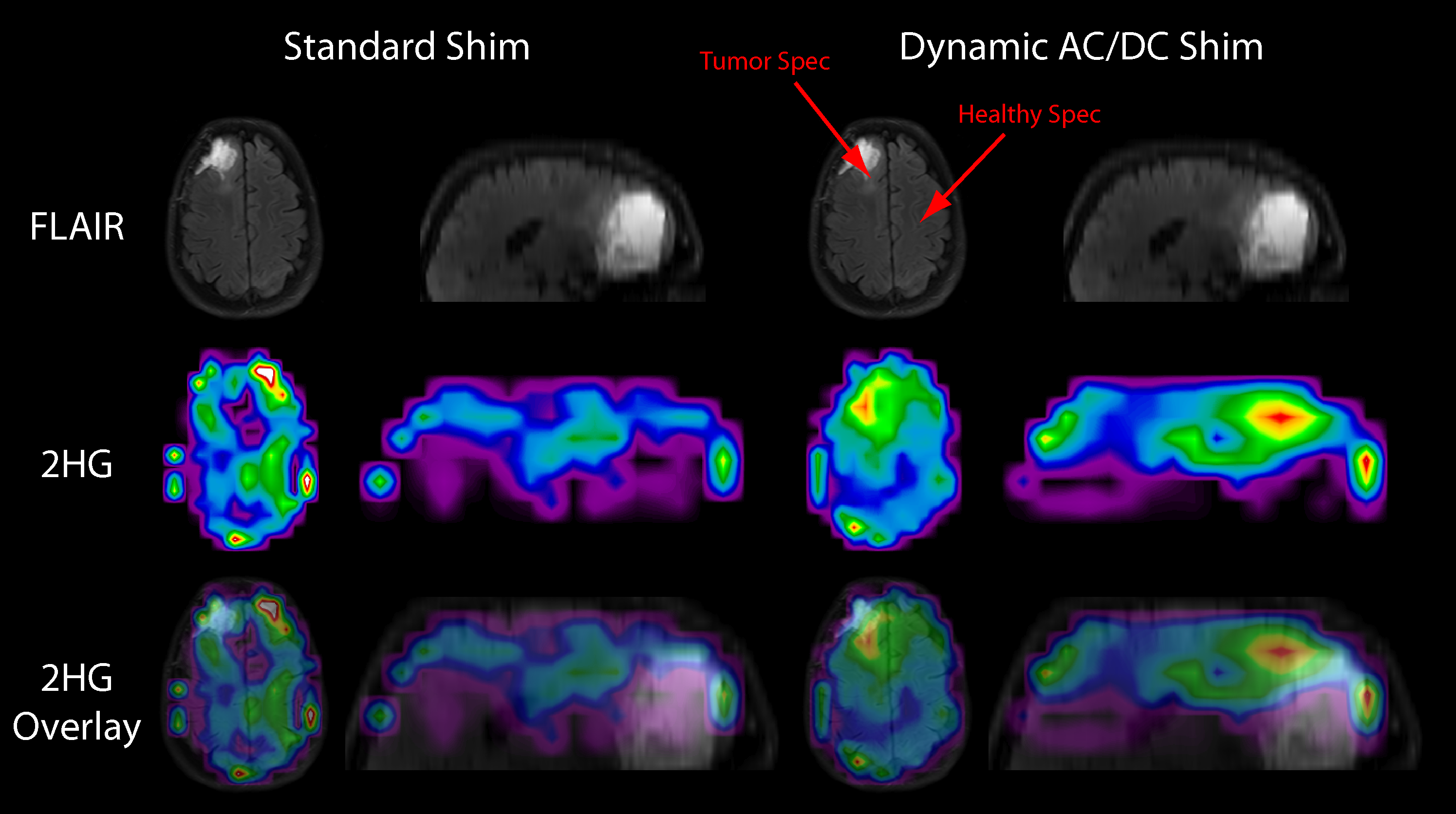
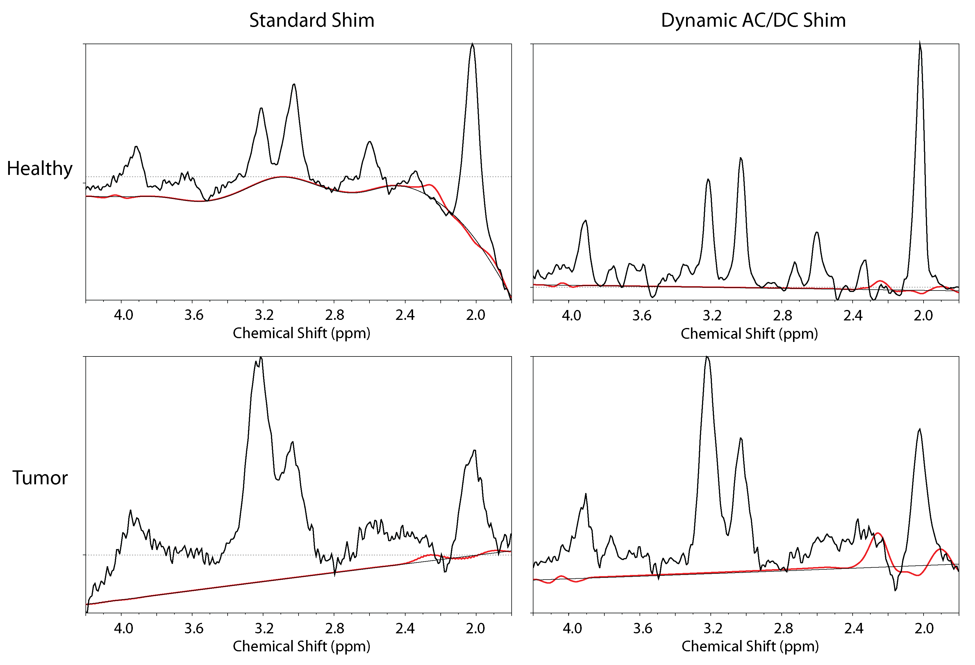
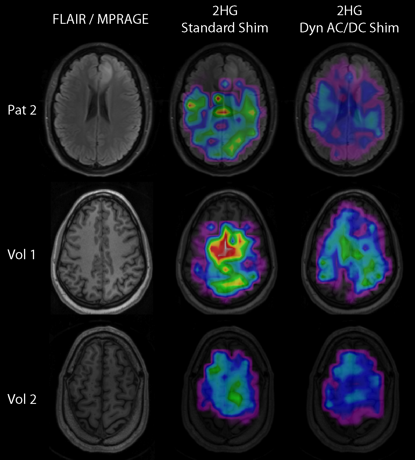
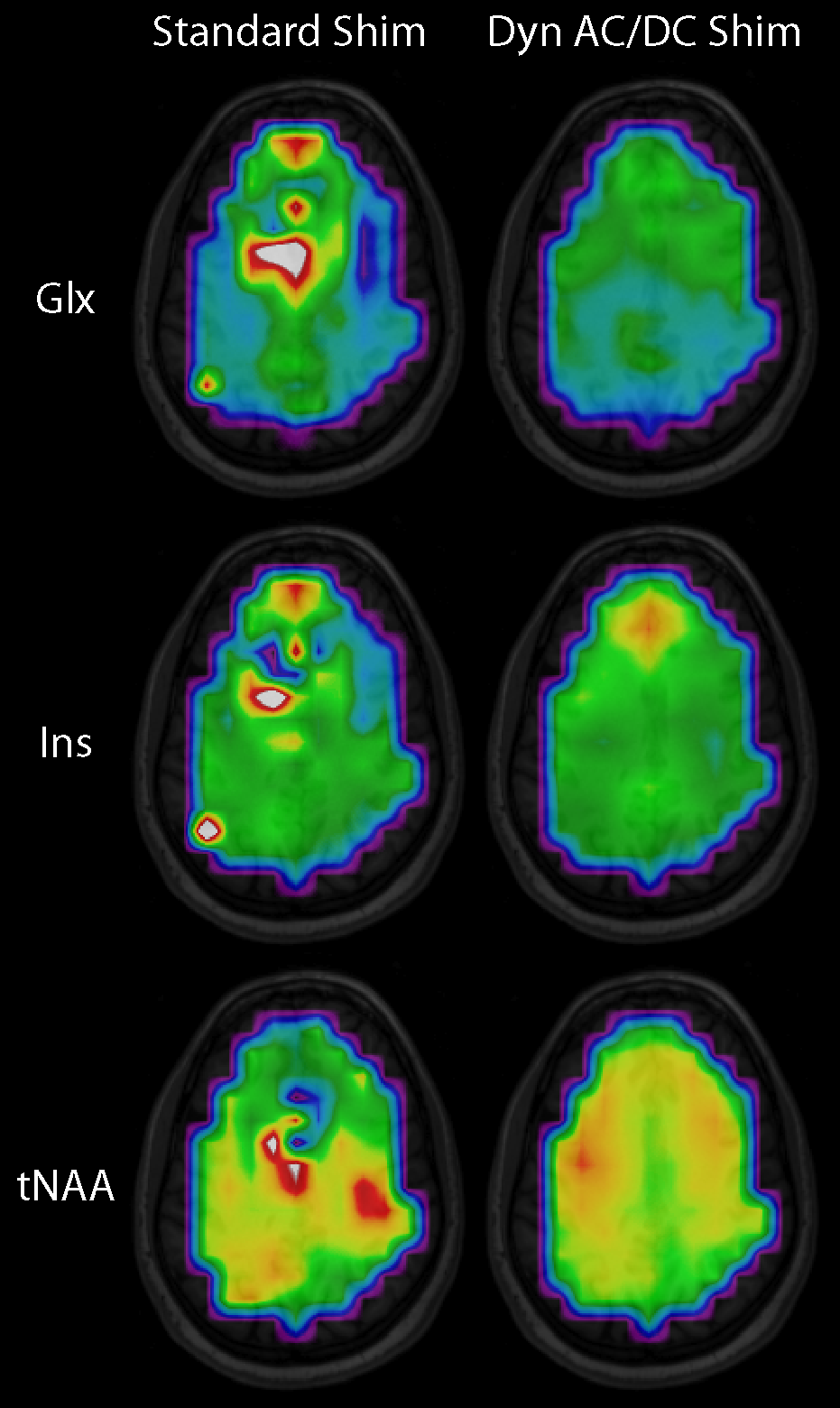
Fig. 1 shows the B0-maps of patient 2, together with the linewidth map, and two example spectra for the standard and the AC/DC condition. Due to the lower B0-inhomogeneity in the AC/DC condition, the linewidth values, as calculated by LCModel, are lower. The sample spectrum additionally shows a cleaner baseline due to the improved lipid suppression. The lipids inside the brain were decreased by 59% over all subjects, the average linewidth decreased by 16%.Fig. 2 shows the 2HG-maps of patient 1. While 2HG is not well fitted close to the tumor in case of the standard shim, a clear hotspot can be seen posterior and superior to the signal-enhanced blood (bright on the FLAIR), where remaining tumor tissue is located. The red arrows indicate the positions of the spectra of Fig. 3. These spectra show a strong improvement in quality when using the AC/DC shim to the standard shim condition. The 2HG-fits are overlaid in red on the spectra.Fig. 4 shows 2HG maps for patient 2 and the two volunteers. No 2HG increases could be detected in the tumor of patient 2 for both shim conditions. In all three subjects, the variability of 2HG is decreased for the AC/DC condition, which is also reflected by a decrease in the standard deviation of 2HG by 58%.Fig. 5 shows that the AC/DC shim also improves the quantification of other metabolites, such as Glx, Ins, and tNAA.Discussion
In this work we showed that 2HG measurements are feasible at low resolutions with the benefit of high SNR, but without the penalties of lipid contamination or high linewidths when using a AC/DC coil. The linewidth and the lipid contamination were both decreased in comparison to a standard shim. This resulted in a strongly improved quantification of 2HG in one patient, and less 2HG variability in 2HG-void regions. The decreased variability reduces the likelihood of false positive 2HG detection. The AC/DC shim also showed improvements in spectral quality and the fitting of other metabolites.Acknowledgements
This work is supported by NIH (1R01CA211080-02) and Austrian Science Fund (J 4110).References
1. Andronesi, O. C. et al. Detection of 2-hydroxyglutarate in IDH-mutated glioma patients by in vivo spectral-editing and 2D correlation magnetic resonance spectroscopy. Sci. Transl. Med. 2012; 4:116ra114.
2. Balss J et al. Analysis of the IDH1 codon 132 mutation in brain tumors. Acta Neuropathol 2008;116:597-602.
3. Andronesi OC et al. Pharmacodynamics of mutant-IDH1 inhibitors in glioma patients probed by in vivo 3D MRS imaging of 2-hydroxyglutarate. Nat Commun. 2018;9:1474.
4. Arango N et al. Dynamically Switched B0 Field Control for Separate Optimization of Tailored Volume Lipid Suppression and B0 Homogeneity for Brain Chemical Shift Imaging at 3T using Multi-Coil Shim Array. ISMRM 2018; 1062.
5. Bogner W et al. 3D GABA imaging with real‐time motion correction, shim update and reacquisition of adiabatic spiral MRSI. Neuroimage 2014;103:290–302
Figures