0946
Rapid High-Resolution Simultaneous Acquisition of Metabolites, Myelin Water Fractions, and Tissue Susceptibility of the Whole Brain Using "SPICY" 1H-MRSI1Beckman Institute for Advanced Science and Technology, University of Illinois at Urbana-Champaign, Urbana, IL, United States, 2Department of Electrical and Computer Engineering, University of Illinois at Urbana-Champaign, Urbana, IL, United States, 3Department of Radiology, The Fifth People's Hospital of Shanghai, Shanghai, China, 4School of Biomedical Engineering, Shanghai Jiao Tong University, Shanghai, China, 5Med-X Research Institute, Shanghai Jiao Tong University, Shanghai, China
Synopsis
We report further advances on ultrahigh-resolution 1H-MRSI without water suppression to enable rapid simultaneous acquisition of brain metabolites, myelin water fractions (MWF) and tissue susceptibility in high spatial resolution. Building on the SPICE (SPectroscopic Imaging by exploiting spatiospectral CorrElation) subspace imaging framework, we extend the SPICE data acquisition scheme with several novel features, including the use of ultrashort-TE (~1.6 ms), very-short-TR (~160 ms), and variable density sampling of (k, t)-space. We reconstruct the spatial distributions of brain metabolites, MWF, and tissue susceptibility using model-based reconstruction methods that incorporate learned spatiospectral features. Experimental results have been obtained which demonstrate that in a single 5-min scan, we can obtain metabolites in a nominal resolution of 2.0×2.4×3.0 mm3 and QSM/MWF in a nominal resolution of 1.8×1.8×1.8 mm3.
Introduction
MR spectroscopic imaging (MRSI) is a unique tool for noninvasive metabolic imaging and it has been used to map the molecular fingerprints of brain function and diseases. Recently, quantitative susceptibility mapping (QSM) and myelin water imaging (MWI) have also been widely used to study brain tissue iron deposition and white matter integrity1-3. Currently, MRSI, QSM and MWI experiments are carried out independently using different acquisition sequences, which often lead to long data acquisition times. This paper reports our success in integrating MRSI, QSM and MWI for brain imaging. This work is built on our recent progress in using the SPICE (SPectroscopic Imaging by exploiting spatiospectral CorrElation) subspace imaging framework for simultaneous QSM and MRSI4. We extend this technique in both data acquisition and processing to enable rapid simultaneous MRSI, QSM and MWI in high spatial resolution. In data acquisition, we further reduce TE/TR and use a variable-density sampling strategy via blipped EPSI trajectories to achieve rapid high-resolution acquisition. In data processing, we reconstruct the spatial distributions of brain metabolites, myelin water fractions (MWF), and tissue susceptibility using model-based constrained reconstruction methods, incorporating pre-learned spatiospectral features. Experimental results have been obtained which demonstrate that in a 5-min scan, we can obtain metabolites in 2.0×2.4×3.0 mm3 and QSM/MWF in 1.8×1.8×1.8 mm3. This capability may significantly enhance the practical utility of 1H-MRSI in various research and clinical applications.Data Acquisition
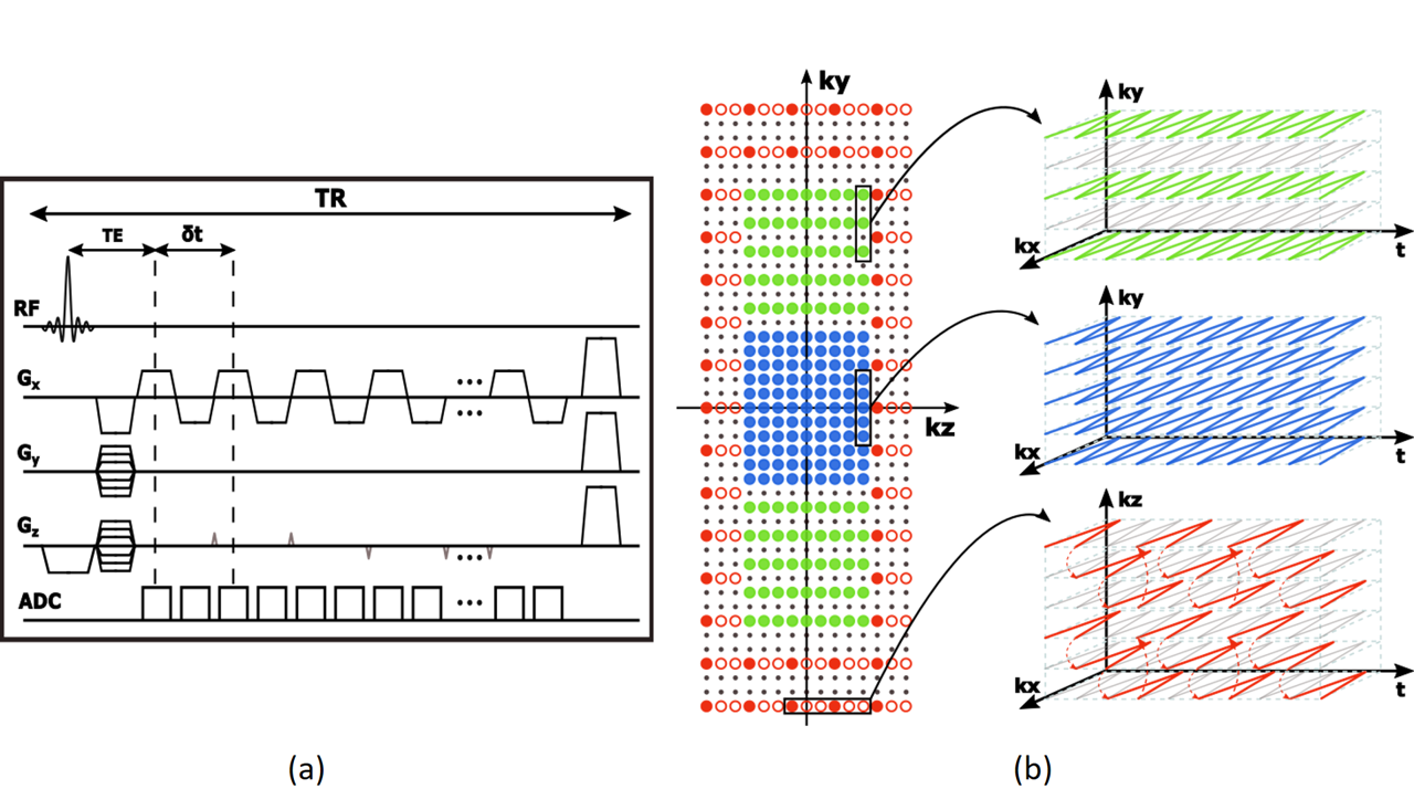
The proposed data acquisition scheme maintains the key features of the SPICE sequence without water/lipid suppression, but extends the technique in several aspects (Fig. 1). Firstly, an ultrashort-TE (~1.6 ms) and very-short-TR (~160 ms) acquisition is adopted, which maximizes the SNR efficiency and preserves the water proton spectroscopic information for MWF. Secondly, a variable-density sampling strategy is used to achieve large k-space coverage and thus high spatial resolution. More specifically, the k-space is partitioned into multiple segments that are sampled with different sampling densities (Fig. 1b); outer k-space is sampled sparsely both in $$$k$$$ and $$$t$$$ using blipped EPSI trajectories. Using this sparse sampling scheme along with ramp sampling, we can achieve a nominal spatial resolution of 1.8×1.8×1.8 mm3 in a 5-min scan.Data Processing
The proposed data processing scheme reconstructs MRSI, MWF and QSM data using model-based constrained reconstruction methods, incorporating both spectral and spatial priors. For spectral constraints, a union-of-subspaces model is adopted, which represents each spectral component (i.e., water, lipids and metabolites) using a low-dimensional subspace. This explicit subspace representation allows effective and efficient incorporation of spectral priors, in the form of spectral basis functions predetermined from quantum simulation and training data4-7. For spatial constraints, a kernel-based model is adopted, which represents the spatial variations in a reproducing kernel Hilbert space, effectively absorbing spatial priors8 (e.g., water side information). Such spatiospectral constraints significantly reduce the degrees-of-freedom, thereby effectively reducing potential artifacts due to sparse sampling and noise.
Tissue susceptibility/MWF mapping: The key issues in reconstructing QSM and MWF are due to sparse sampling and the ill-conditionedness of the problems. We addressed the first issue by interpolating the missing data using pre-determined water/lipid bases. We successfully solved the second problem by integrating the kernel-based representation for spatial variations into the dipole model for QSM reconstruction and the multi-component T2* model for MWF reconstruction.
Metabolite mapping: Two key issues associated with metabolite mapping are: (a) removal
of lipid/water signals, and (b) spatiospectral reconstruction from noisy data. We
have successfully resolved these issues using a union-of-subspaces model, integrating
pre-determined subspace structures and kernel-based spatial representations.
Results
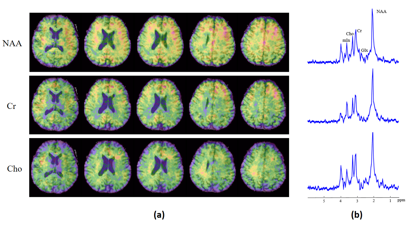
In vivo experiments were carried out to evaluate our new imaging capability. The data were collected from healthy subjects on a 3T scanner with TR/TE = 160/1.6 ms, FOV = 230×230×72 mm3, matrix size = 128×128×42 and echospace = 1.76 ms. We only used the central 96×110×24 k-space encodings for metabolic reconstruction to ensure adequate SNR, yielding a nominal resolution of 2.0×2.4×3.0 mm3. Figure 2 shows some representative MWF and QSM results reconstructed at 1.8 mm isotropic resolution. The metabolite maps reconstructed at 2.0×2.4×3.0 mm3 nominal resolution are presented in Fig. 3. As can be seen, high-resolution metabolite maps and high-quality spatially resolved spectra were successfully obtained. To our knowledge, these are the first experimental results from simultaneous MRSI/QSM/MWF of the brain.
Conclusions
This work reports a new capability for rapid simultaneous acquisition of brain metabolites, MWF and tissue susceptibility in high spatial resolution from a single 5-min 1H-MRSI scan without water suppression. The proposed method is built on the SPICE subspace imaging framework, effectively incorporating learned spatiospectral features for accelerated data acquisition. This new capability may prove useful for many brain imaging applicationsAcknowledgements
This work was supported in part by the following research grants: NIH-R21-EB021013, NIH-R21-EB023413, NIH-R01-EB023704, and NIH-P41-EB022544.References
[1] Wang Y, Liu T. Quantitative susceptibility mapping (QSM): decoding MRI data for a tissue magnetic biomarker. Magn Reson Med. 2015;73(1):82-101.
[2] Borich MR, MacKay AL, Vavasour IM, et al. Evaluation of white matter myelin water fraction in chronic stroke. Neuroimage Clin. 2013;2:569-580.
[3] Vanes LD, Mouchlianitis E, Wood TC, et al. White matter changes in treatment refractory schizophrenia: Does cognitive control and myelination matter? Neuroimage Clin. 2018;18:186-191.
[4] Peng X, Lam F, Li Y, et al. Simultaneous QSM and metabolic imaging of the brain using SPICE. Magn Reson Med. 2018;79(1):13-21.
[5] Lam F, Liang ZP. A subspace approach to high-resolution spectroscopic imaging. Magn Reson Med. 2014;71(4):1349-1357.
[6] Lam F, Ma C, Clifford B, et al. High-resolution 1H-MRSI of the brain using SPICE: data acquisition and image reconstruction. Magn Reson Med. 2016;76(4):1059-1070.
[7] Li Y, Lam F, Clifford B, et al. A subspace approach to spectral quantification for MR spectroscopic imaging. IEEE Trans Biomed Eng. 2017;64(10):2486-2489.
[8] Li Y, Liang ZP. Constrained image reconstruction using a kernel+sparse model. Proc Intl Soc Magn Reson Med. 2018;p. 657.
Figures