0939
Direct Myelin Imaging In Human Brain Using Inversion Recovery Hybrid Encoded Ultrashort Echo Time (IR-HE-UTE) Magnetic Resonance Imaging1Department of Radiology, University of California San Diego, San Diego, CA, United States, 2GE Healthcare, San Diego, CA, United States, 3Department of Neurosciences, University of California San Diego, San Diego, CA, United States, 4Radiology Service, VA San Diego Healthcare System, San Diego, CA, United States
Synopsis
Multiple sclerosis (MS) is the most common immune-mediated demyelinating inflammatory disease, afflicting over 2.3 million people globally. Magnetic resonance imaging (MRI) is the gold standard non-invasive imaging modality to identify MS lesions. Clinically, both CSF-suppressed T2-weighted and T1-weighted images are used for the characterization of MS lesions (FLAIR and MP-RAGE sequences). However, these sequences are not specific for demyelinating lesions and can be challenging to interpret. In this study, we propose a direct myelin imaging technique utilizing IR-prepared hybrid encoded UTE imaging, which provides highly specific volumetric myelin images with scan time less than 7min.
Introduction
Multiple sclerosis (MS) is the most common immune-mediated demyelinating inflammatory disease. Although MRI is the gold standard imaging modality to identify MS-lesions, the clinical sequences are not specific for demyelinating lesions and can be challenging to interpret. Direct imaging of myelin could provide highly specific information to characterize demyelinating lesions in MS. Unfortunately, it is challenging to directly image myelin protons in white matter using MRI due to myelin’s extremely low proton density and rapid signal decay (T2*<0.5ms at 3T)1,2. A breakthrough development demonstrated that direct myelin imaging is feasible using inversion recovery (IR) prepared ultrashort echo time (UTE) MRI3–6. In this study, we explore the efficacy of IR prepared hybrid encoded UTE (IR-HE-UTE) with a novel sampling strategy for direct 3D myelin imaging.Methods
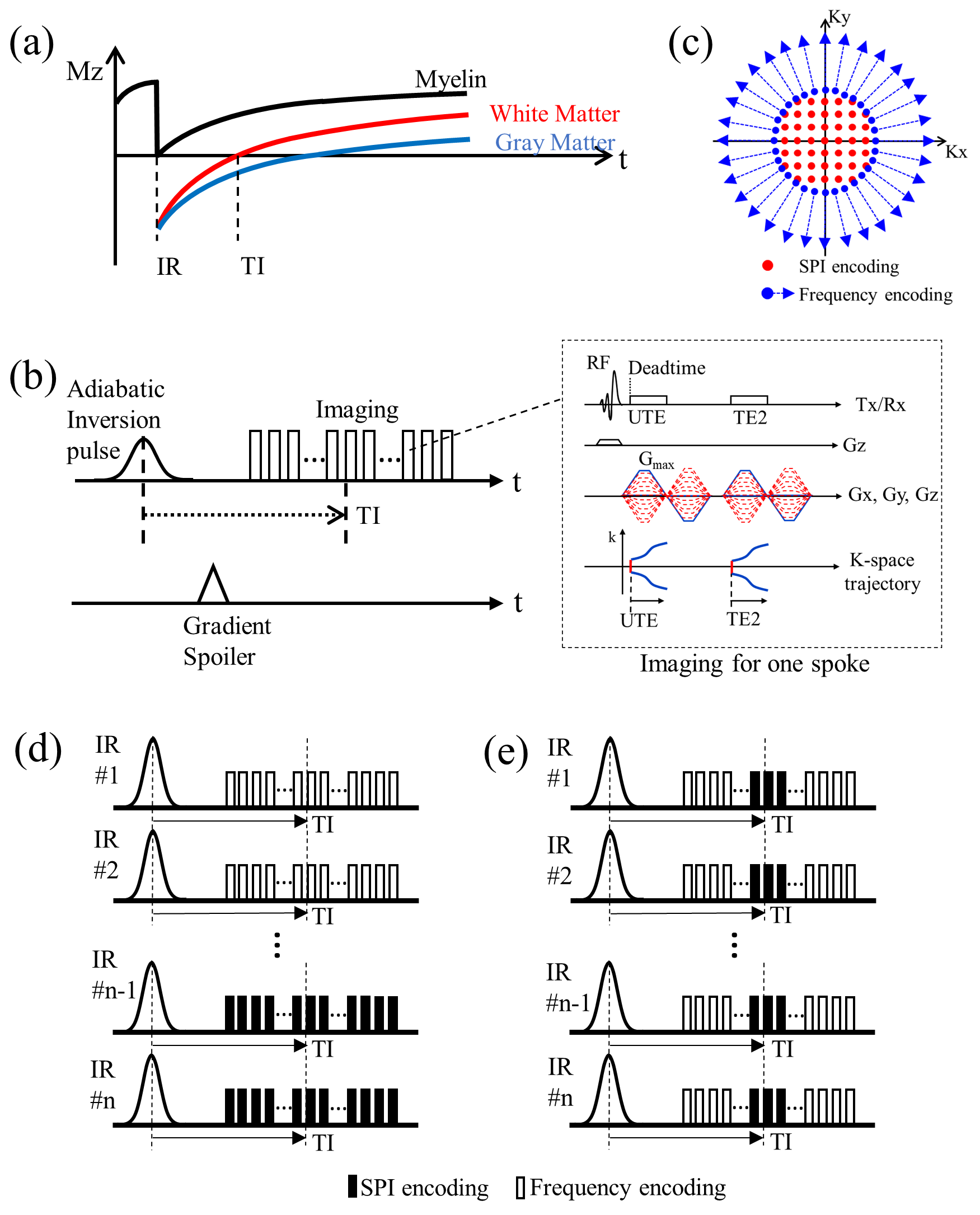
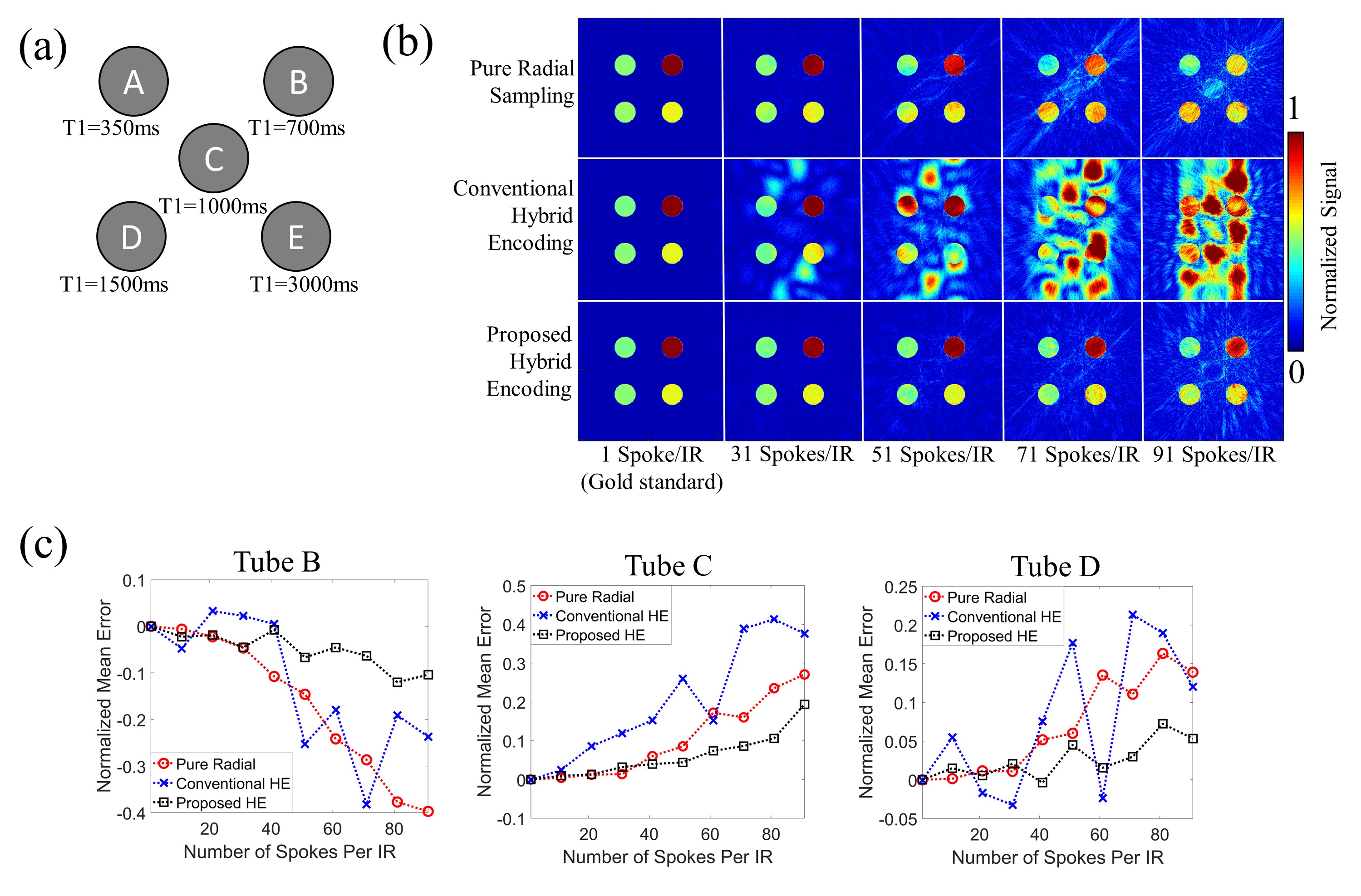
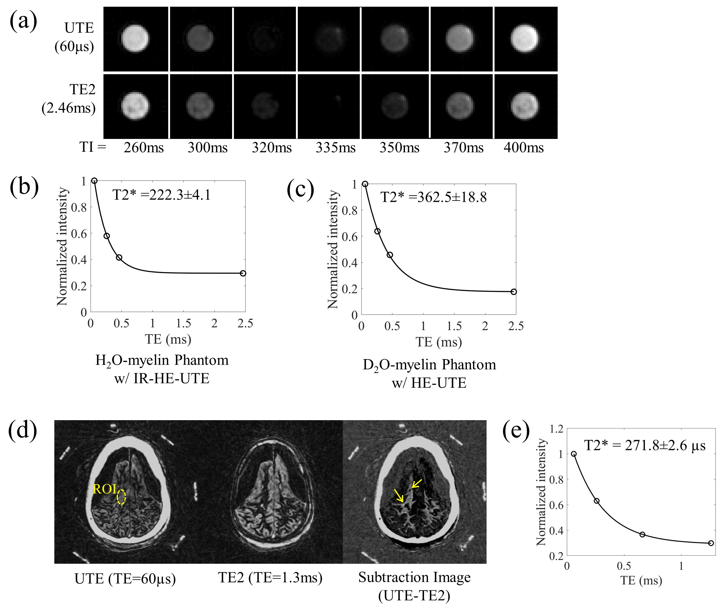
Figure 1-a illustrates the typical T1-recovery curves of tissues in a brain. By selecting TI matched to the nulling point of white matter, the white matter signal can be suppressed. Residual gray matter signal can be suppressed utilizing dual-echo UTE imaging by subtracting the second echo from UTE. After adiabatic inversion preparation, imaging is performed, where multiple spokes are acquired after each IR preparation in order to reduce the scan time for 3D imaging (Figure 1-b). Unfortunately, in the multi-spoke acquisition, image contrast is inevitably degraded due to the spokes acquired at suboptimal TIs, resulting in suboptimal white matter suppression. To address this issue, we propose a strategy benefiting from hybrid encoding (HE)7. Figure 1-c illustrates an example of the HE-sampling pattern. Figure 1-d shows a typical sampling strategy for multi-spoke HE, sequentially performing the radial frequency encoding and the SPI encoding. Figure 1-e shows the proposed sampling strategy where SPI encodings are interleaved to the best time slot near the optimal TI, which allows more efficient white matter suppression with a high degree of multi-spoking since the central k-space encoded by SPI significantly contributes to the image contrast. The proposed IR-HE-UTE was evaluated with computer simulation, myelin phantom, ex-vivo MS brain, in-vivo healthy volunteers, and in-vivo MS patients. For the computer simulation, a digital phantom was generated with T1s selected to cover the typical T1 of myelin, white matter, gray matter, and CSF at 3T (Figure 2-a). Then, multi-spoke IR imaging was simulated to null Tube-C (white matter), using three different encoding schemes (radial frequency encoding, conventional HE with sequential sampling, and the proposed HE with interleaved SPI) with different levels of multi-spoking (1 to 91-spokes/IR). For the phantom experiment, D2O-myelin and H2O-myelin phantoms were prepared by compounding bovine myelin lipid powder with D2O or H2O and imaged in 3T GE-MR750 using a custom-made birdcage-coil. An ex-vivo experiment was performed with a cadaveric MS brain in 3T GE-MR750 using 8-ch-receive-only head-coil. An in-vivo experiment was performed with 8 healthy volunteers and 13 MS patients in 3T GE-MR750 using 12-ch-receive-only HNU coil. The MR imaging parameters can be found in the Figure captions.Results
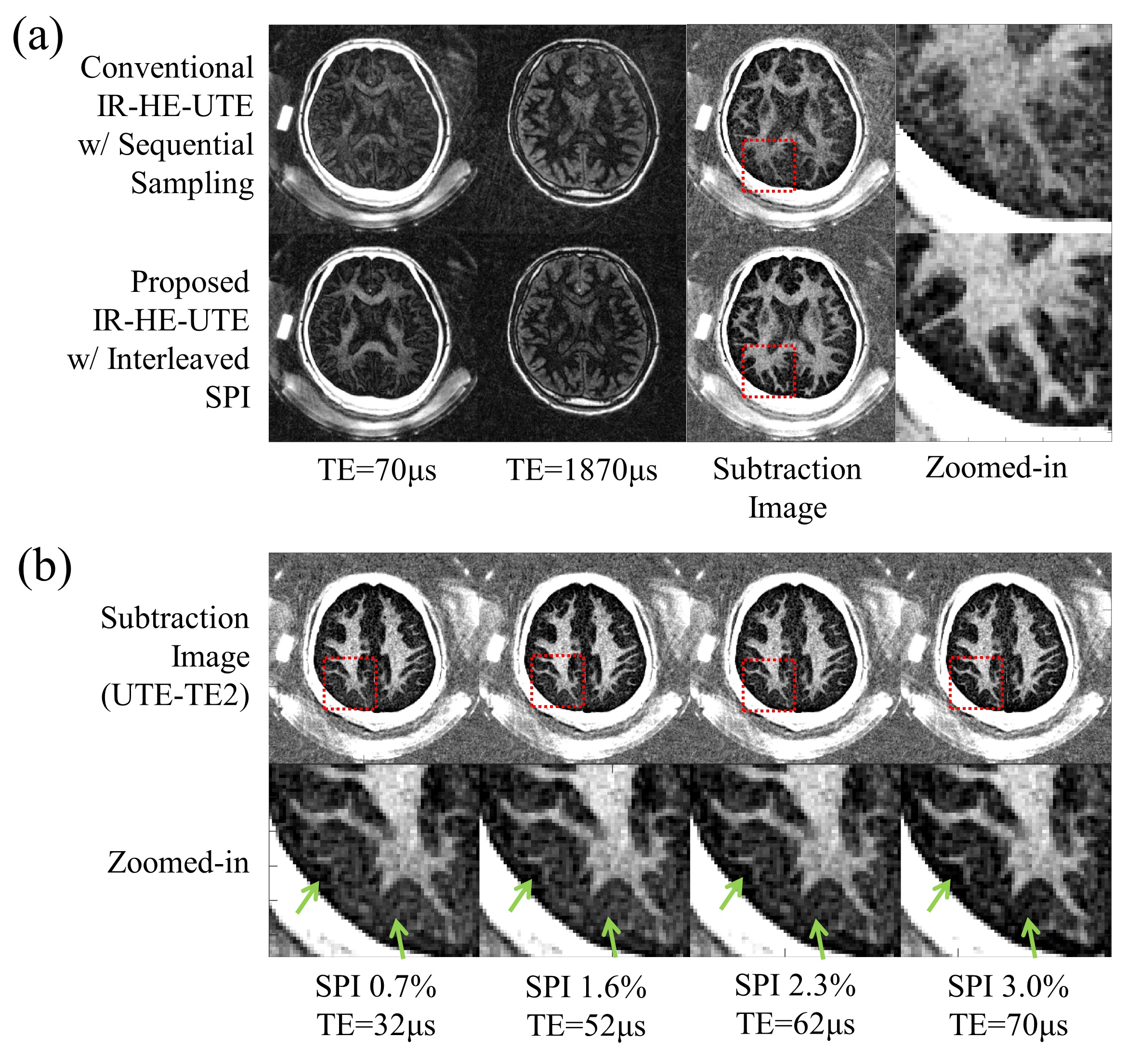
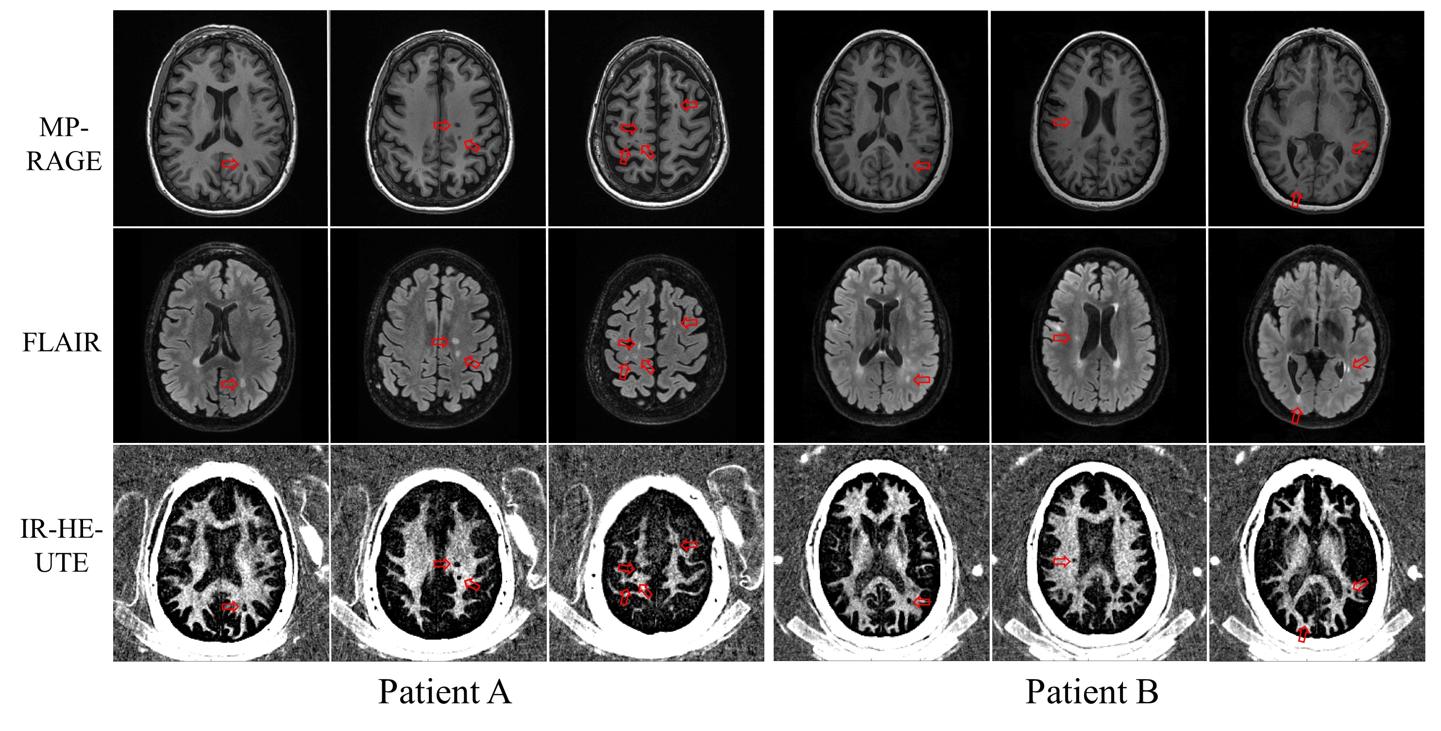
Figures 2-b and 2-c show the result of the computer simulation, where the proposed strategy shows overall better error performance when compared to the conventional methods at a high degree of multi-spoking. Figure 3-a shows the H2O-myelin phantom imaged with the proposed method at different TIs, demonstrating robust suppression of water with TI=335ms at which the estimated T2* was 222.3±4.1µs (Figure 3-b). The estimated T2* of the D2O-myelin phantom was 362.5±18.8µs (Figure 3-c), showing the elevated value presumably due to residual water in the phantom. Figure 3-d shows results with a MS brain, where the demyelinated lesions are clearly seen (yellow arrows). The estimated T2* was 271.8±2.6µs (Figure 3-e), which was consistent with our previous studies (200-350µs)2. Figure 4-a shows the results with a healthy volunteer. As seen, the strategy of interleaving SPI dramatically improves the myelin contrast. Figure 4-b shows four different myelin images by the proposed IR-HE-UTE method, reconstructed with different sizes of the SPI-encoding region. As seen, the background signal tends to be spatially biased with the smaller SPI size, which is well-suppressed with larger SPI size (green arrows)8. Figure 5 shows the IR-HE-UTE and clinical MR images obtained with MS patients. The demyelinated areas can be detected by the proposed IR-HE-UTE imaging as regions where there is loss of the normal myelin signal (red arrows).Discussion and Conclusion
In this study, we have demonstrated the efficacy and feasibility of inversion recovery prepared hybrid encoding for direct myelin imaging in the human brain, which provides highly specific volumetric myelin images, achieving both good image contrast with white matter suppressed near-completely and a clinically feasible scan time of less than 7min.Acknowledgements
The authors acknowledge research support from GE Healthcare, NIH (R01NS092650), and VA Clinical Science and Rehabilitation R&D Awards (I01CX001388 and I01RX002604).References
1. Fan S-J, Ma Y, Chang EY, Bydder GM, Du J. Inversion recovery ultrashort echo time imaging of ultrashort T 2 tissue components in ovine brain at 3 T: a sequential D 2 O exchange study. NMR Biomed. 2017;30:e3767 doi: 10.1002/nbm.3767.
2. Fan SJ, Ma Y, Zhu Y, et al. Yet more evidence that myelin protons can be directly imaged with UTE sequences on a clinical 3T scanner: Bicomponent T2* analysis of native and deuterated ovine brain specimens. Magn. Reson. Med. 2018;80:538–547 doi: 10.1002/mrm.27052.
3. Du J, Ma G, Li S, et al. Ultrashort echo time (UTE) magnetic resonance imaging of the short T2 components in white matter of the brain using a clinical 3T scanner. Neuroimage 2014;87:32–41 doi: 10.1016/j.neuroimage.2013.10.053.
4. Sheth V, Shao H, Chen J, et al. Magnetic resonance imaging of myelin using ultrashort Echo time (UTE) pulse sequences: Phantom, specimen, volunteer and multiple sclerosis patient studies. Neuroimage 2016;136:37–44 doi: 10.1016/j.neuroimage.2016.05.012.
5. Sheth VR, Fan S, He Q, et al. Inversion recovery ultrashort echo time magnetic resonance imaging: A method for simultaneous direct detection of myelin and high signal demonstration of iron deposition in the brain – A feasibility study. Magn. Reson. Imaging 2016;38:87–94 doi: 10.1016/j.mri.2016.12.025.
6. Waldman a, Rees JH, Brock CS, Robson MD, Gatehouse PD, Bydder GM. MRI of the brain with ultra-short echo-time pulse sequences. Neuroradiology 2003;45:887–92 doi: 10.1007/s00234-003-1076-z.
7. Jang H, Wiens CN, McMillan AB. Ramped hybrid encoding for improved ultrashort echo time imaging. Magn. Reson. Med. 2016;76:814–825 doi: 10.1002/mrm.25977.
8. Jang H, Liu F, Bradshaw T, McMillan AB. Rapid dual-echo ramped hybrid encoding MR-based attenuation correction (dRHE-MRAC) for PET/MR. Magn. Reson. Med. 2018;79:2912–2922 doi: 10.1002/mrm.26953.
Figures