0938
Dixon-like multi-contrast GRASE imaging framework: simultaneous acquisition of two T2-weighted spin-echo images and an inverse-T1-weighted stimulated-echo image1Department of Biomedical Sciences and Engineering, National Central University, Taoyuan, Taiwan, 2Department of Computer Science and Information Engineering, National Cheng Kung University, Tainan, Taiwan, 3Department of Biomedical Engineering, University of Arizona, Tucson, AZ, United States
Synopsis
Our novel Dixon-like multi-contrast GRASE imaging framework can simultaneously acquire two distinct T2-weighted and an inverse-T1-weighted images with high scan efficiency. The two single-shot GRASE imaging sequences with phase difference of RF pulses generates signal with spin-echo (SE) signals and stimulated-echo (STE) signals in-phase and 180-degree out-of-phase. A Dixon-like approach can respectively acquire T2-weighted SE-only image and inverse-T1-weighted STE-only images from the data. Multi-contrast images can be efficiently acquired with the proposed framework within clinically feasible scan time.
Introduction
Conventional CPMG imaging sequences generate a series of echo signals from multiple echo pathways, in which multiple contrast information are unseparated. We developed a Dixon-like multi-contrast gradient-and spin-echo (GRASE) imaging1,2 framework, in which two single-shot GRASE scans efficiently acquire 1) a conventional spin-echo (SE) image, 2) an image with SE and stimulated-echo (STE) signal in-phase and 3) an image with SE and STE signal 180-degree out-of-phase. From the in-phase and out-of-phase images, a SE-only image and a STE-only image can be generated with a Dixon-like approach. With the proposed framework, multi-contrast images with two different T2-weightings and a inverse-T1 weighting can be simultaneously acquired within 10 seconds, and thus can provide valuable and instant multi-contrast information for clinical diagnosis.Methods
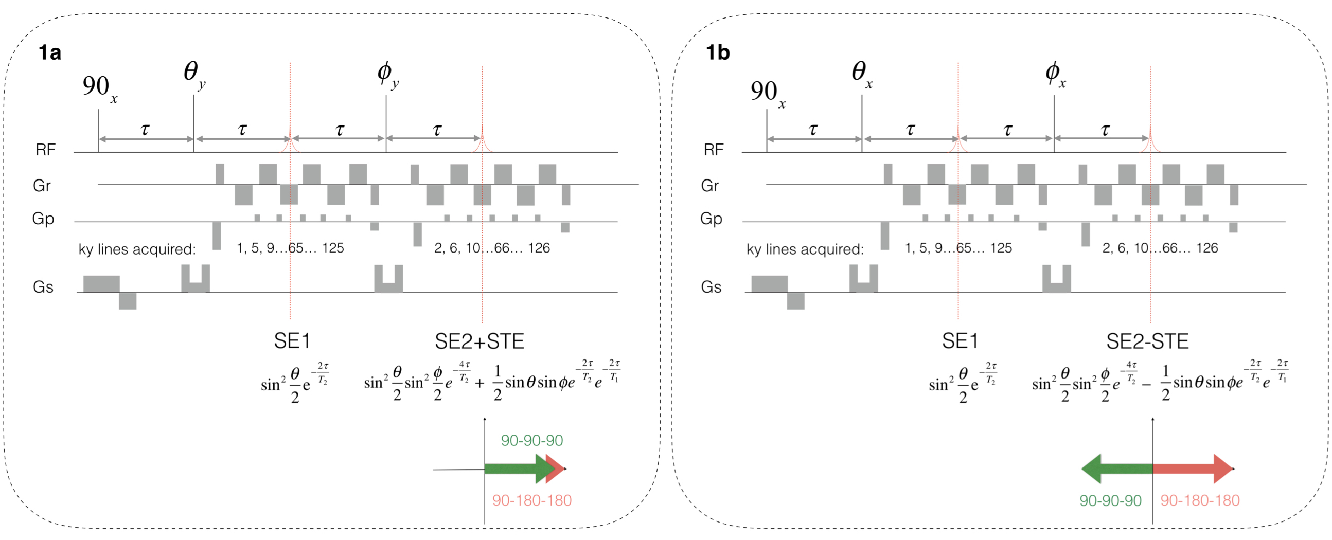
$$$The proposed multi-contrast GRASE imaging sequence for Dixon-like framework, which consists of two single-shot GRASE imaging sequences, is depicted in Figure 1. The first GRASE imaging scan, shown in Figure 1a, includes a 90-degree excitation pulse applied along the x-axis and two refocusing pulses applied along the y-axis. The 90-degree phase difference of RF pulses generate T2-weighted SE signal in the first echo (i.e. SE1), and signal with SE (i.e. SE2) and STE in-phase in the second echo. The second GRASE imaging scan, shown in Figure 1b, includes a 90-degree excitation pulse and two refocusing pulses applied along the x-axis. The zero phase difference of RF pulses also generates SE1 signal in the first echo, while in the second echo generates signal with SE2 and STE 180-degree out-of-phase. Noted that other high-order echo pathways are all eliminated by crusher gradients in the sequence. Based on the extended phase graph theory3,4, the multi-contrast GRASE sequence generates SE1 signal in the first echo with magnitude $$$sin^2(\theta/2)e^{2\tau/T_2}$$$, providing a T2-weighted image. In the second echo, the T2-weighted SE signal is in-phase (Figure 2a) and out-of-phase (Figure 2b) with the inverse-T1-weighted STE signal. The magnitude of the SE signal is $$$sin^2(\theta/2)sin^2(\phi/2)e^{-4\tau/T_2}$$$ and the STE signal is $$$0.5sin(\theta)sin(\phi)e^{-2\tau/T_2}e^{-2\tau/T_1}$$$.
The STE signal, containing inverse-T1 contrast, can be obtained by subtracting the out-of-phase signal from the in-phase signal in the second echo. The SE2 with T2 contrast can be obtained by summing the out-of-phase and in-phase signal in the second echo. This Dixon-like approach provides two distinct T2-weighted images (i.e. SE1 and SE2) and an inverse-T1-weighted image (i.e. STE) from two single-shot GRASE imaging scans. The FAs $$$\theta$$$ and $$$\phi$$$ determine the relative intensity of SE1, SE2 and STE signal. The FAs closed to 180 degree generate strong SE1 and SE2 signal, while resulting in weak STE signal and vice versa. Moreover, the contrast of SE1, SE2 and STE signals depends on the chosen echo time (i.e. TE = 2$$$\tau$$$), which can be determined based on T1 and T2 time constants of the imaging tissues to achieve desirable contrast.
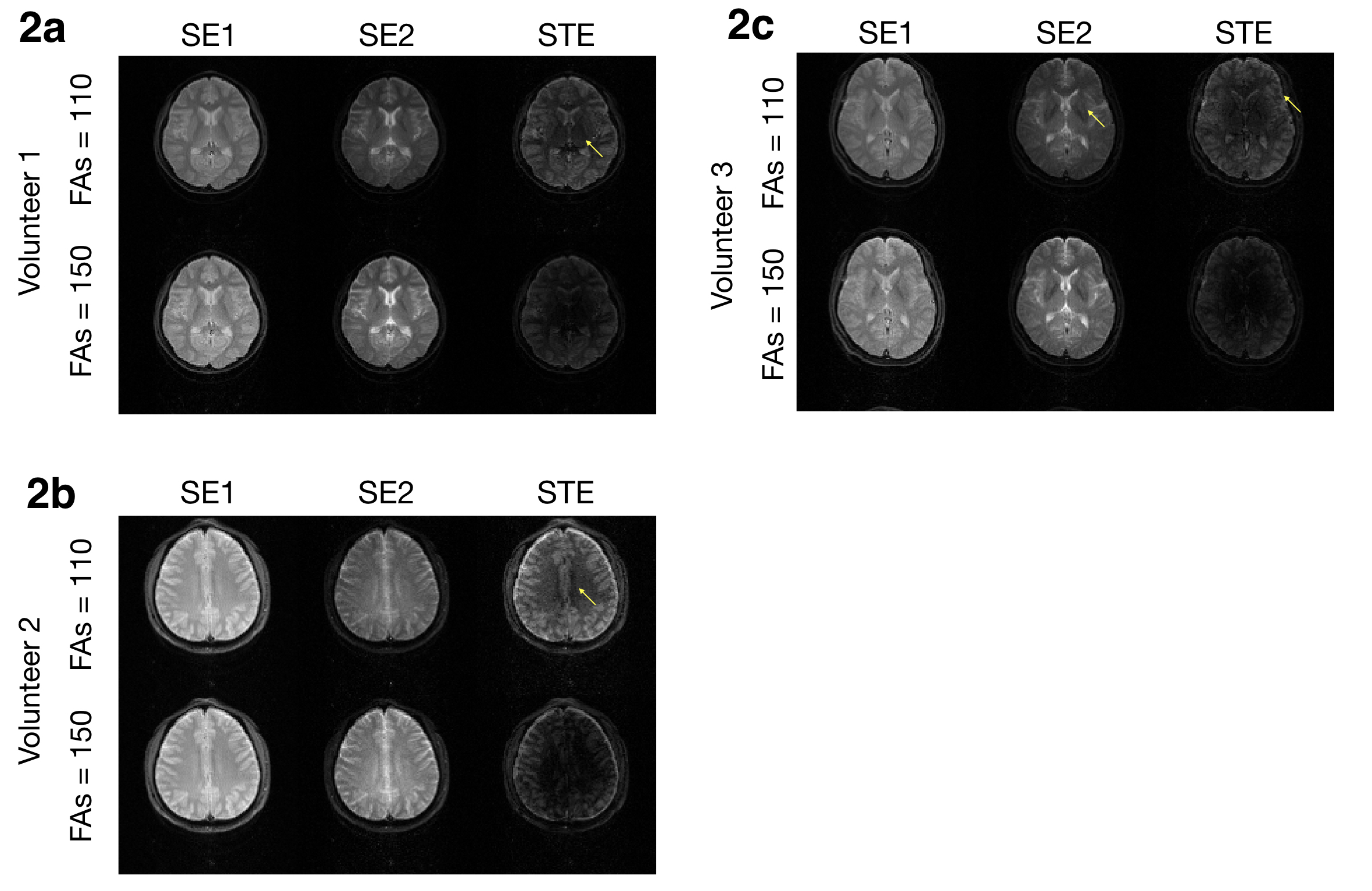
Our new multi-contrast framework was evaluated at a 3 Tesla MR scanner (GE Healthcare) with three healthy volunteers. To assess the performance for different contrast, we performed two Dixon-like multi-contrast GRASE imaging experiments with FAs ($$$\theta$$$ and $$$\phi$$$) = 100 degree and 150 degree. In each experiment, two sets of single-shot GRASE data with two CPMG echoes were acquired using a 8-channel coil, with TE (i.e. 2$$$\tau$$$) = 30 ms, FOV = 24x24 cm2, matrix size = 128x128, slice thickness = 8 mm, and TR = 5s. To reconstruct high-quality images, the N/2 ghost from the odd-even-echo asymmetry of k-space data was removed using a phase cycling method5. The SE1, SE2 and STE images were then reconstructed with the SENSE method6.
Results
Figures 2 shows the experimental results from three volunteers. As shown in the SE1 images, the proposed sequence provides high-quality T2 contrast of brain tissues as expected. The proposed Dixon-like framework generates high-quality SE2 images with stronger T2 weightings and STE images with inverse-T1 weightings. As indicated with yellow arrows in the SE1, SE2, and STE images, three different contrasts can provide complementary information of gray matter, white matter and cerebrospinal fluid for diagnosis, and enable a delineation of small anatomical structures such as external capsule.Discussion and conclusions:
Here we report a novel Dixon-like multi-contrast imaging framework enabled by two single-shot GRASE scans with phase difference of RF pulses . The new framework can simultaneously acquire two distinct T2-weighted images and an inverse-T1-weighted images within a clinically feasible scan time. Experimental results demonstrate the performance of this new technology on efficiently generating high-quality multi-contrast images. We expect the proposed framework can benefit clinical studies requiring high-quality multiple contrast information from challenging patients.Acknowledgements
Support from the Young Scholar Fellowship Program by Ministry of Science and Technology (MOST) in Taiwan with grant MOST107-2636-E-008 -002 and Mind Research and Imaging Center in National Cheng Kung University is acknowledged.References
1. Oshio K, Feinberg DA. GRASE (gradient‐and spin‐echo) imaging: a novel fast MRI technique. Magnetic Resonance in Medicine. 1991;20(2):344-9.
2. Chu ML, Chang HC, Oshio K, Chen NK. A single‐shot T2 mapping protocol based on echo‐split gradient‐spin‐echo acquisition and parametric multiplexed sensitivity encoding based on projection onto convex sets reconstruction. Magnetic Resonance in Medicine. 2018;79(1):383-93.
3. Hennig J, Nauerth A, Friedburg HR. RARE imaging: a fast imaging method for clinical MR. Magnetic Resonance in Medicine. 1986;3(6):823-33.
4. Hennig J. Echoes—how to generate, recognize, use or avoid them in MR‐imaging sequences. Part I: Fundamental and not so fundamental properties of spin echoes. Concepts in Magnetic Resonance. 1991;3(3):125-43.
5. Chen NK, Avram AV, Song AW. Two‐dimensional phase cycled reconstruction for inherent correction of echo‐planar imaging Nyquist artifacts. Magnetic Resonance in Medicine. 2011;66(4):1057-66.
6. Pruessmann KP, Weiger M, Scheidegger MB, Boesiger P. SENSE: sensitivity encoding for fast MRI. Magnetic Resonance in Medicine. 1999;42(5):952-62.
Figures