0937
MESMERISED: Super-accelerated 7T STEAM imaging for quantitative T1 and diffusion MRIFrancisco J. Fritz1, Benedikt A. Poser2, and Alard Roebroeck2
1Cognitive Neuroscience Department, Maastricth University, Maastricht, Netherlands, 2Cognitive Neuroscience Department, Maastricht University, Maastricht, Netherlands
Synopsis
We present the MESMERISED sequence which super-accelerates 7T STEAM imaging by employing both echo-shifting and multiband/simultaneous multislice acceleration. This leads to very high multiplicative acceleration factors and time efficiency for T1w and diffusion imaging. We illustrate this with 8x to 48x accelerated T1 relaxometry data at 1.5mm isotropic and 12x accelerated 3-shell diffusion data up to b = 5000s/mm2 at 1.7mm isotropic. MESMERISED extends possibilities to efficiently probe combined T1, T2 and diffusion contrast for multi-component modeling of relaxometry, diffusion and exchange.
Introduction
There is an increasing interest in quantitative imaging of T1, T2 and diffusion contrast due to a greater robustness against bias fields and artifacts, as well as better biophysical interpretability. However, acquisition time constraints are a challenge, particularly when more than one parameter is desired or combined T1/T2, T1/ADC or T2/ADC measurements are needed. Although ultra-high fields of 7T and above have desirable properties for many MR modalities, the shortening T2s and the high SAR load of inversion and refocusing pulses bring great challenges to quantitative T1, T2 and diffusion imaging. Diffusion-weighted MRI (dMRI) sequences like PGSE1 and SSFP2 have been optimized at 7T to mitigate some of these limitations; however, they remain limited to multiband (MB, a.k.a. SMS) acceleration factors of 2 to 41,3,4. It is well known that STEAM has advantages at higher fields: short TEs are allowable under shorter T2s; the advantage is taken of longer T1s, and it is highly SAR efficient5-7. However, STEAM results in long TRs (and hence acquisition times) which severely restricts the amount of data that can be obtained. In this work, we present the MESMERISED (Multiplexed Echo Shifted Multiband Excited and Recalled Imaging of Steam Encoded Diffusion) sequence to achieve super-accelerated 7T STEAM imaging, by combining echo-shifting and multiband acceleration. This leads to very high multiplicative acceleration factors and time efficiency for 7T T1-weighted (T1w) and diffusion imaging.Methods
Figure 1 shows the MESMERISED sequence in comparison to MB-accelerated STEAM. MESMERISED combines blipped-CAIPI MB-EPI8 with echo-shifting (ES) of multiple stimulated-echoes (STEs). The echo-shifting9,10 is independent of MB acceleration and allows for every high multiplicative ESxMB acceleration factors, particularly under moderate and long TMs. MESMERISED was implemented and tested on one healthy subject on a Magnetom Siemens 7T human MRI system (Siemens Healthineers, Erlangen, Germany). MESMERISED was acquired at 1.8mm isotropic, using ESxMB of 1x3, 2x3 and 4x3 (ES=1 denotes no shift) (GRAPPA-2, FoV 230x230mm2, PF 6/8). Additionally, T1w MESMERISED at 1.5mm isotropic was acquired at variable TM and ES keeping TR constant (GRAPPA-3, FoV 192x192mm2, PF 6/8). Finally, diffusion-weighted images were acquired at 1.7mm isotropic (ESxMB = 4x3, GRAPPA-2, FoV 218x218mm2, PF 6/8) and 2.0mm isotropic (ESxMB = 2x2, GRAPPA-2, FoV 208x208mm2, PF 6/8). TR and TE were kept constant throughout. FLEET GRAPPA ACS lines were obtained (24 for GRAPPA-2 and 36 for GRAPPA-3, flip angle 12deg). To remove the need for SAR-intensive FatSat pulses, “fat-unfocussing” was achieved by appropriate setting of the pulse lengths and BWTP11. Diffusion-weighted data and T1w relaxometry data were distortions corrected with Topup and Eddy tools using FSL v5.0.1112. Relaxometry data and diffusion-weighted data were analyzed using MDT13 with Tensor, Ball&Stickr2, and NODDI diffusion models.Results and discussion
Figure 2 shows a comparison between image and signals obtained using MESMERISED with no ES (ESxMB=1x3 – or MB STEAM, cf. Figure 1A), 2x3 and 4x3 for the spin-echo (SE) and STE which shows that echo-shifted interleaving does not affect the acquired signals and allows them to be acquired at much higher acceleration in a shorter time (see min TR, Figure 2A). Figure 3 shows MESMERISED T1w data at 1.5mm isotropic which illustrates that TR can remain constant across different TMs (and T1-weighting) by increasing ES factor proportionally to TM. This realizes total acceleration factors (ESxMB) of 8 to 48, highlighting the super-acceleration capability of MESMERISED for T1 relaxometry. Figures 3 and 4 show high b-value 2-shell and 3-shell MESMERISED diffusion-weighted data, illustrating that high acceleration (3x4) and b-values (b5000) are possible for 1.7mm isotropic for reduced-SAR pulses which allow a whole brain volume TR of 4.9s (Figure 4). The multi-shell diffusion data is suitable for modeling with standard DTI, biophysical multi-compartment modeling with NODDI, and crossing fiber modeling with the Ball & 2 Sticks model (Figure 5).Conclusions
MESMERISED achieves super-accelerated 7T STEAM imaging by combining echo shifting and multiband/simultaneous multislice acceleration. This leads to very high multiplicative acceleration factors and time efficiency for T1-weighted and diffusion imaging. MESMERISED can probe combined T1, T2 and diffusion contrast with high time efficiency for biophysical modeling of multi-component relaxometry, diffusion, and exchange. Future combination with SAR efficient MB pulses, such as PINS14, multi-PINS3 and pTx-MB4 will allow high data quality at full acceleration without repetition times being limited by SAR.Acknowledgements
No acknowledgement found.References
- Vu A.T., Auerbach E., Lenglet C, Moeller S, Sotiropoulos S.N, Jbabdi S., Andersson J, Yacoub E, Ugurbil K. High resolution whole brain diffusion imaging at 7T for the Human Connectome Project, NeuroImage, 2015; 122: 318-331.
- Lu L, Erokwu B, Lee G, et al. Diffusion-prepared fast imaging with steady-state free precession (DP-FISP): a rapid diffusion MRI technique at 7 T. Magnetic resonance in medicine, 2011; 68(3): 868-873.
- Eichner C, Wald L. L. and Setsompop K. A low power radiofrequency pulse for simultaneous multislice excitation and refocusing. Magnetic resonance in medicine, 2014; 72: 949-958.
- Wu X, Schmitter S, Auerbach EJ, Moeller S, Uğurbil K, Van de Moortele PF. Simultaneous multislice multiband parallel radiofrequency excitation with independent slice-specific transmit B1 homogenization. Magnetic resonance in medicine, 2013; 70(3):630-8.
- Frahm J, Merboldt K.D, Hänicke W, Haase A. Stimulated echo imaging. Journal of Magnetic Resonance. 1985; 64:81-93
- Merboldt KD, Hanicke W, Frahm J. Diffusion imaging using stimulated echoes. Magnetic resonance in medicine. Jun 1991;19(2):233-239.
- Heiland S., Dietrich O., Sartor K. Diffusion-weighted imaging of the brain: comparison of the stimulated- and spin-echo-planar sequence. Neuroradiology (2001) 43: 442-447
- Setsompop K, Gagoski BA, Polimeni JR, Witzel T, Wedeen VJ, Wald LL. Blipped-controlled aliasing in parallel imaging for simultaneous multislice echo planar imaging with reduced g-factor penalty. Magnetic resonance in medicine, 2011; 67(5): 1210-1224.
- Feinberg DA, Moeller S, Smith SM, Auerbach E, Ramanna S, Glasser MF, et al. Multiplexed Echo Planar Imaging for Sub-Second Whole Brain FMRI and Fast Diffusion Imaging. PLoS ONE, 2010; 5(12): e15710
- Gibson, Andrew & M Peters, Andrew & Bowtell, Richard. Echo-shifted multislice EPI for high-speed fMRI. Magnetic resonance imaging, 2006; 24: 433-442.
- Ivanov D, Schäfer A, Streicher M. N., Heidemann R. M., Trampel R. and Turner R. A simple low‐SAR technique for chemical‐shift selection with high‐field spin‐echo imaging. Magnetic resonance in medicine, 2010; 64: 319-326
- Jenkinson M, et al. FSL. NeuroImage, 2012; 62:782-90.8.
- Harms, R.L., Fritz, F.J., Tobisch, A., Goebel, R., Roebroeck, A. Robust and fast nonlinear optimization of diffusion MRI microstructure models. Neuroimage, 2017; 155, 82-96
- Norris D. G., Koopmans P. J., Boyacioğlu R. and Barth M. Power independent of number of slices (PINS) radiofrequency pulses for low‐power simultaneous multislice excitation. Magnetic resonance in medicine, 2011; 66: 1234-1240
Figures

MESMERISED versus MB-STEAM. (A) A single block (TBSTEAM) of the MB-STEAM sequence, consisting of a spin-echo (SE) and stimulated-echo (STE) readout, will acquire MB slices per block, leaving considerable dead time (TD). (B) The MESMERISED sequence utilizes this dead time by echo shifting one or more SBA blocks (here ES factor 3), resulting in full duty cycle and high time efficiency. Stored longitudinal magnetization evolves while SEi is acquired. Subsequently, STEi is read out in the SBB blocks. MESMERISED will acquire ESxMB slices per (slightly longer) TBMSMD. G: diffusion/crusher gradient; S: spoiler gradient; SB: sequence block.

(A) Comparison of MESMERISED images at 1.5 mm isotropic, acquired with MB = 3 at different echo shifts of ES = 1, ES = 2 and ES = 4 but otherwise identical parameters. Left: spin-echo, right: stimulated-echo. Minimal achievable TR (min TR) is indicated to illustrate increased acceleration capability with ES > 1. (B) Corresponding signal line plots (see insert) for SE (left) and STE (right) show no loss in signal fidelity at increasing ES.

MESMERISED T1 relaxometry data at 1.5 mm isotropic acquired with mixing times of 0.25 s, 0.5 s, 0.8 s, 1.2 s, and 1.5 s, at MB=4, and matched TR/TE. TR can remain constant by increasing ES factor proportionally to TM so that total acceleration factor, equal to ES x MB, increases from 8 to 48.
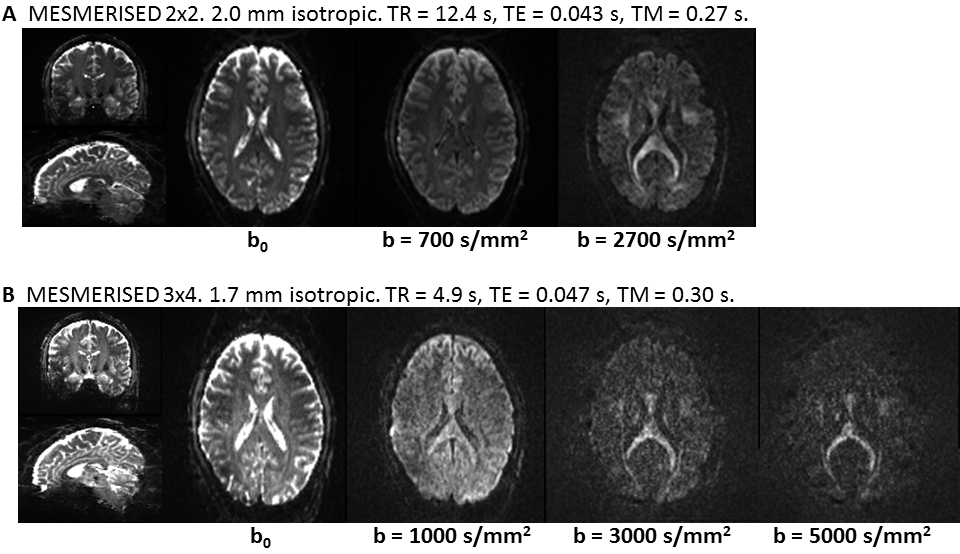
MESMERISED diffusion-weighted data. (A) 2x2 accelerated 2-shell data (27 directions - b700 and 53 directions - b2700) at 2.0 mm isotropic with RF pulses empirically optimised for image quality and fat defocussing (Exc:5ms/BWTP=2.5, Sto/Rec:5ms/BWTP=8). (B) 3x4 accelerated 3-shell data (27 directions - b1000 , 40 directions - b3000 and 40 directions - b5000) at 1.7mm isotropic with SAR optimized pulses (Exc:6ms/BWTP=2.2, Sto/Rec:12ms/BWTP=2.2) running at min TR. Scaling individually adjusted.

Diffusion modeling results for the MESMERISED data in Fig 4. Left: MESMERISED 2x2, FA weighted DEC map from DTI modeling on the b700 shell, and intracellular volume fraction map from NODDI fit using both shells. Right: MESMERISED 3x4, FA weighted DEC map from DTI modeling on the b1000 shell and total stick fraction map from the Ball & 2 Sticks (B&Sr2) crossing fiber model fit.