0936
DiSpect: Displacement Spectrum Imaging of Flow, Diffusion and Tissue Perfusionusing Spin-Labeling and Stimulated Echoes1Department of Electrical Engineering and Computer Sciences, University of California, Berkeley, Berkeley, CA, United States
Synopsis
We present a Fourier-encoding variant of Displacement Encoding with Stimulated Echoes (DENSE). DENSE encodes bulk displacement of spins but fails to capture partial volume displacement or flow. By performing multiple scans with increasing DENSE encoding, we are able to resolve the spectrum of displaced spins within a voxel and differentiate it from static tissue. Our approach opens the possibility of quantitatively measuring complex spin dynamics, such as motion, flow, diffusion and perfusion. We present results in a flow phantom and in-vivo brain. Our results demonstrate sensitivity to slow CSF flow, blood flow, and tissue perfusion.
Introduction
The complex dynamics of flow, diffusion and perfusion are important for the study of many biophysical functions and disease diagnosis. Tools to probe these phenomena1 include diffusion MRI (DWI), arterial spin-labeling (ASL), phase-contrast (PC), and others. Each of these methods sensitizes the magnetization magnitude and/or phase to the desired phenomena to be imaged. However, each technique suffers from shortcomings. For example, DWI and ASL suffer from low SNR and sensitivity to bulk motion. Here we present a new addition to the toolkit of techniques. Our approach is a Fourier-encoding variant of Displacement Encoding with Stimulated Echoes (DENSE)1,2. DENSE encodes bulk displacement of spins in the image phase. Since DENSE is a tagged, stimulated echo approach the displacement phase can be accumulated over a long mixing time, on the order of the T1 relaxation of the tissue. Therefore DENSE found applications in measuring the Cardiac motion between tagging and imaging. DENSE however fails to capture partial volume displacement or blood flow. By performing multiple scans with increasing DENSE encoding, we are able to resolve the spectrum of displaced spins within a voxel and differentiate it from static tissue. In a way, our technique which we name DiSpect (for Displacement Spectrum) is the displacement equivalent to Fourier velocity imaging, while DENSE is the displacement equivalent to velocity phase-contrast. By being able to measure a spectrum of displacements our approach opens the possibility of quantitatively measuring complex inter and intra-voxel spin dynamics, such pulsatile tissue motion, flow, diffusion and even perfusion that occurs over the evolution time of T1 relaxation. Here, we demonstrate DiSpect MRI in a complex flow phantom and example of in-vivo brain.Methods
DiSpect (Fig. 1a), starts with DENSE, a mixing duration is tuned to accommodate the spin displacement until an RF excitation pulse is applied. Conventional imaging acquisition samples the displacement encoded signals. To obtain pure displacement phase modulation, phase cycling has to be applied in the DENSE module1. Fourier encoding of the displacement is done by phase-encoding increments of the displacement encoding gradients. The displacement spectrum of each voxel is obtained by a Fourier transformation along the displacement encoding dimension after a phase correction. Fig. 1b shows the different displacement spectra we expect for different tissue dynamics. To fully capture the 3D displacement spectrum Multi-dimensional encoding with appropriate displacement resolution and range should be used to avoid aliasing.
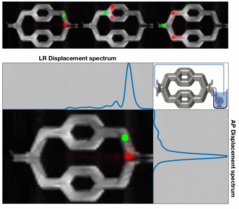
Our 3D-printed flow phantom was imaged using a GRE-based DiSpect method on Siemens Trio 3T system: Two displacement encoding directions were acquired with spatial resolutions of 3×3 mm2, 64x40 image matrix, mixing duration of 400 ms and displacement encoding range of 150 mm with 50 encodes along the LR direction and 90 mm with 30 encodes along the AP direction. To further explore the potential of DiSpect MRI for perfusion studies, an in-vivo brain scan was performed using DiSpect EPI method with pulse-ox triggering: Three displacement encoding directions-LR, AP and SI-were acquired with spatial resolution 2×2 mm2, displacement encoding range of 4 mm with 32 encodes along each direction and a mixing duration of 500 ms.
Results and Discussion
Fig.2 shows that DiSpect can be used to trace the fluid pathway in a flow phantom. Two displacement encoding directions, LR and AP , were acquired in order to trace the in-plane water flow in the tube. What is remarkable about DiSpect is its ability to trace multiple contributions of displaced spins. It is important to note, that unlike phase-contrast (PC) which provides information on where spins are going, DiSpect provide information on where they came from.
Fig.3 shows the potential of DiSpect for perfusion studies. Three colored maps can be generated by analysing the asymmetry of displacement spectrum in the brain (Fig 1b) from three displacement encoding directions: LR, AP, and SI (Superior to Inferior). By computing the energy in these three maps, a displacement asymmetry energy is generated. We hypothesize that this is an indicator for flow and perfusion in tissues. Unlike ASL3, which employs a time delay between the application of the labeling pulse and image acquisition, in order to allow for the labeled bolus to flow into the target tissue in the imaging region, DiSpect encodes the displacement immediately after the displacement encoding gradient (several ms). Therefore, DisPect benefits from less T1 relaxation signal loss as a result of the reduced perfusion mixing time compared to the PLD of ASL MRI. More work is currently on-going to explore the potential use of DiSpect for perfusion studies.
Conclusions
The displacement spectrum imaging (DiSpect) MRI using spin-labeling and stimulated echoes can obtain different displacement spectra for different tissue dynamics, such as motion, flow, diffusion and perfusion.Acknowledgements
We thank the following funding sources: National Institutes of Health (NIH) grants: R01EB009690, U01EB025162, R01EB026136, R01HL136965; Bakar Fellowship.References
1. Aletras, A. H., Ding, S., Balaban, R. S., & Wen, H. DENSE : Displacement Encoding with Stimulated Echoes in Cardiac Functional MRI, 1999, 252,: 247–252.
2. Epstein, F. H., Gilson, W. D. Displacement-encoded cardiac MRI using cosine and sine modulation to eliminate (CANSEL) artifact-generating echoes. Magnetic Resonance in Medicine, 2004, 52: 774–781.
3. Alsop, D. C., Detre, J. A., Golay, X., et al. Recommended implementation of arterial spin-labeled Perfusion mri for clinical applications: A consensus of the ISMRM Perfusion Study group and the European consortium for ASL in dementia. Magnetic Resonance in Medicine, 2015, 73(1), 102–116
Figures