0932
Template-based field map prediction for whole-body shimming applications1United Imaging Healthcare, Shanghai, China, 2UIH America, Houston, TX, United States
Synopsis
For a whole-body scan, shimming are required to be carried out in multiple bed positions to provide homogeneous field for imaging different body parts. Thus, techniques to shorten the shimming time are required. This work presents a rapid B0 field prediction method for whole-body shimming based on a large field map database of different body parts. A range of applications could benefit from this technique.
Purpose
This work presents a rapid B0 field prediction method for whole-body shimming based on a large field map database of different body parts. Moreover, it demonstrates there is similarity of the field distribution in the corresponding body parts between different subjects. For routine whole-body imaging, this method accelerates the static shimming stage across multiple bed positions.Introduction
In routine magnetic resonance imaging protocols, static shimming is implemented to providing a homogeneous field. The conventional shimming procedures start from acquiring field map with the default vendor calibrated shim for an oil phantom, tune-up shim, because of the lack of prior knowledge of B0 field distribution in the sample. For a whole-body scan, multiple bed positions are needed for imaging different body parts (e.g., brain, chest, abdomen and extremities). Thus, the time for shimming is multiplied. If the shim solutions are not converged, multiple iterations are required which further prolong the scan time and significantly shorten acquisition time compared with conventional field map. Previously it has been shown that a fast template-based field map prediction method could produce optimal shims for brain imaging without measuring B0 field.1 In this work, we extend the scope and examine the performance of shimming using the template-based method on different body parts.Methods
The experiments were performed on a simultaneous whole-body PET/MR scanner (Unites Imaging, Shanghai, China) in Zhongshan Hospital, Shanghai, China. A low-resolution whole-body field map database (FOV=12.5mm*15.6mm*15.6mm, Slices=32) was acquired from different subjects as part of clinical studies (head image=274, chest image=127, buttock image =128, leg image =116). The procedure of predicting the field map of different body parts are show in Figure 1:
- Register the structure of a specific body part to a reference structural template by using a MATLAB embedded 3D image registration algorithm.
- Transform field maps to the reference by applying the registration transformation generated in Step 1.
- Average all the registered field maps to generate a field map template for this specific body part.
- Register an input structure image to the reference structure and the field map template is transformed to the space of the input image to generate a predicted field map.
The performance of static shimming using the method was evaluated on the field map database in simulation with a leave-one-out cross validation. For comparison, we calculated the optimal static shim based on measured field maps of specific body parts for reference and the averaged of the shim, termed the fixed shim.
Results
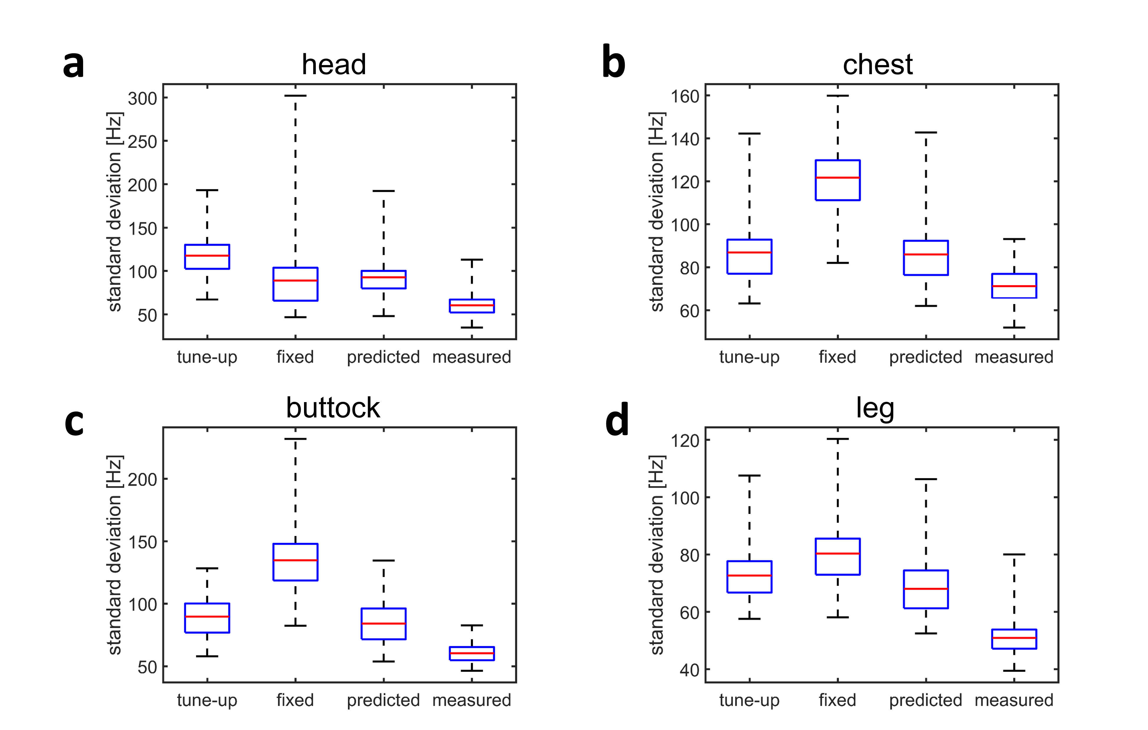
Figure 2 shows the structure of different body parts and the corresponding generated field map template. As can been seen from the figure, most prominent field heterogeneity occurs in the brain. Field distribution in different body parts is highly structured.
Comparison of the residual field standard deviation after shimming using different methods are shown in Figure 3. Overall, the proposed prediction method outperforms the tune-up shim and the fixed shim. It demonstrates that the field map template produces extra field distribution information even for different body parts. However, shimming using the prediction method does not perform as good as the measured field maps. Interestingly, for the brain region, the fixed shim yields better results that the prediction method. This shows that there are some field information are better estimated by the fixed shim in the brain.
Discussion
In whole-body imaging, the prediction method is capable of providing a rapid prediction of the field distribution of different body parts using a field map database and body structure information. Furthermore, the template-based prediction method can be used as alternative to routine static shimming for some applications which are not demanding on highly homogeneous field. Additionally, the prediction method can be further improved by incorporating deep learning techniques.
Conclusion
The template-based prediction method has potential to provide quick and good whole-body shimming. A range of clinicalapplications could benefit from it.
Acknowledgements
No acknowledgement found.References
1. Shi Y, Vannesjo SJ, Miller KL and Clare S. Template‐based field map prediction for rapid whole brain B0 shimming. Magn Reson Med. 2018:80:171-180.Figures