0929
Model-Based Single-Shot EPI Reconstruction with Sparsity Regularization1Department of Radiology, Mayo Clinic, Rochester, MN, United States, 2Department of Radiologic Technology, Faculty of Associated Medical Sciences, Chiang Mai University, Chiang Mai, Thailand
Synopsis
Echo planar imaging (EPI) is widely used
clinically for its speed, but is known to be sensitive to non-idealities like
B0 field inhomogeneity, eddy currents, and gradient nonlinearity. Such
non-idealities are not typically managed during image reconstruction, resulting
in geometrically distorted images. Post-processing corrections (e.g., image-based
interpolation) usually tend to degrade resolution. In this work, a
comprehensive model-based reconstruction framework that prospectively and
simultaneously accounts for non-idealities in accelerated single-shot EPI
acquisitions is proposed. Sparsity regularization is also incorporated to
mitigate noise amplification. The proposed algorithm is demonstrated on
brain MRI data acquired on a compact 3T MRI system.
Overview
Echo-planar-imaging$$$^1$$$ (EPI) is widely used clinically for its speed, but is known
to be sensitive to hardware non-idealities including B0 field-inhomogeneity$$$^2$$$, eddy-currents$$$^3$$$, and gradient nonlinearity$$$^{4}$$$ (GNL).
Such non-idealities are not typically managed during image reconstruction,
resulting in geometrically distorted images. To correct such distortion,
image-based post-processing methods$$$^{5-6}$$$ (e.g., interpolation) are commonly
applied, but these tend to degrade resolution and provide only partial corrections. Hybrid reconstruction models that leverage
image-based corrections within an iterative framework have also been proposed,
with some success$$$^{7-10}$$$. In this work, a comprehensive model-based iterative
reconstruction (MBIR) framework that prospectively and simultaneously accounts
for multiple non-idealities in accelerated single-shot-EPI acquisitions is
proposed. Sparsity regularization is also incorporated to mitigate noise
amplification. Following technical
description of the framework setup, associated calibration procedures, and a
nonlinear optimization strategy for solving it, the practical benefits of the
proposed algorithm are demonstrated on brain MRI data acquired on a
high-performance, compact 3T-MRI system. Comparisons against conventional
reconstruction and correction pipelines are also presented.
Methods
Comprehensive EPI Signal Model: Let $$$TE$$$, $$$\Delta t$$$, and $$$T$$$ denote the echo, dwell, and echo-spacing times of a single-shot EPI acquisition. Neglecting $$$T_2^*$$$ and presuming pre-correction of eddy-current and gradient delay effects$$$^3$$$, the signal measured during readout $$$m \in[0,M)$$$ of phase-encoding line $$$n\in[0,N)$$$ can be modeled as:$$g_c[m,n]=\int_{\Omega _x}\int_{\Omega_y}s_c(x,y)f(x,y)e^{-j\Delta\omega_0(x,y)\tau[m,n]}e^{-j((-1)^nk_x[m]\eta_{x}(x,y)+k_y[n]\eta_{y}(x,y))}dxdy+\epsilon[m,n]\qquad(1)$$where $$$f$$$ is the target signal, $$$k_x$$$ and $$$k_y$$$ are sampling maps, $$$s_c$$$ is the sensitivity profile for coil $$$c\in[0,C)$$$, $$$\Delta\omega_0$$$ is the B0-field map, $$$\tau[m,n]=TE+(m-\frac{M-1}{2})\Delta{t}+(n-\frac{N-1}{2})T$$$ denotes sampling-time, $$$\eta_{x}(x,y)$$$ and $$$\eta_{y}(x,y)$$$ are GNL maps$$$^{11}$$$, and $$$\varepsilon$$$ is Gaussian noise. Note that Eq.1 accommodates ramp-sampling, parallel imaging, and partial Fourier acceleration.
As EPI uses high readout-bandwidth, $$$\Delta{t}<<T$$$ and off-resonance primarily manifest along its phase encoded (i.e., blipped) direction -- thus $$$\Delta{t}\approx{0}$$$ can be assumed. Letting $$$u(x,y)=f(x,y)e^{i\Delta\omega_0(x,y)TE}$$$, discretizing$$$^{12}$$$ and time-segmenting$$$^{13}$$$ (Eq.1) yields:$$g_c[m,n]=\sum\limits_{l=0}^L\Phi_l(n)\left\{\sum\limits_{p=0}^{P-1}\sum\limits_{q=0}^{Q-1}s_c[p,q]u[p,q]e^{-j\Delta\omega_0[p,q][\gamma_{l}-\frac{N-1}{2}]T}e^{-j((-1)^nk_x[m]\eta_{p}[p,q])+k_y[n]\eta_{q}(p,q))}\right\}+\epsilon[m,n]\qquad(2)$$where $$$\Phi_l$$$ is a spectral window, $$$\gamma_{l}$$$ denotes the center of segment $$$l$$$, and $$$p\in[0,P)$$$ and $$$q\in[0,Q)$$$ are pixel indices. Defining $$$W_l=diag\left\{e^{-i\Delta\omega_0[p,q][\gamma_l-\frac{N-1}{2}]T}\right\}$$$ and $$$S=[diag\left\{s_0\right\}\cdots{diag}\left\{s_{C-1}\right\}]^{T}$$$, Eq.2 abstracts to:$$g=(I\otimes\sum\limits_{l=0}^L\Phi_lFW_l)Su+\epsilon=Au+\epsilon\qquad(3)$$where $$$F$$$ is a Fourier transform. If a phase reference ($$$\theta$$$) for $$$u$$$ is available, the complex target reduces to $$$u=diag\left\{e^{j\theta}\right\}v=P(\theta)v$$$, where $$$v\in\mathbb{R}$$$$$$^{12}$$$.
EPI Image Reconstruction: The target image $$$u$$$ in (Eq.2) can be estimated via regularized least squares estimation:$$\mathop{\min}_{v\in\mathbb{R}}\left\{J(v)\triangleq\beta{R(v)}+\|AP(\theta)v-g\|_2^2\right\}\qquad(4)$$where $$$\beta>0$$$ is a regularization parameter. Here, we adopt joint redundant wavelet sparsity regularization:$$R(v)=\left\|\sum\limits_{k\in\xi}(\delta^{T}_k\otimes\Psi\Gamma{_k}v)\right\|_{1,2}\qquad(5)$$where $$$\delta$$$and $$$\otimes$$$ are Kronecker's delta and product, $$$\Psi$$$ is the Daubechies-4-wavelet, $$$\Gamma{_k}$$$ is a shift operator over neighborhood $$$\xi$$$, and $$$\|\cdot\|_{1,2}$$$ is the matrix-1,2 norm. As Eq.4 is convex, it can be solved using FISTA$$$^{14}$$$ with composite splitting$$$^{15}$$$. For efficiency, $$$F$$$ is implemented as a type-III non-uniform fast Fourier transform (NUFFT)$$$^{16}$$$. Vendor-provided coil sensitivity profiles, and ramp-sampling and $$$GNL$$$ information were incorporated. B0 field maps were estimated from a dual-echo GRE calibration scan using a graph cut-based procedure$$$^{16-17}$$$. The phase reference, $$$\theta$$$, was obtained by minimizing Eq.4 without phase constraints.
Data Acquisition and Processing: Two healthy volunteers were imaged on a compact 3T MRI system: (G$$$_{max}$$$=80mT/m, SR=700T/m/s) under an IRB-approved protocol, with $$$C=32$$$ or $$$C=8$$$ head-only receiver-only RF head coils$$$^{18-21}$$$. Following field map, coil sensitivity, and eddy current calibration, single-shot EPI acquisitions were performed with the parameters in Table 1. Prior to MBIR, eddy current correction was performed in k-space using vendor-provided tools. MBIR was performed in Matlab, using a 2X-oversampled NUFFT with width=5 Kaiser-Bessel kernel, L=N time segments, and a $$$7\times{7}$$$ shift window. FISTA was executed for 30 iterations. The regularization parameter was chosen manually ($$$\beta=0.02$$$). For comparison, vendor reconstructions were saved and post-processed using standard intensity-corrected image interpolation for distortion correction$$$^{22}$$$.
Results
Fig. 1 demonstrates the distortion correction performance of the proposed MBIR strategy against the vendor reconstruction, with and without post-processing. Registration error maps are shown with respect to a spin warp reference; large geometric mismatches are indicated with yellow ellipses. Fig. 2 shows enlargements of the Fig. 1 images to demonstrate the relative noise performance of MBIR. Both conventional post-processing and MBIR effectively correct coarse distortion, with the latter providing modest improvements; however, MBIR offers reduced noise amplification relative to both the original and post-processed vendor reconstructions. This advantage is particularly apparent in low-contrast structures like the caudate head, as indicated in Fig. 3.Discussion
We have developed a comprehensive MBIR strategy for single-shot EPI that prospectively accounts for off-resonance, GNL, ramp-sampling, and parallel imaging and partial Fourier-type acceleration, and leverages wavelet-based sparsity regularization for improved noise performance. The advantages of this approach were demonstrated relative to the conventional vendor-reconstruction, with and without post-processing. Although demonstrated here for anatomical imaging, the proposed framework could benefit any EPI-based application, such as diffusion or elastography. Like other MBIR strategies, advancement opportunities include development and use of tailored optimization strategies and automated regularization parameter selection -- these will be investigated in future work.Acknowledgements
This work was supported by NIH U01 EB024450.References
1) Mansfield P. Multi-planar image formation using NMR spin-echoes. J Phys C. 1977;10:L55–L58.
2) Jezzard P, Balaban RS. Correction for geometrical distortion in echo planar images from bo field variations. Magn Reson Med. 1995;34:65–73.
3) Ahn CB, Cho ZH. Analysis of eddy currents induced artifacts and temporal compensation in nuclear magnetic resonance imaging. IEEE Trans Med Imaging 1991; 10: 47–52.
4) David C. Newitt, Ek T. Tan, Lisa J. Wilmes, et al. Gradient Nonlinearity Correction to Improve Apparent Diffusion Coefficient Accuracy and Standardization in the American College of Radiology Imaging Network 6698 Breast Cancer Trial. J Magn Reson Imaging 2015; 42(4): 908–919.
5) AnderssonJLR., Skare S., Ashburner, J. How to correct susceptibility distortions in spin-echo echo-planar images: application to diffusion tensor imaging. NeuroImage 2003; 20(2): 870–888.
6) Glover GH, Pelc NJ. Method for correcting image distortion due to gradient nonuniformity. US Patent # 4,591,789, May 27, 1986.
7) Yarach U., In MH, Chatnuntawech I, et al. Model-based iterative reconstruction for single-shot EPI at 7T. Magn Reson Med 2017; 78: 2250-2264.
8) Mengye L, Yilong L, and Ed XW, Improved Parallel Imaging Reconstruction of EPI using Inversely Distortion Corrected FLASH as Calibration Data. Proceeding in ISMRM 2018. (Abstract#3509)
9) Usman M, Kakkar L, Shmueli K, Arridge S, Atkinson A. An integrated model-based framework for the correction of signal pile-up and translational offsets in prostate diffusion MRI. Proceeding in ISMRM 2018. (Abstract#1639)
10) Zahneisen B, Aksoy M, Maclaren J, Wuerslin C, Bammer R. Extended hybrid-space SENSE for EPI: Off-resonance and eddy current corrected joint interleaved blip-up/down reconstruction. NeuroImage 2017; 153: 97-108.
11) Tao S, Trzasko JD, Shu Y, et al. Integrated Image Reconstruction and Gradient Nonlinearity Correction. Magn Reson Med 2015; 74(4): 1019-1031.
12) Fessler JA. Model‐based image reconstruction for MRI. IEEE Signal Process Mag 2010; 27:81–89.
13) Sutton B, Noll D, Fessler JA. Fast, iterative image reconstruction for MRI in the presence of field inhomogeneities. IEEE Trans Med Imaging 2003; 22: 178–188.
14) Beck A, and Teboulle M. A fast iterative shrinkage-thresholding algorithm for linear inverse problems, SIAM Journal on Imaging Sciences 2009; 2(1): 183–202
15) Huang J, et al. Composite splitting algorithms for convex optimization. Computer Vision and Image Understanding 2011; 115(12): 1610-1622.
16) Tao S, Trzasko JD, Shu Y, Huston J 3rd, Johnson KM, Weavers PT, Gray EM, Bernstein MA. Non-Cartesian MR image reconstruction with integrated gradient nonlinearity correction. Med Phys 2015; 42: 7190–7201.
17) Kolmogorov V, and Zabih R. What energy functions can be minimized via graph cuts?, IEEE Trans. Pattern Anal. Mach. Intell 2004; 26: 147–159.
18) Foo TK, Laskaris E, Vermilyea M, Xu M, Thompson P, Conte G, Van Epps C, Immer C, Lee SK, Tan ET. Lightweight, compact, and high-performance 3 T MR system for imaging the brain and extremities. MRM 2018. doi: 10.1002/mrm.27175.
19) Weavers PT, Shu Y, Tao S, Huston J, 3rd, Lee SK, Graziani D, Mathieu JB, Trzasko JD, Foo TK, Bernstein MA: Technical Note: Compact three-tesla magnetic resonance imager with high-performance gradients passes ACR image quality and acoustic noise tests. Med Phys 2016, 43(3):1259-1264.
20) Weavers PT, Tao S, Trzasko JD, Frigo LM, Shu Y, Frick MA, Lee SK, Foo TK, Bernstein MA. B0 concomitant field compensation for MRI systems employing asymmetric transverse gradient coils. Magn Reson Med 2018 79, 1538-1544.
21) Tao S, Weavers PT Trzasko JD, Shu Y, Huston J , Lee SK, Frigo LM, Bernstein MA. Gradient Pre-emphasis to Counteract First-Order Concomitant Fields on Asymmetric MRI Gradient Systems. Magn Reson Med. 2017; 77:2250-2262.
22) Holland D, Kuperman JM, Dale AM. Efficient Correction of Inhomogeneous Static Magnetic Field-Induced Distortion in Echo Planar Imaging. Neuroimage 2010; 50: 175-183
Figures
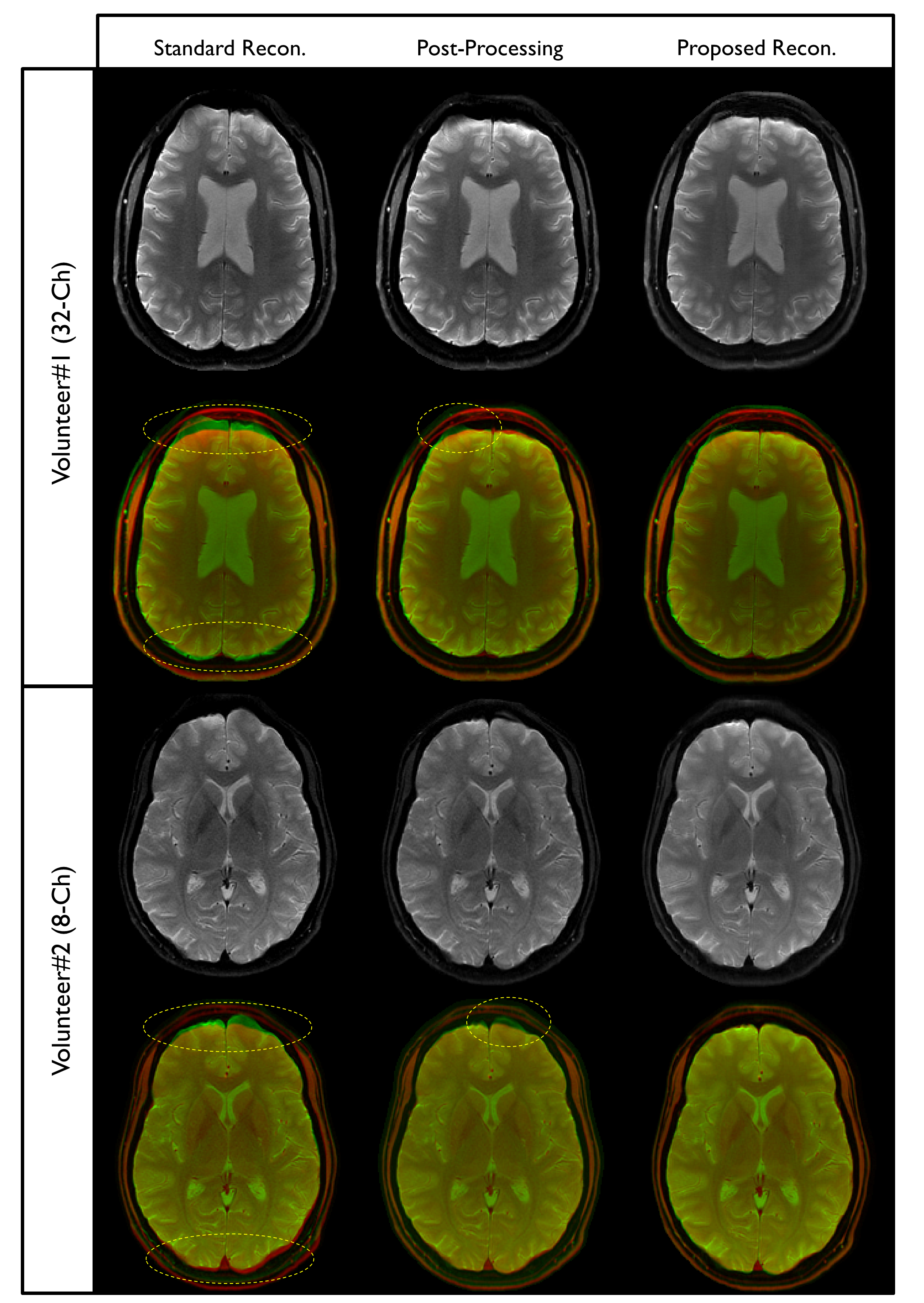
Fig. 1. (1st column) The images obtained by the vendor-provided reconstruction. (2nd column) Vendor reconstruction results post-processed with intensity-corrected image-domain interpolation. (3rd column) Results from the proposed MBIR strategy.
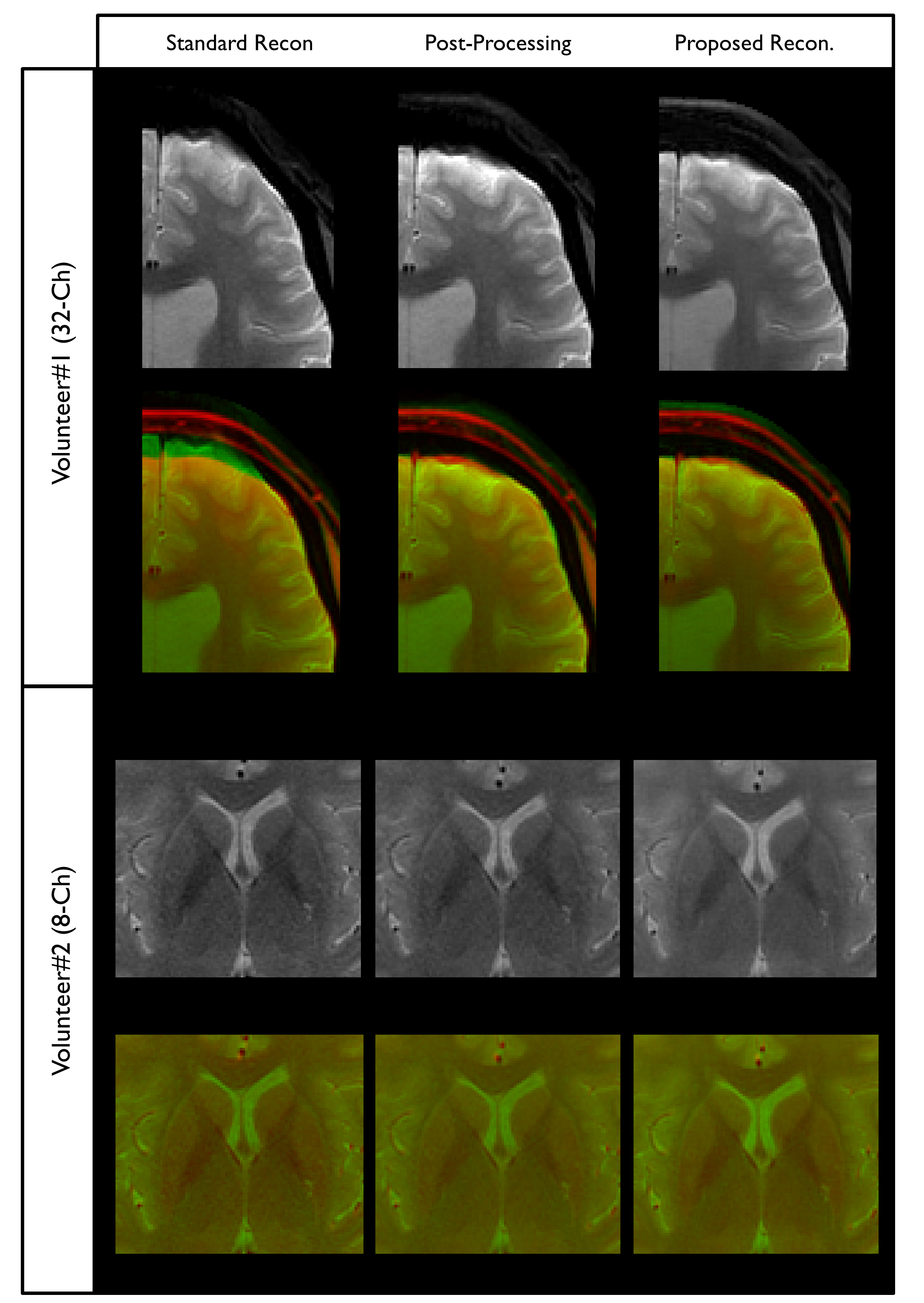
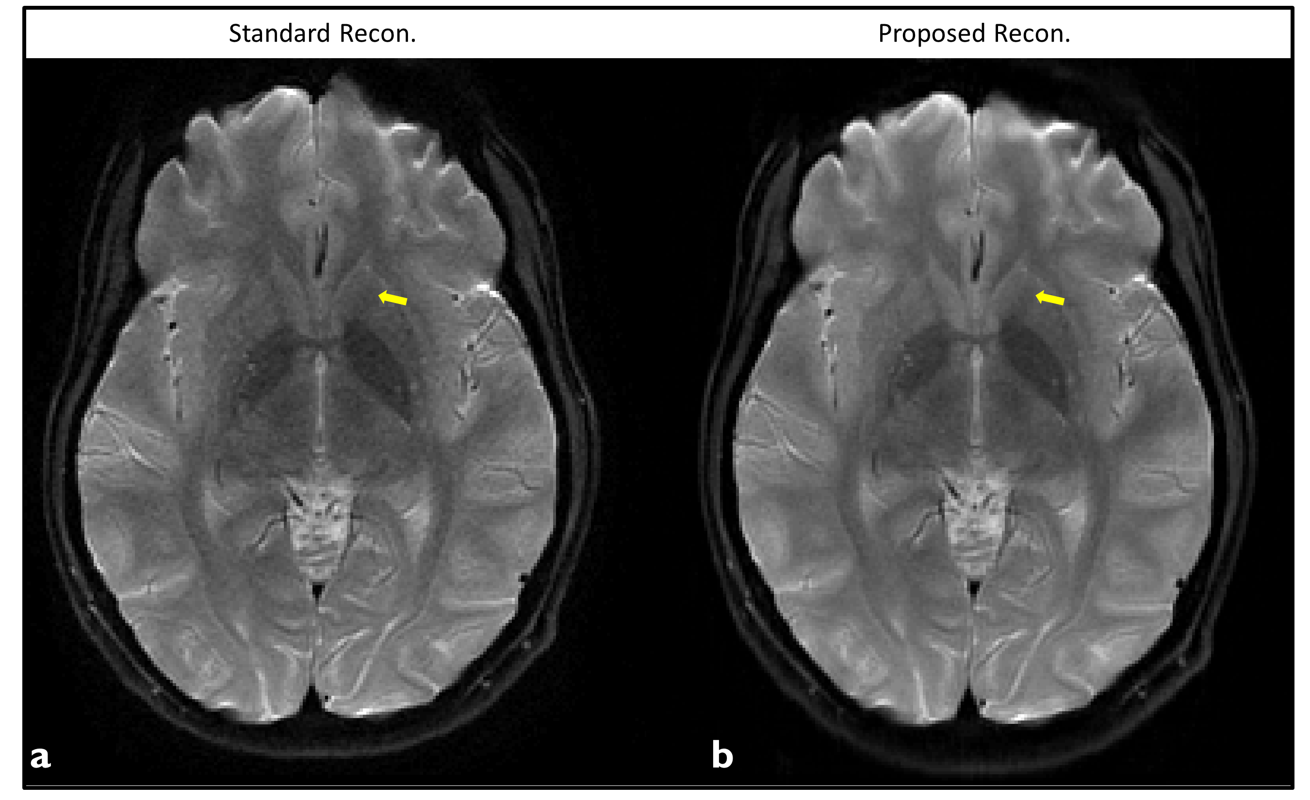
Fig. 3. A healthy volunteer with an image obtained by the standard reconstruction (a) and the proposed reconstruction (b). The yellow arrows identify the lateral margin of the caudate head which is better visualized with the proposed (b) compared to the standard reconstruction (a).